Jackson M.J. Micro and Nanomanufacturing
Подождите немного. Документ загружается.

352 Micro- and Nanomanufacturing
3 10 20 30
Negative Bias Time (minute)
(a)
N 0
n
-
b
i a s
•>.^'
..^-^^.
FV^rfi^
•S^v^yui^^^^^^^^'gg^y.g^^g:'
HjH
<•<
Bi^
iil
(b)
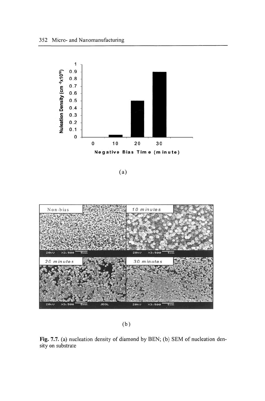
Fig. 7.7. (a) nucleation density of diamond by BEN; (b) SEM of nucleation den-
sity on substrate
Diamond Microcutting Tools 353
2100
O
o
¥
I 1800
d>
a
E
0)
1500
1900-'C
1700''C
5 10 15 20
Filament distance (mm)
25
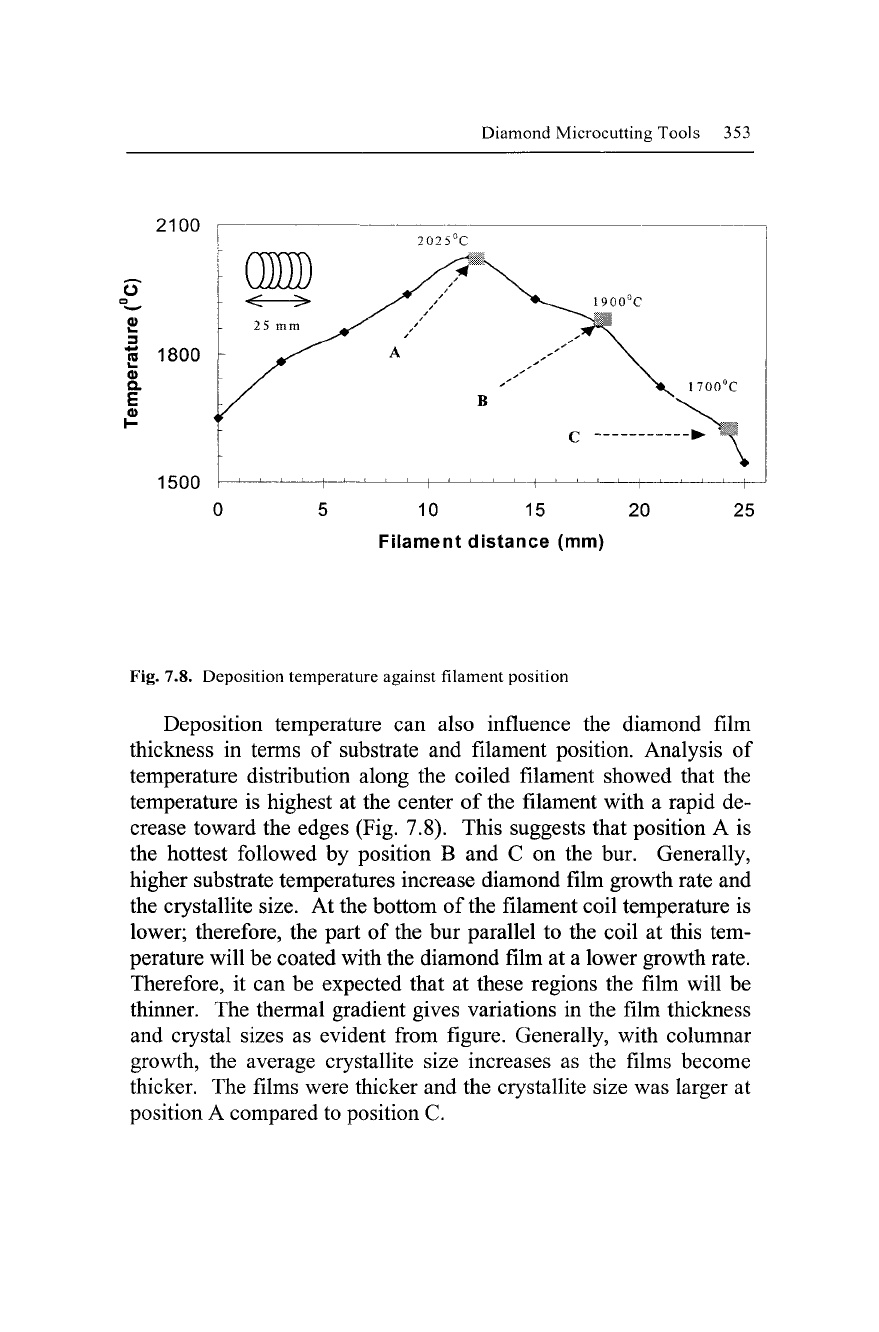
Fig. 7.8. Deposition temperature against filament position
Deposition temperature can also influence the diamond film
thickness in terms of substrate and filament position. Analysis of
temperature distribution along the coiled filament showed that the
temperature is highest at the center of the filament with a rapid de-
crease toward the edges (Fig. 7.8). This suggests that position A is
the hottest followed by position B and C on the bur. Generally,
higher substrate temperatures increase diamond film growth rate and
the crystallite size. At the bottom of the filament coil temperature is
lower; therefore, the part of the bur parallel to the coil at this tem-
perature will be coated with the diamond film at a lower growth rate.
Therefore, it can be expected that at these regions the film will be
thinner. The thermal gradient gives variations in the film thickness
and crystal sizes as evident from figure. Generally, with columnar
growth, the average crystallite size increases as the films become
thicker. The films were thicker and the crystallite size was larger at
position A compared to position C.
354 Micro- and Nanomanufacturing
Filament voltage
CH4/H2
Micro tool
Bias
voltage
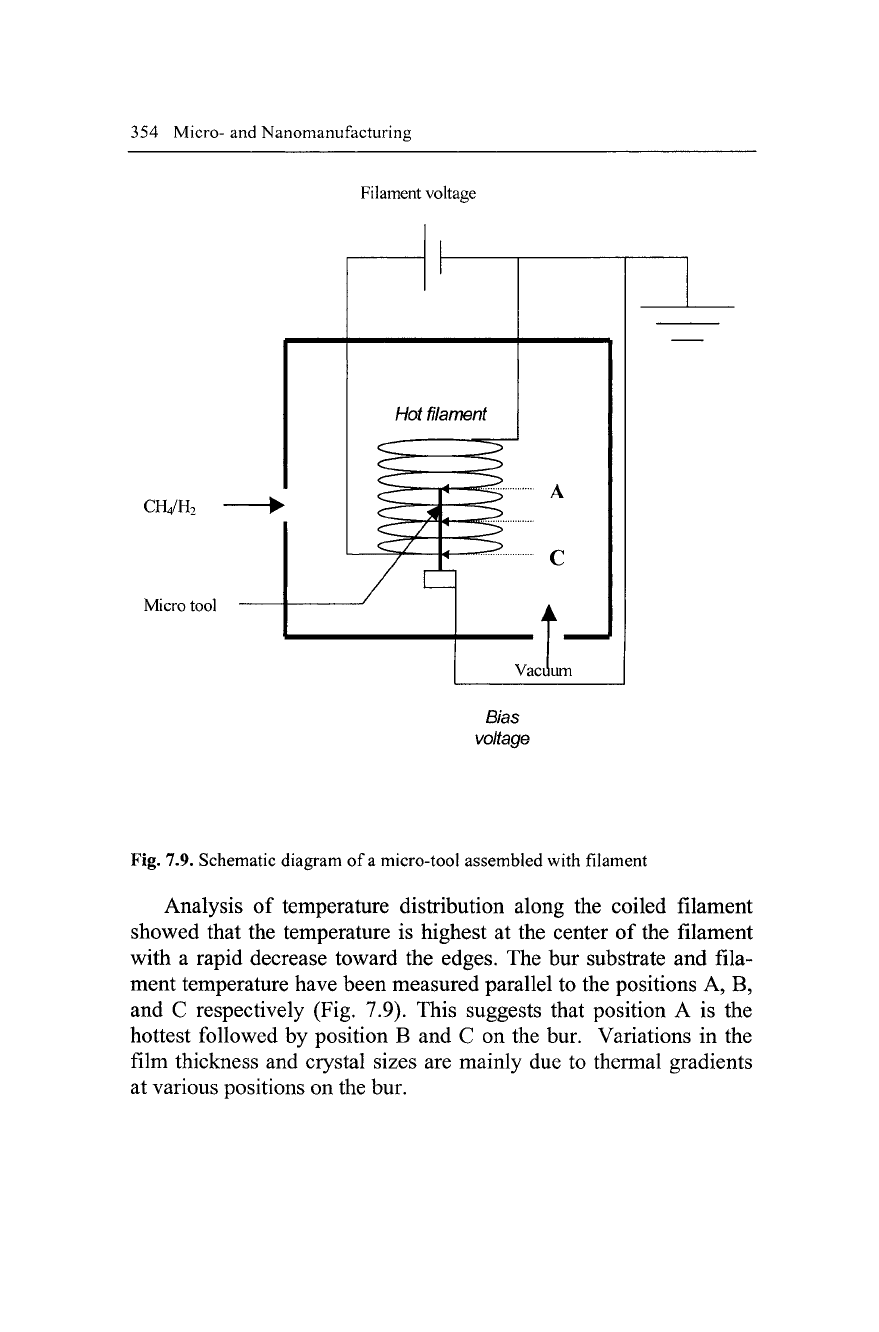
Fig. 7.9. Schematic diagram of
a
micro-tool assembled with filament
Analysis of temperature distribution along the coiled filament
showed that the temperature is highest at the center of the filament
with a rapid decrease toward the edges. The bur substrate and fila-
ment temperature have been measured parallel to the positions A, B,
and C respectively (Fig. 7.9). This suggests that position A is the
hottest followed by position B and C on the bur. Variations in the
film thickness and crystal sizes are mainly due to thermal gradients
at various positions on the bur.
Diamond Microcutting Tools 355
/
Diamond
Coiled nianicnt
Cross-section of
ihc tool
Cutting teeth
g[^;^^j Ctitling area
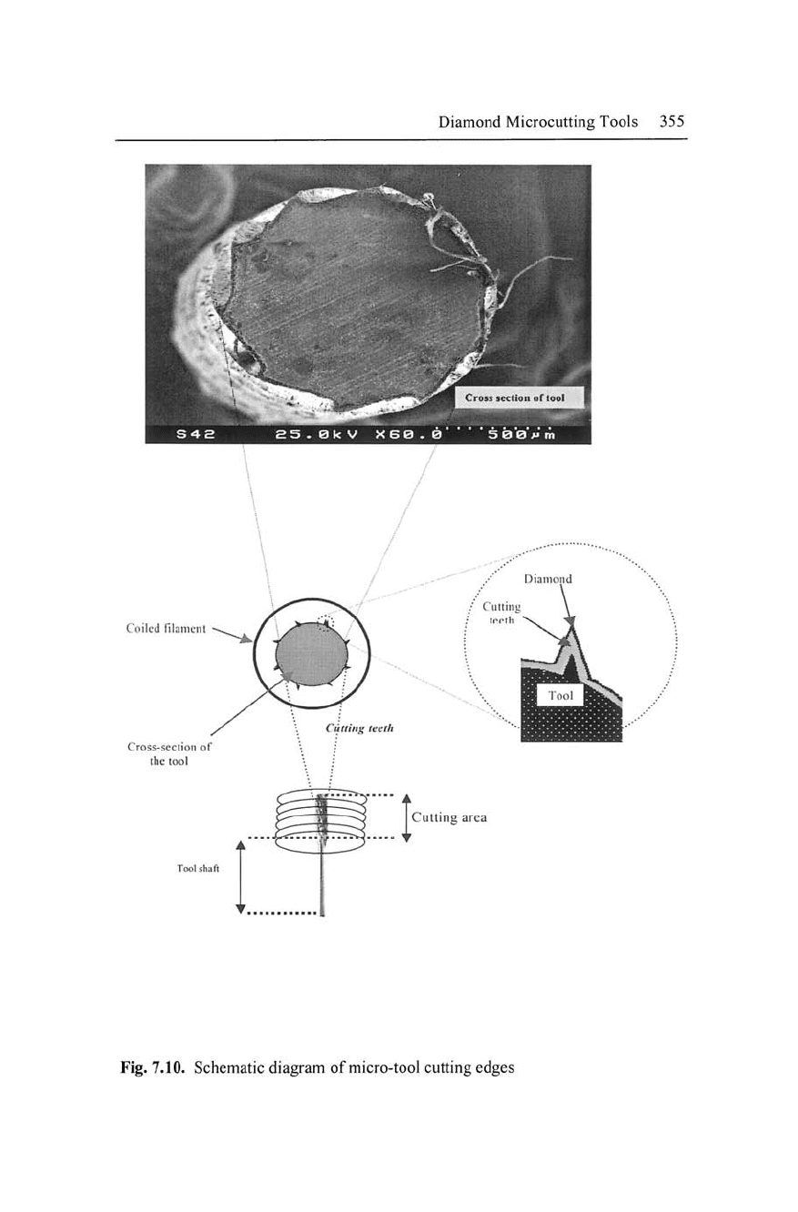
Fig. 7.10. Schematic diagram of micro-tool cutting edges
356 Micro- and Nanomanufacturing
7.9 Deposition on Three-Dimensional
Substrates
7.9.1 Diamond Deposition on Metallic (Molybdenum)
Wire
It is difficult to deposit CVD diamond onto cutting tools, which gen-
erally have a 3-D shape and possess complex geometry and sharp
edges,
using a single step growth process [76]. The cylindrical shape
wire,
which has complex geometry, can be used as a model applica-
tion for deposition of diamond on cutting tools such as microdrills
and micro-tools. The molybdenum (Mo) wires are deposited with
CVD diamond by modified vertical filament approach. After deposi-
tion time of
5
hours is continuous films of
5 |Lim
thick CVD diamond
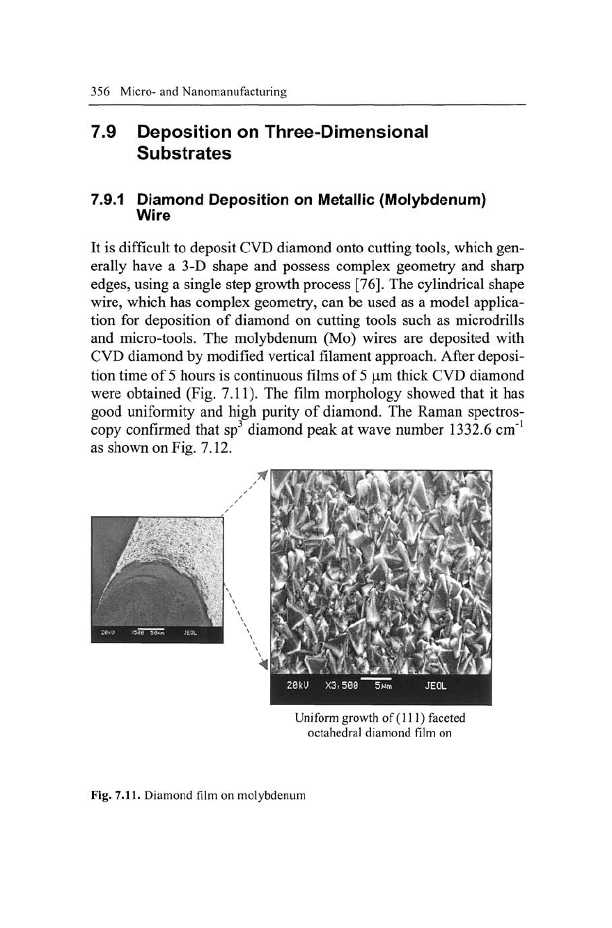
were obtained (Fig. 7.11). The film morphology showed that it has
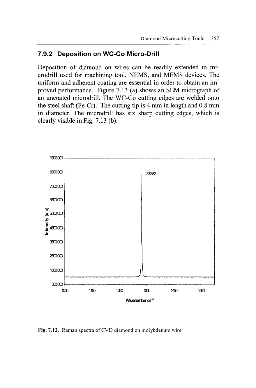
good uniformity and high purity of diamond. The Raman spectros-
copy confirmed that sp^ diamond peak at wave number 1332.6 cm"^
as shown on Fig. 7.12.
-1
n
i|p§
i^M^la^^'Qyliib^ttb 1
IPI
iM'il
^SnKrw
^^^^^^^^^^^^^^Q^^^^^^^^^^^^^^H
Uniform growth of (111) faceted
octahedral diamond film on
Fig. 7.11. Diamond film on molybdenum
Diamond Microcutting Tools 357
7.9.2 Deposition on WC-Co IVIicro-Drill
Deposition of diamond on wires can be readily extended to mi-
crodrill used for machining tool, NEMS, and MEMS devices. The
uniform and adherent coating are essential in order to obtain an im-
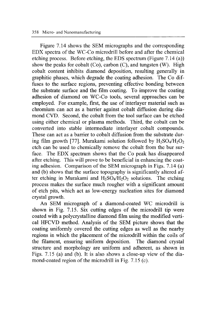
proved performance. Figure 7.13 (a) shows an SEM micrograph of
an uncoated microdrill. The WC-Co cutting edges are welded onto
the steel shaft (Fe-Cr). The cutting tip is 4 mm in length and 0.8 mm
in diameter. The microdrill has six sharp cutting edges, which is
clearly visible in Fig. 7.13 (b).
9800.000
88DQ000
laOQOOO
soaooo
1080
1193
1293
1380
V\£MeruTiDercnnr^
1490 1550
Fig. 7.12. Raman spectra of CVD diamond on molybdenum wire
358 Micro-and Nanomanufacturing
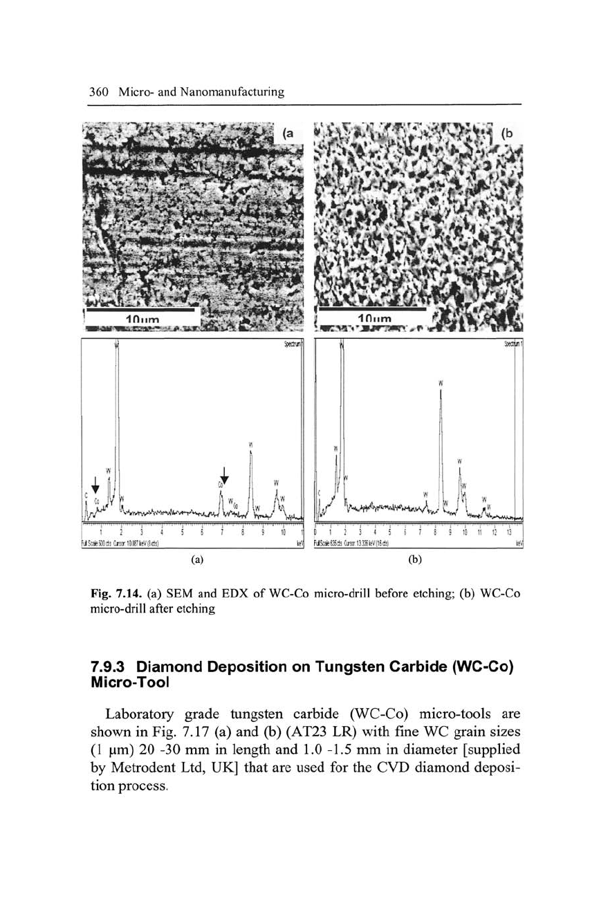
Figure 7.14 shows the SEM micrographs and the corresponding
EDX spectra of the WC-Co microdrill before and after the chemical
etching process. Before etching, the EDS spectrum (Figure 7.14 (a))
show the peaks for cobalt (Co), carbon (C), and tungsten (W). High
cobalt content inhibits diamond deposition, resulting generally in
graphitic phases, which degrade the coating adhesion. The Co
dif-
fuses to the surface regions, preventing effective bonding between
the substrate surface and the film coating. To improve the coating
adhesion of diamond on WC-Co tools, several approaches can be
employed. For example, first, the use of interlayer material such as
chromium can act as a barrier against cobalt diffusion during dia-
mond CVD. Second, the cobalt from the tool surface can be etched
using either chemical or plasma methods. Third, the cobalt can be
converted into stable intermediate interlayer cobalt compounds.
These can act as a barrier to cobalt diffusion from the substrate dur-
ing film growth [77]. Murakami solution followed by H2SO4/H2O2
etch can be used to chemically remove the cobalt from the bur sur-
face.
The EDX spectrum shows that the Co peak has disappeared
after etching. This will prove to be beneficial in enhancing the coat-
ing adhesion. Comparison of the SEM micrograph in Figs. 7.14 (a)
and (b) shows that the surface topography is significantly altered af-
ter etching in Murakami and H2SO4/H2O2 solutions. The etching
process makes the surface much rougher with a significant amount
of etch pits, which act as low-energy nucleation sites for diamond
crystal growth.
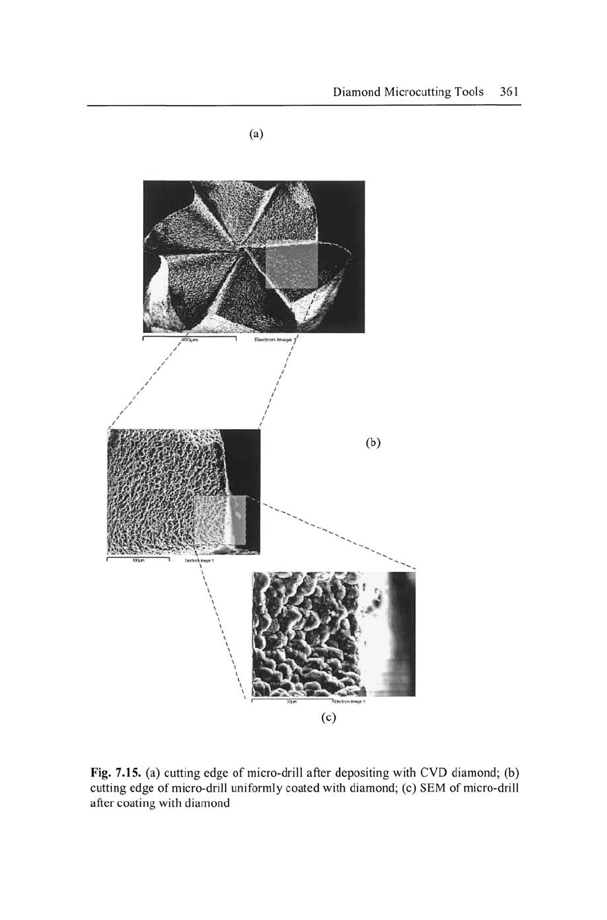
An SEM micrograph of a diamond-coated WC microdrill is
shown in Fig. 7.15. Six cutting edges of the microdrill tip were
coated with a polycrystalline diamond film using the modified verti-
cal HFCVD method. Analysis of the SEM picture shows that the
coating uniformly covered the cutting edges as well as the nearby
regions in which the placement of the microdrill within the coils of
the filament, ensuring uniform deposition. The diamond crystal
structure and morphology are uniform and adherent, as shown in
Figs.
7.15 (a) and (b). It is also shows a close-up view of the dia-
mond-coated region of the microdrill in Fig. 7.15 (c).
Diamond Microcutting Tools 359
Fig. 7.13. Tip and cutting edge of micro-drill
Typically the crystallite sizes are of the order of 5-8 |Lim. The
visibly adherent diamond coatings on the WC-Co microdrills consist
of mainly (111) faceted diamond crystals. The design of the filament
and substrate in the reactor offer the possibility of uniformly coating
even larger diameter cylindrical substrates.
Raman analysis was performed in order to evaluate the diamond
carbon-phase quality and film stress in the deposited films. The
Raman spectrum in Fig. 7.16 shows a single peak at 1335 cm"^ for
the tip of the diamond-coated microdrill. The Raman spectrum also
gives information about the stress in the diamond coatings. The dia-
mond peak is shifted to a higher wave number of 1335 cm"^ than that
of natural diamond peak 1332 cm"^ indicating that stress, which is
compressive in nature, exists in the resultant coatings [78].
360 Micro- and Nanomanufacturing
''• iminiu|.n..,..., , ,
,^
I
2 3
MSafeSDOdiOnorlOganeVQcl:)
5
6 J 3 9 10 1|
PL
^'^J
^^'•XVV.UYUJ
D
1 2 3 4 5 6 7
Fiifc^ES5dsCtfarn3Sl;V[l6d5)
9
10 11 12 t3
(a)
(b)
Fig. 7.14. (a) SEM and EDX of WC-Co micro-drill before etching; (b) WC-Co
micro-drill after etching
7.9.3 Diamond Deposition on Tungsten Carbide (WC-Co)
IVIicro-Tool
Laboratory grade tungsten carbide (WC-Co) micro-tools are
shown in Fig. 7.17 (a) and (b) (AT23 LR) with fine WC grain sizes
(1 |j,m) 20 -30 mm in length and 1.0 -1.5 mm in diameter [supplied
by Metrodent Ltd, UK] that are used for the CVD diamond deposi-
tion process.
Diamond Microcutting Tools 361
(a)
(c)
Fig. 7.15. (a) cutting edge of micro-drill after depositing with CVD diamond; (b)
cutting edge of micro-drill uniformly coated with diamond; (c) SEM of micro-drill
after coating with diamond