Czichos H., Saito T., Smith L.E. (Eds.) Handbook of Metrology and Testing
Подождите немного. Документ загружается.

238 Part B Chemical and Microstructural Analysis
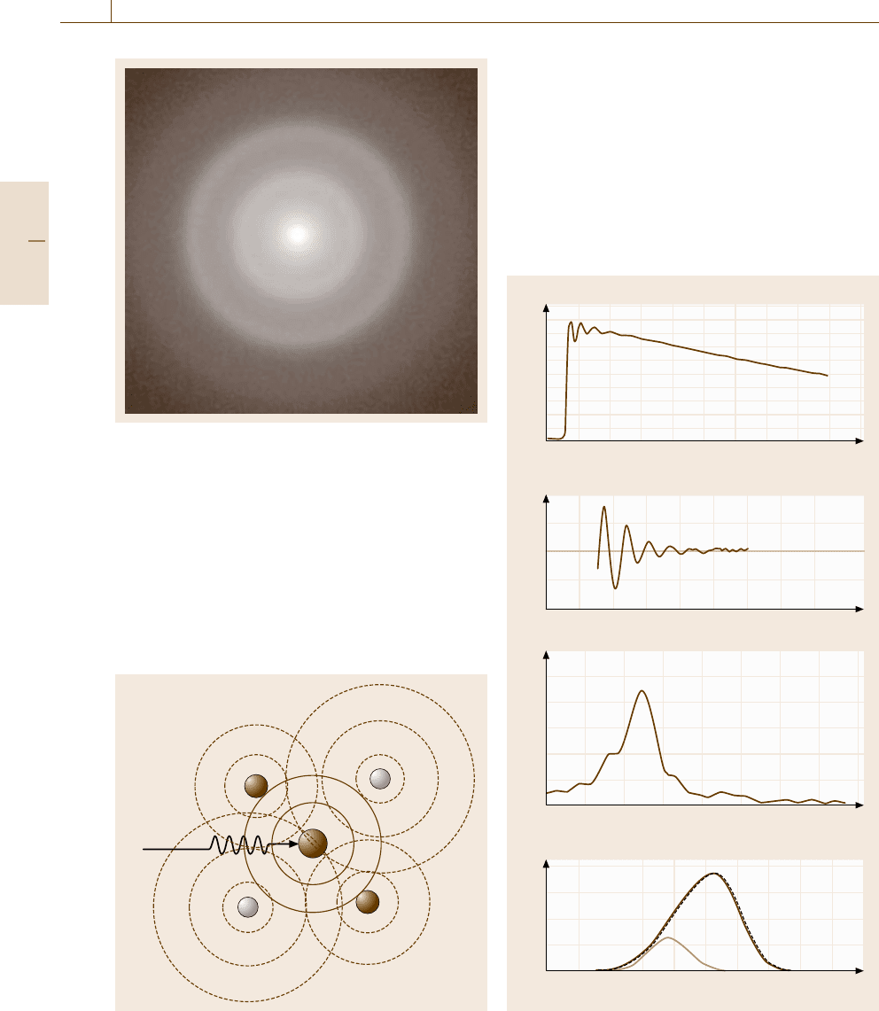
Fig. 5.45 Halo pattern of selected area electron diffraction
from an amorphous carbon film
quantity r[ρ(r) −ρ
0
] is given by the inverse Fourier
transformation of the quantity on the left, which is mea-
surable in diffraction experiments.
For multicomponent solids, the similarly defined
partial distribution functions are deduced from diffrac-
tion patterns if one uses techniques such as anomalous
dispersion that allow one to enhance the atomic scat-
tering factor f
j
of only a specific element by tuning
e
–
hξ
Fig. 5.46 Interference of core-excited electron waves scat-
tered by surrounding atoms giving rise to EXAFS
the wavelength of the x-rays. The atomic scattering fac-
tor for neutrons is generally quite different from that
for x-rays (Sect. 5.1.1). So coupling neutron diffraction
with x-ray diffraction, we could extend the range of ele-
ments that can be studied by using diffraction methods.
Extended X-Ray Absorption Fine Structure
(EXAFS)
X-ray absorption occurs when the photon energy ex-
ceeds the threshold for excitations of core electrons
a)
b)
c)
d)
2
1
0
0.3
0.2
0.1
0
0.1
0
–0.1
20
10
0
9.6 9.8 10.0 10.2 10.4 10.6
41216
Δν(k)t
k(Å
–1
)
206
632
a-Mg
70
Zn
30
ln(I
0
/I)
E(keV)
r (Å)
r (Å)
Partial RDF (r)
Zn–Mg
Zn–Zn
|(r)|
8
48
Fig. 5.47a–d Analysis of short-range order by extended
x-ray absorption fine structure. After [5.35]
Part B 5.2

Nanoscopic Architecture and Microstructure 5.3 Lattice Defects and Impurities Analysis 239
to unoccupied upper levels. The core absorption spec-
trum has a characteristic fine structure extending over
hundreds of eV above the absorption edge. The undu-
lating structures within ≈ 50 eV of the edge is referred
to as x-ray absorption near-edge structures (XANES) or
near-edge x-ray absorption fine structures (NEXAFS),
whereas the fine structures beyond ≈ 50 eV are called
the extended x-ray absorption fine structures (EXAFS).
The EXAFS is brought about by interference of the
excited electron wave primarily activated at a core
with the secondary waves scattered by the surround-
ing atoms (Fig. 5.46). Therefore, the EXAFS contains
information on the local atomic arrangement, neigh-
bor distances and the coordination numbers, around the
atoms of a specific element responsible for the core
excitation absorption. Figure 5.47a shows an experi-
mental x-ray absorption spectrum near the zinc K-edge
of an amorphous Mg
70
Zn
30
alloy, and Fig. 5.47bthe
oscillatory part of the EXAFS obtained by subtracting
the large monotonic background in (a). Assuming ap-
propriate phase shifts on electron scattering, we obtain
Fig. 5.47c, a Fourier transform of a function modified
from (b), from which we deduce Fig. 5.47d, the par-
tial distribution function around Zn atoms. The area of
deconvoluted peaks in (d) gives the coordination num-
ber for the respective atom pair. The element-selectivity
of the EXAFS method provides a unique tool that en-
ables one to investigate the local order for different
atomic species in alloys, regardless of the long-range
order or the crystallinity and amorphousness of the
sample.
X-Ray Photoemission Spectroscopy (XPS)
Though x-ray photoemission spectroscopy (XPS)is
surface sensitive, it may be used for studies of very
short-range order around atoms. The energy of the pho-
toelectrons measured relative to the Fermi level of the
sample directly reflects the energy of the core elec-
trons. The core level is slightly affected by how much
electronic charge is distributed on the nucleus in the
solid (chemical shift): the more negatively charged, the
higher the core level and hence the smaller the pho-
toelectron energy. The spectral peak position and its
intensity in XPS thus indicate the character of chemical
bonding or the degree of chemisorption to the selec-
tively probed atoms.
Raman Scattering
Since the principle of Raman scattering has been
described already (Sect. 5.1.2), we only mention an ap-
plication of Raman scattering to studies of structural
order in crystalline and amorphous solids. From the
energy of Raman-active optical phonons, the material
phases present in the sample can be determined. The
selection rule in infrared absorption based on the trans-
lational symmetry of the crystal allows only optical
modes near k ≈0 in the Brillouin zone. Similarly the
translational symmetry restricts the Raman scattering to
limited modes. The presence of defects or disorder in
crystals breaks the translational symmetry of the lattice,
so that translationally forbidden Raman modes become
active and detectable as an increase of the extended
lattice phonon signal. Raman scattering is one of the
standard methods for structural assessment of graphitic
or amorphous carbon [5.36]. This is owing to the fact
that the visible spectral region, the most widely used
for Raman scattering experiments, is electronically res-
onant with graphitic crystals, enhancing the scattering
signals. Two broad spectral bands, called G and D, re-
spectively corresponding to crystalline and disordered
graphitic phases are well separated for quantitative anal-
ysis of disorder.
5.3 Lattice Defects and Impurities Analysis
This section deals with methods for the study of atom-
istic structural defects and impurities in regular lattices.
Section 5.3.1 covers point defects including vacancies,
interstitial atoms, defect complexes, defect clusters, and
impurities. Section 5.3.2 deals with extended defects in-
cluding dislocations, stacking faults, grain boundaries,
and phase boundaries.
The sensitivity (or field of view) of the methods de-
scribed here is not necessarily high (wide) enough for
detecting a low density of defects, so in many cases the
defects must be intentionally introduced into the sample
by some means. For intrinsic point defects such as va-
cancies and self-interstitials, the most common method
is to irradiate the sample with high energy particles such
as MeV electrons and fast neutrons. For dislocations, the
sample may have to be deformed plastically. However,
one should always pay careful attention to the possibil-
ity that the primary defects may react to form complexes
with themselves or impurities and the plastically de-
formed samples may contain point defects as well.
Part B 5.3
240 Part B Chemical and Microstructural Analysis
5.3.1 Point Defects and Impurities
Diffraction and Scattering Methods
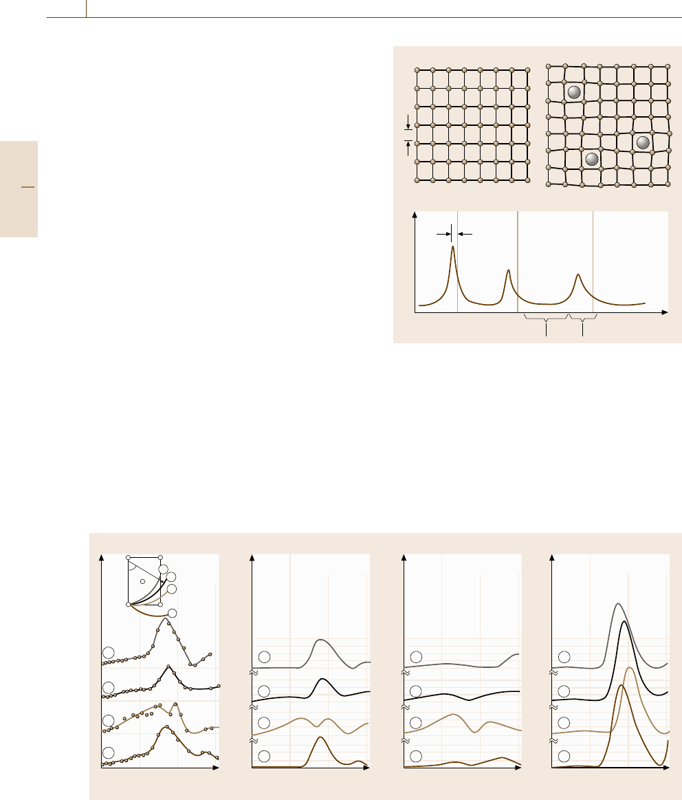
X-Ray Diffuse Scattering. The lattice distortion in-
duced by the presence of point defects and impurities,
as illustrated in Fig. 5.48a, causes a diffuse scattering
in x-ray diffraction as well as a shift of the diffraction
peaks. The diffuse scattering from such imperfect crys-
tals generally has two components (Fig. 5.48b): Huang
scattering, which appears near the diffraction peaks and
represents a long-range strain field around the defects
and impurities, and Stokes–Wilson scattering, which
extends the between diffraction peaks and represents
a short-range atomic configuration and hence its dis-
tribution in the reciprocal space reflects the symmetry
of the strain field associated with the defect and im-
purity. Figure 5.49 shows the Stokes–Wilson scattering
from an electron-irradiated Al sample [5.37] experi-
mentally deduced by carefully subtracting a background
of different origins one or two orders of magnitude
larger than the signal. The experimental profiles meas-
ured along different trajectories in the reciprocal space
agree most with the theoretical curves predicted by self-
interstitials split along the 100direction.
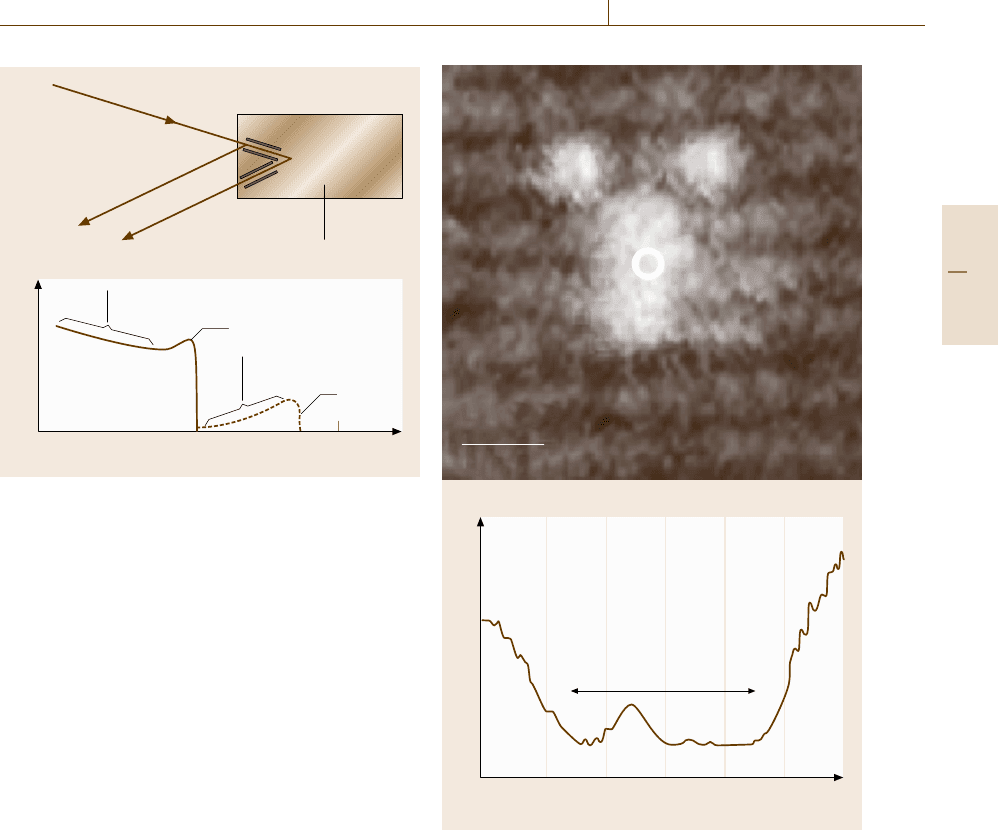
Rutherford Back Scattering. When light ions such as
H
+
and He
+
in energy of a few MeV are incident
on a solid, some of them are classically scattered by
atoms in the solid, reflected backward and emitted from
the sample, which is called Rutherford back scattering
(Fig. 5.50a). These backscattered ions lose an energy
20
10
0
10
0
10
0
10
0
Cross section (arb. units)
15 30 45 60
Cross section (arb. units)
15 30 45 60
20
10
0
10
0
10
0
10
0
Cross section (arb. units)
15 30 45 60
20
10
0
10
0
10
0
10
0
Cross section (arb. units)
15 30 45 60
Scattering angle
ε
Scattering angle
ε
ε
022 222
0
200
111
Experi-
mental
Scattering angle
ε
Scattering angle
ε
〈100〉-split
Octahedral Tetrahedral
4
3
2
1
4
3
2
1
3
2
1
4
3
2
1
4
3
2
1
4
Fig. 5.49 Structural determination of selfinterstitial in Al by x-ray diffuse scattering (after [5.37, p. 87])
a) b)
c)
a
I(K)
Δa
(K)
Stokes–Wilson scattering Huang scattering
Fig. 5.48 (a) A perfect lattice and (b) a lattice distortion
induced by point defects reflected in the x-ray diffraction
(c) as shifts of Bragg peaks, Stokes–Wilson scattering for
the short-range elastic fields and Huang scattering for the
long-range fields
depending on the atoms with which the ions collide
and the collision parameters: the heavier the atoms,
the larger the energy loss in an elastic collision. Fig-
ure 5.50b shows a schematic diagram of the energy
distribution of the backscattered ions. If the sample con-
Part B 5.3
Nanoscopic Architecture and Microstructure 5.3 Lattice Defects and Impurities Analysis 241
a)
b)
E
0
H
+
He
+
E
Channeling
sample
Back scattered ions
Scattered from
deep positions
Matrix
Diffusion profile
Heavy
impurity
Energy of back scattered ions
E
kE
0
E
0
Fig. 5.50 (a) Light ions with primary energy E
0
are inci-
dent along a channel direction and inelastically scattered
backward (Rutherford back scattering) with a reduced en-
ergy E. (b) The energy distribution of back scattered ions.
When heavy impurities are present in the channels, a dis-
tribution arises for smaller losses
tains impurity atoms much heavier than the atoms in the
matrix, an additional component due to these impurities
appears between the primary ion energy E
0
and the en-
ergy kE
0
(k < 1) representing the energy of an ion that
has collided only once with a matrix atom. This pro-
vides a means to measure the depth profile of diffusant
atoms from the surface. Furthermore, in single crys-
tal samples, ions can penetrate, without scattered, over
an anomalously large distance along specific crystallo-
graphic directions called channels. If an interstitial atom
is present in the channel, the number of backscattered
ions incident in the channeling directions is increased.
The out-going backscattered ions also may feel the
channeling effect, which enables the method of double
channeling that allows determination of the crystallo-
graphic position of the interstitial atoms in the lattice.
Microscopic Methods
Scanning Tunneling Microscopy (STM). One of the
microscopic methods currently available for direct
observations of point defects is scanning tunneling
microscopy (STM). As explained in Sect. 5.1.2, the tun-
neling current reflects the local density of states (LDOS)
of electrons at the tip position above the surface. There-
fore, if the wave functions associated with defects even
–1.5 –1.0 –0.5 0.0 0.5 1.0 1.5
(dI/dV)/(I/V)
As-related defect
in LT-GaAs
Sample bias (V)
E
g
1nm
a)
b)
Fig. 5.51 (a) Scanning tunneling microscopic image of an
arsenic-related point defect beneath the surface and (b)
the local density of states measured at the defect posi-
tion [5.38]
beneath the surface extend out of the surface, we can im-
age the defect contrasts using STM. Figure 5.51 shows
an STM image obtained for an arsenic-related point de-
fect located below the surface [5.38]. Defect studies by
STM are in the early stages of promising applications
for various material systems.
High-Angle Annular Dark-Field STEM (HAADF-STEM).
An ambitious approach for rather indirect observa-
tions of point defects is high-angle annular dark-field
STEM (HAADF-STEM). The progress of improve-
Part B 5.3

242 Part B Chemical and Microstructural Analysis
ment of spherical aberration has achieved successful
atomic-scale imaging of impurity atoms [5.39]andva-
cancies [5.40] in each atomic column. Nevertheless,
elaborate image simulations are necessary for decisive
conclusions to be drawn.
Spectroscopic Methods
Relaxation Spectroscopy.
Mechanical Spectroscopy. When the lattice distortion
induced by defects and impurities is anisotropic, such
as the strain ellipsoid depicted in Fig. 5.52a, the stress-
induced reorientation (ordering) of the anisotropic
distortion centers can be detected as the Snoek relax-
ation (peak), which allows one to evaluate the λ tensor
(Fig. 5.52a) and the relaxation time. The equilibrium
anelastic strain ε
(eq)
a
is given by
ε
(eq)
a
=
C
0
v
0
σ
3kT
⎡
⎣
p
λ
(p)
2
−
1
3
p
λ
(p)
2
⎤
⎦
(5.26)
with
λ
(p)
=
α
(p)
1
2
λ
1
+
α
(p)
2
2
λ
2
+
α
(p)
3
2
λ
3
, (5.27)
where C
0
is the concentration of the anisotropic distor-
tion centers, v
0
is the molar volume, and α
(p)
1
, α
(p)
2
and
α
(p)
3
are the direction cosines between the stress axis
and the three principal axes of the λ tensor. A typical
application is to carbon, nitrogen and oxygen intersti-
tial atoms in bcc metals which occupy one of three
equivalent interstitial sites (octahedral sites) inducing
tetragonal distortions in different crystallographic di-
rections. The concept of the Snoek relaxation is also
applied to the hydrogen internal friction peak in amor-
phous alloys.
A pair of differently sized solute atoms in the solid
solution in the nearest-neighbor configuration can be an
a) b)
λ
1
λ
2
λ
3
Fig. 5.52 (a) The strain ellipsoid, (b) hydrogen atoms in
a bent specimen
anisotropic distortion center. For such cases, the anelas-
tic relaxation (peak) associated with the stress-induced
reorientation (ordering) of the anisotropic distortion
centers is referred to as the Zener relaxation (peak).
The Zener relaxation provides a tool for the study
of atomic migration in substitutional alloys, without
radioisotopes, at temperatures much lower than in con-
ventional diffusion methods.
When the lattice distortion induced by an applied
stress is not homogeneous, a stress-induced redistribu-
tion of interstitial impurities takes place resulting in an
anelastic strain (Fig. 5.52b). Such relaxation is referred
to as the Gorsky relaxation and provides a tool for the
study of long-range migration of interstitial impurities.
The relaxation time τ and the relaxation strength Δ
E
for
a specimen of rectangular section are given by
τ =d
2
/π
2
D (5.28)
and
Δ
E
=(C
0
v
0
E/9kTβ)(tr λ)
2
, (5.29)
where D is the diffusion coefficient of the impurity, β
is the number of interstitial sites per host atom, and d
and E are the thickness and the Young’s modulus of
the specimen, respectively. This method is applied to
hydrogen diffusion because of the rapid diffusion rate.
Dielectric or Magnetic Relaxation. Quite similar re-
laxation processes occur in the dielectric or magnetic
response of defects and impurities. Tightly bound pairs
in ionic crystals have in some cases, such as in F
A
centers (pairs of anion vacancy and isovalent cation im-
purity atom) in alkali halides, an electric dipole moment
along the bond orientation [5.41]. Therefore, respond-
ing to the application and the removal of an electric
field, the dipole moment reorients by a movement of
constituent atoms, which is detected as a dielectric re-
laxation. Magnetic aftereffects are also brought about
by movements of point defects and impurities. Since
interstitial atoms in bcc metals diffuse in response to
an external magnetic field through magnetostriction, es-
sentially by the same mechanism as the Snoek effect,
the Snoek relaxation can be studied by measurements
of the magnetic relaxation.
Noise Spectroscopy. Relaxation and fluctuation repre-
sent two aspects of statistical behaviors of a physical
quantity q that is under stochastic random forces [5.42,
43]. Debye-type relaxation behavior, which is charac-
terized by a time constant τ
r
with which the quantity
Part B 5.3
Nanoscopic Architecture and Microstructure 5.3 Lattice Defects and Impurities Analysis 243
responds to a stepwise stimulation, is described by an
autocorrelation function
ϕ
q
(τ) =
q(t)q(t +τ)
∝exp
(
−τ/τ
r
)
. (5.30)
Generally, the autocorrelation function in the time
domain is related to the fluctuation power spectrum
S
q
(ω) in the frequency domain through the Wiener–
Khintchine theorem
S
q
(ω) =4
∞
0
ϕ
q
(τ)cosωτ dτ. (5.31)
Therefore, from measurements of the fluctuation
power spectrum without intentional stimulation, we can
deduce the relaxation time. The fluctuations may be de-
tected in the form of electrical noise. The source of the
noise varies from case to case. In semiconductors con-
taining impurities or defects forming deep levels in the
band gap, we may observe a generation–recombination
(g–r) noise arising from the fluctuations of carrier den-
sity on random exchange of carriers between the deep
levels and the band states. Since the fluctuation or re-
laxation time constant is determined, in many cases, by
the rate of thermal activation of carriers from the deep
level to the band, measurements of the noise intensity as
a function of temperature give spectroscopic informa-
tion about the depth of the deep level from the relevant
band edge. Since such noise becomes more significant
as the number of carriers becomes smaller, noise spec-
troscopy is useful for studies of small systems such as
nanostructures.
Infrared Absorption Spectroscopy (IRAS). The optical
photon energy that IRAS detects is determined by the
atomic mass and the strength of the bonds with the
surrounding atoms. Therefore, the local vibrational fre-
quency of impurity atoms of mass similar to the matrix
easily overlap with the lattice phonon band and form
a resonance mode that is hard to detect in IRAS due
to the strong background. Since the local vibrational
modes (LVM) associated with intrinsic point defects be-
have similarly, few studies have been reported for IRAS
experiments of LVM of intrinsic point defects. (This is
also due to the fact that simple intrinsic point defects
are usually easily reconstructed to secondary defects
such as complexes with impurities.) Instead, IRAS ex-
periments on light elements such as oxygen in Si have
been intensively carried out and the experimental data
documented. Hydrogen is an exceptionally light ele-
ment which is used indirectly to detect point defects,
such as self-interstitials and vacancies in Si for exam-
ple [5.44], through the LVM associated with hydrogen
defect complexes. The detection limit of impurities by
IRAS is ≈0.01%.
Raman Spectroscopy. Since Raman scattering is a two-
photon process, the scattering signal is generally very
weak, as mentioned in Sect. 5.1.2. Therefore, Raman
scattering has so far rarely been used for studies of lo-
cal vibrations associated with defects at low density.
However, when resonant Raman scattering is used, the
detection limit could be reduced down to several ppb
under good conditions if the fluorescence or lumines-
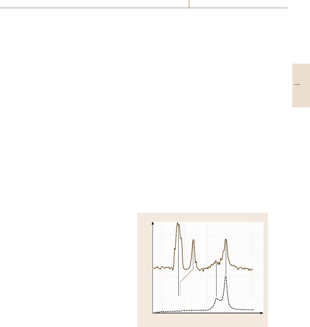
cence background is low. Figure 5.53 demonstrates the
power of resonant Raman scattering, which enables
detection of the LVM of As-related point defects in
GaAs [5.45]. Generally, for LVM to be detected clearly
by Raman scattering, the vibrational energy of the LVM
must be well separated from those of the resonance
modes (the local mode in resonance with the lattice
phonon bands) which have relatively large intensities.
The F-center (a neutral anion vacancy trapping an elec-
tron) in KI [5.46] is such an exceptional case in which
the masses of potassium and iodine are so different that
the gap between the acoustic and the optic bands is
wide enough to accommodate the gap mode induced by
defects.
4000
3000
2000
1000
Raman intensity (units)
150 200 250 300 350
Raman shift (cm
–1
)
LT-GaAs at 200K
Local
vibrational
modes
(TO)(LO)
Nd-YAG
(1064 nm)
He–Ne
(514.5 nm)
Fig. 5.53 Defect-selective resonant Raman spectra of
a low-temperature grown GaAs crystal measured at 200 K
by using two different excitation lasers. The wavelength
of the Nd:YAG laser (1064 nm) is resonant with the intra-
center electronic excitation of the As-related EL2 centers.
(Courtesy of Dr. A. Hida)
Part B 5.3
244 Part B Chemical and Microstructural Analysis
Visible Photoabsorption and Photoluminescence (PL).
In contrast to IRAS and Raman scattering, photoab-
sorption and photoluminescence (PL) in the visible to
near-infrared region are widely used for studies of de-
fects in nonmetallic solids. If the sample yields strong
PL, the sensitivity can be as high as ≈ 0.1 ppm. The
spectrum of PL is complementary to the photoabsorp-
tion spectrum, with both representing electronic dipolar
transitions. Defects in semiconductors and insulators
often introduce electronic states in the band gap. The
classic examples are color centers in alkali halides.
When the electronic state is deep in the gap, the elec-
tronic wave function tends to be localized in space. In
such cases, the electronic energy is sensitive to the local
arrangement of atoms at the defect. This effect is re-
ferred to as strong electron lattice coupling, the degree
of which is reflected in the optical transition spectra.
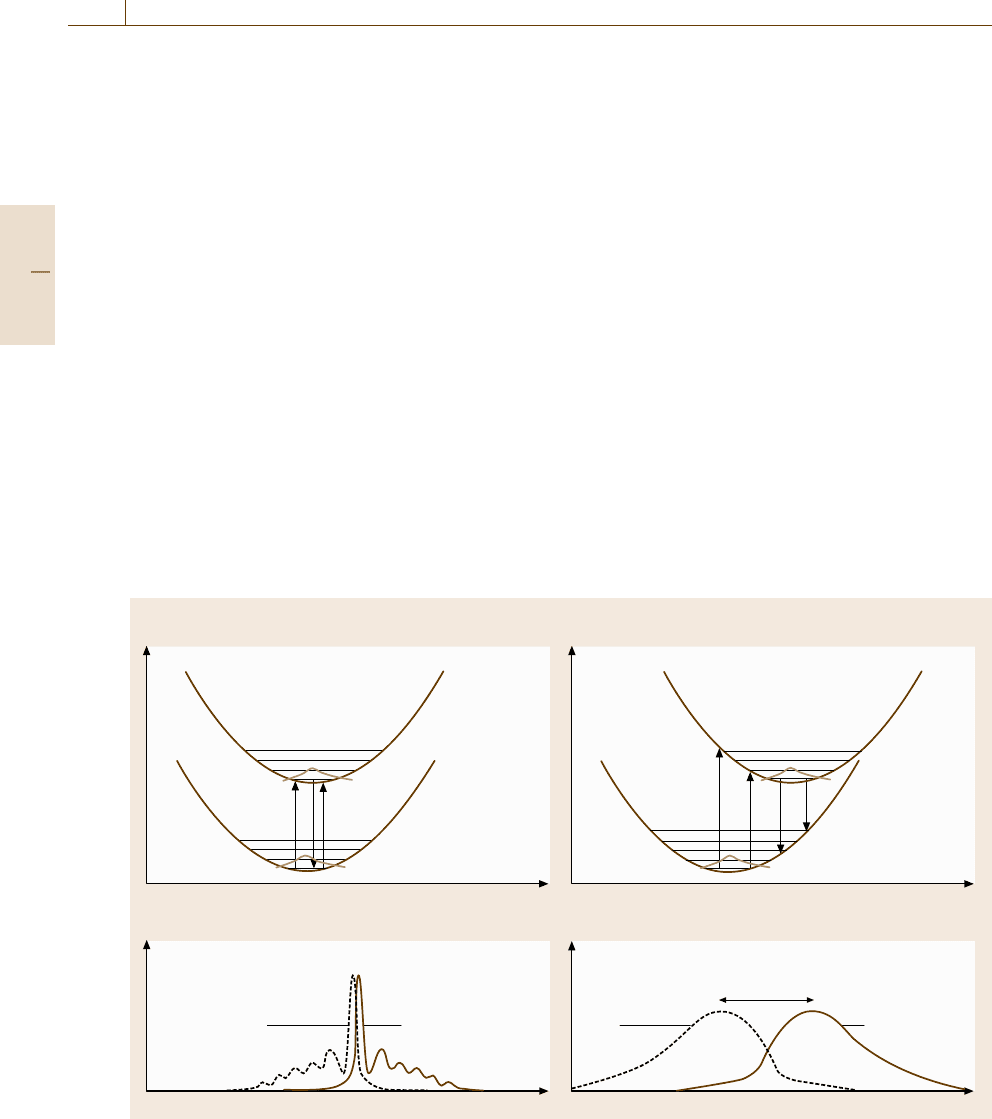
Figure 5.54a,b illustrates the situations by using config-
uration coordinate diagrams: each curve indicates the
(adiabatic) potential energy of the system, consisting of
the electronic energy and the nuclear potential energy,
drawn as a function of the nuclei positions in the sys-
tem that are expressed by one-dimensional coordinate
a)
Adiabatic potential
Excited state
Ground state
Configuration coordinate
b)
Adiabatic potential
Excited state
Ground state
Configuration coordinate
c)
Intensity
Photon energy
d)
Intensity
Photon energy
Photo-
luminescence
Photo-
absorption
Photo-
luminescence
Photo-
absorption
Stokes shift
Fig. 5.54a–d Configuration coordinate diagrams and corresponding spectra of photoabsorption and photoluminescence
(PL) for two cases in which electron–lattice coupling is weak (a),(c) and strong (b),(d). Note that the absorption and PL
spectra are almost mirror symmetric, regardless of the electron–lattice coupling
(configuration coordinate). The lower curves represent
the adiabatic potentials for the electronically ground
state while the upper curves the adiabatic potentials for
an electronically excited state. The ladder bars indi-
cate the vibrational states in each electronic state. If the
electron–lattice coupling is weak (Fig. 5.54a), the stable
configurations do not differ much between the ground
state and the excited state. In this case, the optical tran-
sitions take place with spectra characterized by a sharp
zero-phonon line reflecting transitions with no phonon
involved, as shown in Fig. 5.54c. If the electron–lattice
coupling is strong (Fig. 5.54b), the stable configura-
tions differ considerably between the ground state and
the excited state. The consequences are a broad band-
width and a large Stokes shift (red shift) as shown in
Fig. 5.54d.
Generally, the dipolar optical transition is not as ef-
ficient as competitive nonradiative processes, including
Auger processes, multiphonon emission processes, and
thermally activated opening of different recombination
channels. The last process is common in many systems,
so samples for PL measurements must usually be cooled
to a low temperature (≤20 K) to obtain a detectable
Part B 5.3
Nanoscopic Architecture and Microstructure 5.3 Lattice Defects and Impurities Analysis 245
PL intensity. The sample cooling is also beneficial for
both photoabsorption and PL measurements to resolve
the spectral fine structures.
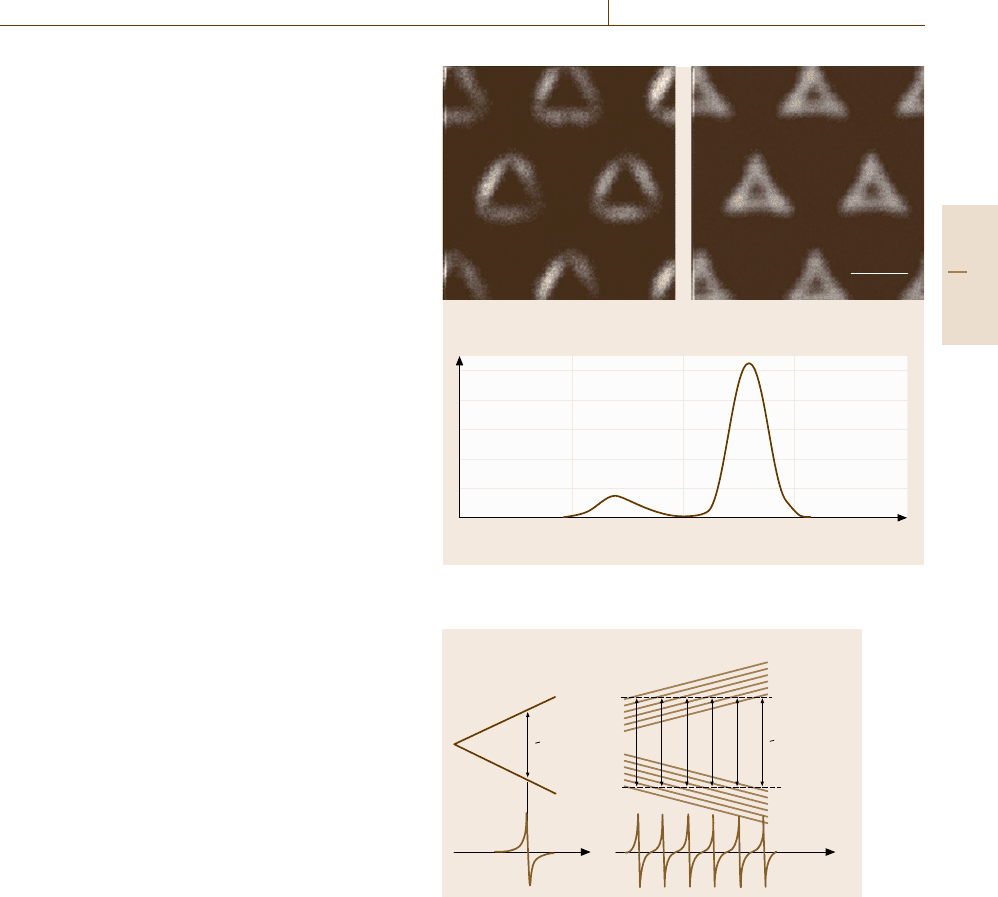
Scanning electron microscopy using cathodolumi-
nescence as the signal (SEM-CL) allows one to observe
the distribution of radiative centers in semiconducting
crystals. Figure 5.55 shows monochromatic SEM-CL
images of semiconductor quantum dots [5.47] from
which we can determine from where in the dots each
luminescence arises. Similar images can be obtained
by STEM equipped with a light collecting system. An
advantage of STEM-CL is that one can also obtain crys-
tallographic information about the defect.
Electron Paramagnetic Resonance (EPR). The second
term −2SJ
e
S in (5.19) represents the spin–spin ex-
change interaction that is of fundamental importance
in magnetic (ferromagnetic and antiferromagnetic) ma-
terials. In paramagnetic substances where the exchange
interaction is absent, the effective spin Hamiltonian rel-
evant to electron paramagnetic resonance (EPR)is
H = B
0
γ
e
S−λ
2
SS+SAI . (5.32)
Here the term −λ
2
SS, though formally similar to
−2SJ
e
S, now represents the magnetic dipole interac-
tion between electronic spin S and orbital momentum L
(spin–orbit coupling) of magnitude λ(L ·S). For the sys-
tems with S =1/2 encountered in most cases, this term
is absent. Still, however, the effective magnetogyric ra-
tio γ
e
≡γ
e
(1 −λ) is modified from that of an isolated
electronic spin γ
e
=μ
B
g
e
(μ
B
≡e/2m
e
c: Bohr magne-
ton) to
γ
e
≡γ
e
(1 −λ) =μ
B
g
e
(1 −λ) ≡μ
B
g . (5.33)
Since a static magnetic field B
0
induces a Zee-
man splitting of unpaired electrons much larger than
that of nuclear spins, a measurable microwave ab-
sorption is observed when the microwave frequency
becomes resonant with the Larmor frequency. Unlike
Fourier-transform NMR (Sect. 5.4.2), an EPR spectrum
is measured by sweeping the external magnetic field
applied to the sample placed in a microwave cavity
resonant at a fixed frequency of ≈ 10 GHz. Usually
the signal is detected by a magnetic field modulation
technique so that the resonance field is accurately deter-
mined as the fields at which the signal crosses the zero
base line (Fig. 5.56a).
The symmetry of the defect can be determined
by the dependence of the electronic g-tensor or the
corresponding resonance field on the direction of the
1.30 1.35 1.40 1.45 1.50
Photon energy (eV)
1.37 eV 1.43 eV
5 μm
CL intensity (arb. units)
Fig. 5.55 Monochromatic SEM-CL images of semiconductor
quantum dots and the Cl spectrum (after [5.47])
a) b)
S
z
+
1
–
2
–
1
–
2
B
0
B
r
hω
hω
S
z
+
1
–
2
–
1
–
2
I
z
+ 5/2
+ 1/2
– 3/2
+ 3/2
– 1/2
– 5/2
– 3/2
+ 1/2
+ 5/2
– 5/2
– 1/2
– 3/2
B
0
Fig. 5.56a,b Spin resonance of isolated electrons (a) and
electrons interacting at the hyperfine level with a nucleus
with I =5/2 (b)
crystallographic axis with respect to the direction of the
external magnetic field. Some point defects in semicon-
ductors form electronic levels in the band gap and could
change their charge states according to the Fermi level.
If the levels are degenerate with respect to the orbitals
due to the point symmetry of the center, a symmetry-
breaking lattice distortion (Jahn–Teller distortion) may
occur depending on the charge state so as to lift the
degeneracy and consequently lower the electronic en-
Part B 5.3
246 Part B Chemical and Microstructural Analysis
Mg
2+
Fig. 5.57
Oxygen vacancy
in MgO
ergy. An historical example in which the EPR method
shows its power is seen in structural determination of
vacancies in Si in different charge states [5.48].
The chemical environment of the center or the lat-
tice position of the defect can be identified from the
knowledge of hyperfine structures arising from the last
term SAI in (5.32) that originates in the magnetic
dipole–dipole interaction between the electronic spin
and the nuclear spins over which the electron cloud is
mostly distributed. Figure 5.56b illustrates how the hy-
perfine structure is brought about by the interaction of
the electron with a nucleus of I = 5/2. As a concrete
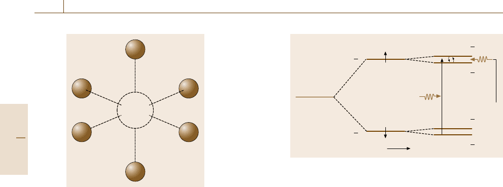
example [5.49], we consider oxygen vacancies in ionic
MgO crystals as shown in Fig. 5.57. The vacancy of
the oxygen ion O
2−
is double-positively charged and
EPR insensitive because all the electrons are paired.
But if it traps an electron and becomes single-positively
charged, EPR signals arise from the unpaired S =1/2
spin. Since no nucleus is present at the center of the
vacancy, the hyperfine interaction is due only to the sur-
rounding six equidistant Mg
2+
ions. Among abundant
isotopes, only the
25
Mg nucleus (10.11% abundance)
has nonzero nuclear spin of I = 5/2. Therefore, the rel-
ative intensity of hyperfine signals is determined by
the occupation probability of
25
Mg ions among the
six Mg ion sites. For example, the probability that
all the Mg sites are not occupied by
25
Mg ions is
(0.8989)
6
=52%, so 52% of the vacancies should show
no hyperfine splitting, as shown in Fig. 5.56a. Similarly
the probability of finding one
25
Mg ion (I =5/2) in
the neighborhood is 6×(0.8989)
5
×(0.1011) =35.6%,
so that 35.6% of vacancies should exhibit (2I +1) =6
hyperfine splitting signals, as illustrated in Fig. 5.56b
following the selection rule in EPR Δs
z
=±1and
ΔI
z
= 0. The probability of finding two
25
Mg ions
(I = 5) is (6!/4!2!)×(0.8989)
4
×(0.1011)
2
= 10%, so
B
m
s
= –
1
2
m
s
= +
1
2
m
N
= +
1
2
m
N
= +
1
2
Δm
N
=0
Δm
N
= ± 1
m
N
= –
1
2
m
N
= –
1
2
Micro-
wave
Fig. 5.58 Electron nuclear double resonance (ENDOR)in
I = 1/2 nuclei
that 10% of vacancies should exhibit (2I +1) =11 hy-
perfine splitting signals. The intensity of the hyperfine
signals in this case differs depending on the degener-
acy of the spin configurations: only one configuration
(5/2, 5/2) contributes to the I
z
= 5 signal, two con-
figurations, (5/2, 3/2) and (3/2, 5/2), to the I
z
= 4
signal, three configurations, (5/2, 1/2), (3/2, 3/2) and
(1/2, 5/2), to the I
z
= 3 signal, and so on, resulting in
the relative intensity ratio 1 :2 :3 :4 :5 : 6 : 5 : 4 :3 :
2 :1, which is verified experimentally.
The electron–nuclear interaction represented by the
term SAI in (5.32) can be extended to include indi-
rect interactions
k
SA
k
I
k
with nuclei k beyond the
nearest neighbors, giving rise to the super-hyperfine
structures around each hyperfine signals. One major
drawback of EPR spectroscopy is the low resolution
(Δν ≈ 10 MHz) compared to NMR (Δν ≈ 50 kHz),
which is a disadvantage in the analysis of crowded
or overlapped signals. Due to the electron–nuclear in-
teraction, the EPR intensity of each super-hyperfine
structure changes when nuclear magnetic resonance oc-
curs in the nucleus responsible for the super-hyperfine
EPR signal (Fig. 5.58). This EPR-detected NMR spec-
troscopy is called electron nuclear double resonance
(ENDOR)[5.50] which also improves the low sensi-
tivity of conventional NMR [5.51]. ENDOR is used
to investigate the spatial extent of the electron cloud
probed by EPR.
In semiconductors, unpaired spins as sparse as 10
14
to 10
16
cm
−3
can be detected by EPR provided that the
density of free carriers is below 10
18
cm
−3
so that the
microwave can penetrate the sample well. In metals, the
presence of free electrons limits the penetration depth
below the sample surface due to the skin effect. It should
be stressed that EPR signals can be detected only when
unpaired electrons are present.
Part B 5.3

Nanoscopic Architecture and Microstructure 5.3 Lattice Defects and Impurities Analysis 247
Optically Detected Magnetic Resonance (ODMR).
Optically detected magnetic resonance (ODMR)is
applicable to studies of electronic states in which a mag-
netic field lifts the degeneracy of energy levels differing
only in magnetic spin. ODMR is a double resonance
technique that allows highly sensitive EPR measure-
ments and facilitates the assignment of the spin state
responsible for the magnetic resonance. The most com-
mon experimental schemes are either detecting circular
polarized photoluminescence (PL) or magnetic circular
dichroic absorption (MCDA) in response to magnetic
resonance. Figure 5.59 illustrates a simple case in which
the ground state and an excited state are spin degener-
ate in the absence of a magnetic field. When a static
magnetic field is applied, these states are split to Zee-
man levels. Due to the selection rule, optical transitions
(PL and photoabsorption) occur either between the ex-
cited down-spin state and the ground up-spin state with
a right-hand circular polarization (σ
+
), or between the
excited up-spin state and the ground down-spin state
with a left-hand circular polarization (σ
−
). In the re-
laxed excited state in which the occupancy of the
up-spin level is higher than that of the down-spin level,
the σ
−
PL is stronger in intensity than σ
+
PL.How-
ever, if we apply an electromagnetic field in resonance
with a spin flip transition between the excited Zee-
man levels, the occupancy of the down-spin excited
state becomes somewhat increased, which is detected
by an increase of the σ
+
emission at the expense of
the σ
−
emission. Thus, the PL-detected magnetic reso-
nance or magnetic circular polarized emission (MCPE)
measurements (Fig. 5.59a) allow the detection of EPR
in the electronically relaxed excited state. MCPE is also
possible when the nonradiative recombination is spin-
dependent. An example is found in studies of dangling
bonds in amorphous Si :H solids [5.52] and of relaxed
excited state of F-centers in alkali halides [5.50]. MCPE
can be more sensitive than ordinary EPR as long as
the PL intensity is high. However, the concentration of
the centers studied successfully by MCPE is limited by
the concentration quenching effect, a decrease of PL
intensity due to an energy transfer mechanism (cross re-
laxation) starting to operate between nearby centers in
close proximity (Sect. 5.4.3).
Similarly, ODMR based on magnetic circular
dichroic absorption (MCDA) shown in Fig. 5.59b facil-
itates the assignment of the ground state spin. MCDA
has advantages over MCPE in its applicability to non-
luminescent samples. Other signals used in ODMR
include such spin-dependent quantities as spin-flip
Raman scattering light, resonant changes of electric
Excited state
EPR Microwave
EPR
B
σ
+
σ
–
B
Microwave
Ground state
Excited state
Ground state
σ
+
σ
–
a)
b)
Fig. 5.59 (a) Magnetic circular polarized emission (MCPE)
and (b) Magnetic circular dichroic absorption (MCDA)
current affected by the spin state of the defect cen-
ters that determines the carrier lifetime. Especially the
last electrically detected magnetic resonance (EDMR)
technique allows one to detect selectively a very few
paramagnetic centers that are located along the narrow
current path [5.7].
Muon Spin Rotation (μ
+
SR). A muon is a mesonic par-
ticle which is created during the decay of a π-meson
generated by a high energy particle accelerator. The
muon is a Fermion having spin 1/2, whose mass is 207
times the electron mass. There are two types of muon,
μ
+
and μ
−
, differing in the electric charge +e and −e,
respectively. The muon disintegrates to an electron (or
positron) and two neutrinos in a mean lifetime of ≈2 μs
emitting a γ -ray with a characteristic angular distribu-
tion relative to the spin direction, as shown in Fig. 5.60a.
Therefore, the rotation of spins under a static magnetic
field during the lifetime can be observed as a signal os-
cillating at the Larmor frequency detected by a γ-ray
counter placed in an appropriate direction (Fig. 5.60b).
Since the negatively charged μ
−
are strongly attracted
to nuclear ions, they behave as if forming a nulcleus
of atomic number Z −1. In contrast, since the posi-
tively charged μ
+
are repelled by nuclear ions in solids,
they migrate along interstitial sites or become trapped
at vacancies, behaving like a light isotope of protons.
In ferromagnetic solids, μ
+
feel an internal magnetic
field specific to the site on which the μ
+
spend their
Part B 5.3
