Czichos H., Saito T., Smith L.E. (Eds.) Handbook of Metrology and Testing
Подождите немного. Документ загружается.

268 Part B Chemical and Microstructural Analysis
that allow chromatography to become useful as a pre-
process for more sophisticated analysis.
Electrophoresis utilizes the difference in the drift
mobility of ionic molecules in a stationary phase under
an electric field. Electrophoresis using gel as the station-
ary phase is called gel electrophoresis (GE), and that
using aqueous solution in a capillary is called capillary
electrophoresis (CE). CE has advantages over GE be-
cause of reduced problems with Joule heating and the
action of the capillary itself as the pumping system.
Circular Dichroism (CD)
Circular dichroism (CD) arises from the helicity or
chirality of molecules that exhibit optical absorption
due to electronic excitations at ultraviolet to visible
wavelengths. CD spectra of chiral proteins, peptides
and nucleic acids have distinct structures and are sen-
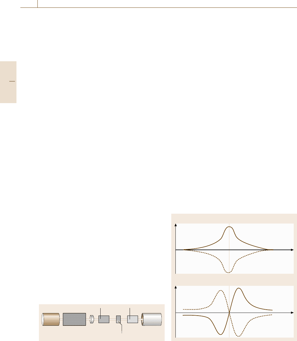
sitive to conformational changes. Figure 5.96 shows
a schematic diagram of experimental setup. Linearly
polarized light can be regarded as a superposition of
left- and right-hand circularly polarized light. Optically
active substances such as chiral molecules transmit
the opposite circularly polarized light with slightly
different absorption coefficients. As a consequence,
the linearly polarized light incident on the sample
becomes elliptically polarized light without changing
the main polarization axis. The degree of elliptic-
ity (CD) depends on the difference in the absorption
coefficients.
In contrast, optical rotation arises from differences
in the index of refraction that cause a difference of
the rotation angle between the opposite components
of circularly polarized light. The optical rotary dis-
persion (ORD) spectrum, the dependence of rotation
angle on wavelength, is related to the CD spectrum
by a Kramers–Kronig transformation. The ORD curve
changes sign at the extremum peak of the CD spectrum,
known as the Cotton effect, as illustrated in Fig. 5.97.
Owing to the fact that CD/ORD curves exhibit fea-
tures that are characteristic to the molecular structures,
CD/ORD measurements provide structural information
about the molecule. In proteins, for example, CD sig-
Wavelength λ
Polarizer Analyzer
Light
source
Mono-
chromator
Sample Optical
detector
Fig. 5.96 Experimental setup of circular dichroism (CD)
and optical rotary dispersion (ORD) measurements
nals are more sensitive to α-helical residues than those
of random coils and β-sheets. Experimental data are fit-
ted to reference model spectra to determine the amounts
(composition) of the constituent secondary structure of
proteins. The reliability of composition analysis is en-
hanced by the use of proteins of known structure as the
basis set. More sophisticated analysis of molecular po-
sitions, conformation and absolute configuration can be
conducted with detailed knowledge of the influence of
these factors on the Cotton effect.
Samples are usually prepared as a solute in an opti-
cally nonactive solution. CD/ORD measurements allow
quantitative analysis since the signal intensity is propor-
tional to the molecular density in the solution. Structural
analysis, however, is only possible at densities that yield
an optical absorbance of 1–2 at the absorption maxi-
mum, where the Cotton effect is observed, in order to
avoid signal weakening due to excessive absorption.
Although simple CD and ORD are observed only
in optically active molecules, similar dichroic effects
are also induced in achiral substances under a mag-
netic field applied parallel to the measuring light beam.
Magnetic circular dichroism (MCD) combined with
photoabsorption measurements is most commonly used
to analyze chromophoric groups in which the magnetic
field lifts the degeneracy of optically excited energy
levels differing only in magnetic spin (Sect. 5.3.1). In
biology, MCD is also applicable to metalloproteins, pro-
Elipciticy
Rotation angle
Wavelength μ
Wavelength μ
μ
max
μ
max
Fig. 5.97 Schematic spectra of circular dichroism (CD)
and optical rotary dispersion (ORD)
Part B 5.4
Nanoscopic Architecture and Microstructure 5.5 Texture, Phase Distributions, and Finite Structures Analysis 269
tein molecules that contain a metal substance necessary
for a certain reaction to take place.
Fluorescence Resonant Energy Transfer (FRET)
Fluorescence resonant energy transfer (FRET) occurs
when the distance between a fluorescent donor dye
molecule and a photoabsorbing acceptor dye molecule
is close enough for the electronic excited energy of
the donor molecule to be transferred to the acceptor
molecule nonradiatively. Since the efficiency of FRET
depends on the inverse sixth power of the intermolecu-
lar distance, it is useful for determining the proximity of
the two molecules within 1–10 nm, which is a distance
range of particular importance in biological macro-
molecules. For FRET to take place, the fluorescence
spectrum of the donor and the absorption spectrum of
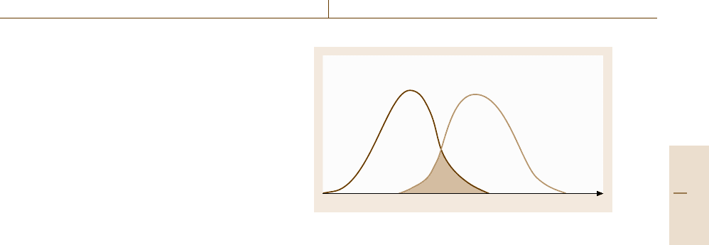
the acceptor must overlap, as shown in Fig. 5.98. Based
on this technique, the approach of two molecules can
be detected by quenching the donor fluorescence, and
hv
Acceptor
absorption
Donor
fluorescence
Fig. 5.98 Spectral overlap necessary for fluorescence res-
onant energy transfer (FRET)
one can study the conformation change of a single
molecule by detecting FRET under a laser scanning
confocal microscope (LSCM) (Sect. 5.1.2)oratotal
internal reflection fluorescence microscope (TIRFM)
(Sect. 5.1.2)[5.78].
5.5 Texture, Phase Distributions, and Finite Structures Analysis
This section deals with materials that are inhomoge-
neous on a relatively large scale with respect to their
composition, structure, and physical properties. Poly-
crystals are said to have a texture when they have
a nonrandom distribution of grain orientations. The
phase distribution or element distributions may play
an important role in the macroscopic properties of the
material. Another issue addressed is solid structures
with finite three-dimensional size that may have their
own functions, such as biological cells, nanoparticles,
etc. In the last subsection, we describe some of the
recent progress in stereology, an emerging field of three-
dimensional analysis of materials.
5.5.1 Texture Analysis
Textures evolve by various mechanisms. Plastic de-
formation of crystals by glide motion of dislocations
proceeds on preferential crystallographic slip planes,
usually low-index planes with the largest spacing. If
polycrystalline wires are drawn through a die, the wires
have a texture (wire texture) such that the slip planes
tend to align parallel to the wire axis, because otherwise
each grain would be subject to further deformation. The
texture of polycrystalline materials is of technological
importance for various physical properties, mechanical
properties such as strength and elastic constants, mag-
netic permeability, flux pinning in superconductors of
the second kind, and so on.
Texture Analysis by X-Ray Diffraction
The best established technique for assessing textures is
x-ray diffraction. In powder diffraction, each diffrac-
tion ring consists of fine spots which correspond to
crystallites in different orientations. If the crystallites
are randomly oriented, the spots or the diffraction in-
tensity is uniformly distributed around the circle. If
textures are present, however, one observes an inhomo-
geneous distribution of diffraction spots or diffraction
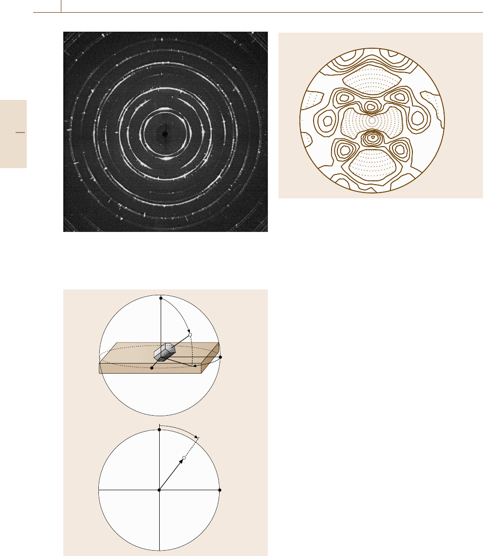
arcs, as shown in Fig. 5.99. In modern experiments, the
data are collected by using a diffractometer equipped
with a four-circle goniometer (Fig. 5.37) that scans all
orientations by a combinatorial rotation around the Eu-
lerian ω–φ–χ axes. For thick or large-grained samples,
neutron diffraction may offer an alternative to x-ray
diffraction, if a facility is accessible. The merit of
diffraction techniques is the ease with which good sta-
tistical data can be acquired.
The texture of materials is expressed by several
different representations. The pole figure (Fig. 5.100)
presents the orientational distribution of a specific pole
(crystallographic (0001) axis in Fig. 5.100a) with re-
spect to the sample orientation, which is displayed on
a stereographic projection with the surface normal at
Part B 5.5
270 Part B Chemical and Microstructural Analysis
Fig. 5.99 X-ray diffraction pattern from a mechanically
drawn Al wire. Exposure time 30 s using x-ray source Mo
K
α
(50 kV, 40 mA) recorded on an imaging plate placed
10 cm from the sample (Courtesy of Rigaku Co)
a)
b)
Normal
direction
Rolling
direction
Transverse
direction
(0001)
β
α
Rolling direction
Normal
direction
Transverse
direction
β
α
(0001)
Fig. 5.100a,b A presentation of the texture (a) by the
stereographic pole figure in
(b)
Levels:
0.7
1.0
1.4
2.0
2.8
4.0
4.7
RD
TD
F
max
: 4.7
Stereographic
projection
{111}
Fig. 5.101 An experimental pole figure of an aluminum
sheet rolled and annealed. After [5.79]
the origin, a specific direction along the sample surface
(e.g., the rolling direction in rolled metals) at the north
pole, and the transverse direction in the horizontal direc-
tion. Figure 5.101 shows an experimental pole figure of
an aluminium sheet thermally annealed after mechani-
cal rolling [5.79, p. 102]. The four crystallographically
equivalent (111) poles are distributed in some dominant
directions. Also widely used is the inverse pole figure
that presents the orientation distribution of the specimen
coordinate system with respect to the crystal coordinate
system, the latter being reduced to a standard stereo-
graphic triangle. Whichever the case, however, the pro-
jection of the three-dimensional orientation distribution
onto a two-dimensional figure causes a loss of infor-
mation. The orientation distribution function (ODF)is
a three-dimensional presentation thus devised for a full
description of texture. The ODF can only be obtained by
calculations from a data set of several pole figures. For
details, see the textbook by Randle and Engler [5.79].
Texture Analysis by SEM Electron Channeling
Pattern (ECP)
The disadvantage of diffraction techniques for the anal-
ysis of texture is the lack of direct information about the
grain size. Local crystal orientations may be determined
by microscopic techniques that also reveal the dimen-
sion of each grain directly [5.80]. TEM is a routine
method to conduct such measurements, but the samples
are limited to thin films. The electron channeling pat-
tern (ECP), which can be obtained by scanning electron
microscopy (SEM), is a more convenient approach for
studies of texture using bulk samples.
Part B 5.5
Nanoscopic Architecture and Microstructure 5.5 Texture, Phase Distributions, and Finite Structures Analysis 271
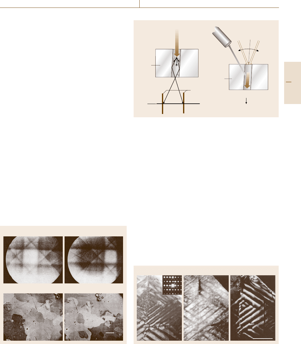
SEM-ECP corresponds to the Kikuchi lines
(Sect. 5.1.2) or bands observed in TEM. Figure 5.102a il-
lustrates how the Kikuchi lines are formed. As men-
tionedinSect.5.1.2, the Bloch wave with its largest
amplitude on the atomic column (Fig. 5.102b) interacts
more strongly with the atoms and is more likely to be
scattered inelastically, losing coherency with the direct
wave. As shown in Fig. 5.102a, some of the electrons
thus inelastically scattered in various directions will be
reflected by lattice planes satisfying the Bragg condi-
tion and, if the specimen is moderately thick, propagate
along the channel direction with an anomalously small
absorption coefficient. The electrons are projected onto
the back focal plane of the objective lens as a pair
of traces of the diffraction cones. Line pairs (Kikuchi
lines) or bands formed in this way indicate the orien-
tation of the crystal. In a similar way to the Kikuchi
lines in TEM, if the electron beam in SEM is incident
to a crystalline sample in an orientation along a chan-
neling direction (Fig. 5.102b), the electrons penetrate
deeply into the sample and have less chance to be back
scattered and detected by a back-scattering electron de-
tector. If one rocks the beam direction as in Fig. 5.102b
and synchronously displays a two-dimensional map of
the intensity of back-scattered electrons in the SEM
screen as a function of the beam orientation, one ob-
tains patterns as shown in Fig. 5.103a,b. Since the
SEM-ECP pattern reflects the crystalline orientation
of the sample position at which the beam is rocked,
beam scanning with the beam direction fixed gives
a) b)
c) d)
100μm
1
2
1
2
Fig. 5.103a–d Electron channeling patterns (a),(b) and
images
(c),(d) at different tilt angles. After [5.17]
a) b)
Back-scattered
electron detector
Channeling
Reduction of
back-scattered electrons
Bulk
sample
Thin
sample
θ
B
Beam
rocking
Back
focal
plane
Kikuchi
lines
Fig. 5.102a,b Formation of Kikuchi lines in TEM (a) and electron
channeling pattern in SEM (b)
ECP images, as shown in Fig. 5.103c,d. From such
crystallographic orientation contrasts, one can deter-
mine the orientation distribution and the size of each
grain.
5.5.2 Microanalysis of Elements and Phases
Dark-Field TEM Imaging
Dark-field (DF) TEM images (Sect. 5.1.2)formedby
a diffracted beam of a specific diffraction vector provide
the most standard technique for microscopic analysis of
coexisting multiphases. Figure 5.104 shows a bright-
field image of a metallic compound of Nb
3
Te
4
and
the corresponding DF images obtained for two differ-
ent diffraction vectors. The lateral resolution is similar
to that of BF-TEM images as long as the sample is
thin enough for the overlap of different phases to be
avoided.
0.5 μm
b) c)
a)
Fig. 5.104 (a) Bright-field and (b),(c) dark-field images of multi-
phase Nb
3
Te
4
(courtesy of M. Ichihara)
Part B 5.5
272 Part B Chemical and Microstructural Analysis
Electron Probe Microanalysis (EPMA)
Another common method to investigate element dis-
tribution in solid materials is scanning electron mi-
croscopy operated in the x-ray fluorescence mode,
usually referred to as an electron probe microanalyzer
(EPMA). On the impingement of electrons in the energy
range of 1–40 keV, the samples emit x-rays at wave-
lengths characteristic of the elements contained. The
emitted x-ray is usually detected with an energy dis-
persive x-ray (EDX) solid-state detector placed above
the specimen (Fig. 5.105). Scanning the beam gives us
a map of the element distribution with a spatial reso-
lution of a few nm to 100 μm. The technique becomes
more sensitive as the mass number increases.
Similar measurements could be conducted using
TEM. In ordinary SAD, however, it is difficult to re-
duce the size of a selected area to smaller than several
tens of nm, whereas in CBED the beam size can be re-
duced down to a subnanometer scale. The best choice
is to use a focused beam in STEM that allows one to
conduct CBED analysis as well as mapping element
distribution over the scanned area. For heavy elements,
STEM-EDX has advantages over the following tech-
niques, though there are technical problems with the
energy resolution (≈ 150 eV) and the time for data
acquisition.
Electron impingements induce the emission of
Auger electrons whose energies are characteristic of
specific elements (Fig. 5.67). Since the energy of Auger
electrons is so small that the escape distance is as short
as ≈ 1nm (Fig.5.59), Auger electron spectroscopy
(AES) is overly sensitive to the surface.
Energy Loss Analysis
Energy filtering TEM (EFTEM)isaTEM or STEM
equipped with an energy filter passing only electrons
of a specific energy that are used to construct an im-
age of the spatial distribution of the corresponding
elements. The energy filter may be placed inside the op-
tical column of the TEM (in-column type) or in front
of the camera chamber (post-column type). EFTEM al-
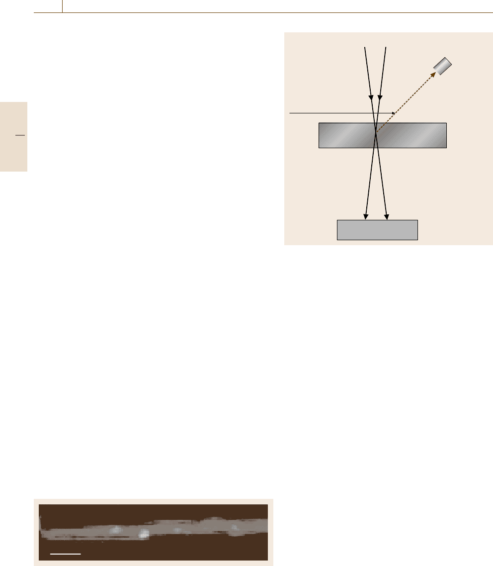
Fig. 5.106 STEM-EELS image of Gd (white) atoms in a carbon
(gray) nanotube. Scale bar =3nm.After[5.81]
Focused electron beam
Beam scanning
E
0
–ΔE
E
0
Energy
dispersive
x-ray
detector
Fluorescent x-ray
Sample
Electron energy
loss spectrometer
Fig. 5.105 Element analysis by an electron probe
lows spectrum imaging of the element distribution with
an energy resolution of ≈ 30 eV and a spatial resolu-
tionofdownto≈ 1 nm at 200 kV and ≈ 0.5nm at
300 kV.
In contrast to EFTEM, the spatial resolution in
STEM-EELS, if equipped with a field emission gun,
is much higher owing to the fact that STEM generally
does not need an objective lens to image the aberration
that governs the spatial resolution of TEM micrographs.
A disadvantage of STEM-EELS is the long acquisi-
tion time due to the scanning nature of the microscopy,
but the use of a parallel EELS (PEELS) detector and
the great assistance of a computer have advanced the
progress of STEM-EELS as a tool able to identify even
a single atom in small molecules [5.81], as demon-
strated in Fig. 5.106. STEM-EELS has advantages over
STEM-EDX for light elements.
Micro-Raman Scattering
Raman scattering measurements (Sects. 5.1.2, 5.2.3,
and 5.3.1) can be coupled with optical microscopes to
investigate the local distribution of particular phases
with a spatial resolution of ≈1 μm. The diffraction limit
of OM is now overcome by the use of scanning near-
field optical microscopy (SNOM) (Sect. 5.1.2), in which
the enhancement of the optical field by surface plas-
mon resonance at the metallic tip allows the detection of
Raman spectra for even a single molecule [5.82] under
good conditions.
Part B 5.5

Nanoscopic Architecture and Microstructure 5.5 Texture, Phase Distributions, and Finite Structures Analysis 273
Scanning Nano-Indenter
Materials often encountered may be in the form of
thin films or nanoscale omplexes of multiple phases.
One may need to conduct mechanical tests for very
thin films grown on a substrate. The difference in ma-
terial phase may reflect the mechanical strength. The
scanning nano-indenter is a modern version of the
micro-hardness tester with which the local mechanical
strength of crystallites smaller or thinner than ≈1 μm
can be measured with a lateral resolution of several hun-
dreds nanometres from the indent size in situ observed
by a microscope similar to an AFM and STM. The ex-
tremely tiny indenter can be driven into and retracted
from the sample surface at a controlled rate, so that one
can also deduce the local elastic modulus.
5.5.3 Diffraction Analysis of Fine Structures
Tiny objects such as precipitates, crystallites, and fine
particles smaller than ≈ 100 nm can be investigated by
not only microscopic but also diffraction and spectro-
scopic techniques.
Small-Angle Scattering Analysis of Tiny Phases
The Guinier–Preston (GP) zone, an extremely tiny pre-
cipitate with a thickness of only one atomic layer
and lateral size of several atomic distances, is directly
observable by HRTEM at present; it was originally
discovered in x-ray diffraction as a source of diffuse
scattering. In Guinier’s approximation, in which a par-
ticle is a Gaussian sphere
ρ
(
r
)
=ρ
0
exp
−
r
2
a
2
(5.37)
with a representing the particle size, the diffraction in-
tensity or the Fourier transform of (5.37) will be
I(K) =ρ
2
0
a
2
π exp
−
a
2
2
K
2
. (5.38)
The magnitude of (5.38) is small except for small |K|
values or small scattering angles, which means that the
primary beam is diffusely scattered within a small an-
gle. In terms of the radius of gyration R
g
, the root mean
square of the mass-weighted distances of all subvol-
umes in a particle from the center of mass,
I(K) ∝exp
−R
2
g
K
2
/3
. (5.39)
Thus, from the slope of the so-called Guinier plot,
log I(K) versus K
2
, we can evaluate a measure of the
particle radii R
g
experimentally.
Line-Broadening Analysis
of Crystallite Size and Stress
The broadening of x-ray beams occurs not only in the
primary beam but also in diffracted beams when the
crystallites or domains are small [5.83]. This arises from
the fact that the diffraction intensity depends on the in-
terference function, which reflects the external shape of
the crystal (Sect. 5.1.1).
More generally, the line broadening is induced by
causes other than the finite crystal size. The main causes
are instrumental errors and the strain distribution. The
instrumental broadening due to spatial, angular and
wavelength distribution of the incident x-ray beam is
experimentally evaluated by using a standard sample,
usually a large and perfect single crystal such as Si. The
line broadening due to lattice strain is in most cases due
to lattice defects such as dislocations.
As a measure of the line broadening, we conve-
niently employ the integral breadth β, which is defined
as the width of a rectangle having the same area A and
height I
0
as the line profile of interest, or β ≡ A/I
0
.For
Gaussian broadening, the relevant breadth β
s
of sample
origin in our concern is estimated by
β
2
s
=β
2
m
−β
2
i
, (5.40)
where β
m
and β
i
are the experimental and instrumen-
tal breadths, respectively. The separation of the lattice
strain component is achieved by plotting β
s
versus sin θ
for various diffractions. From the Bragg condition (5.1),
the line broadening in terms of the diffraction angle 2θ
due to a distribution of net-plane spacing Δd is given
by
Δθ = tan θ
Δd
d
. (5.41)
When there is another contribution from the finite crys-
tal, the experimentally measured integral breadth β
s
is
generally given by
β
s
(
g
)
=aε tan θ
g
+β
p
(5.42)
where ε ≡ Δd/d represents the strain and β
p
the size
contribution in our concern. Since the integral breadth
β
p
is related to the average size of crystallites L by
Scherrer’s formula [5.84, p. 102]
β
p
(g) =
λ
L cos θ
g
, (5.43)
(5.43) is written as
β
s
(
g
)
cos θ
g
=aε sin θ
g
+
λ
L
. (5.44)
Part B 5.5
274 Part B Chemical and Microstructural Analysis
Willamson–Hall plot
λ
L
sinθ
g
β
s
(g) cosθ
g
∝ε
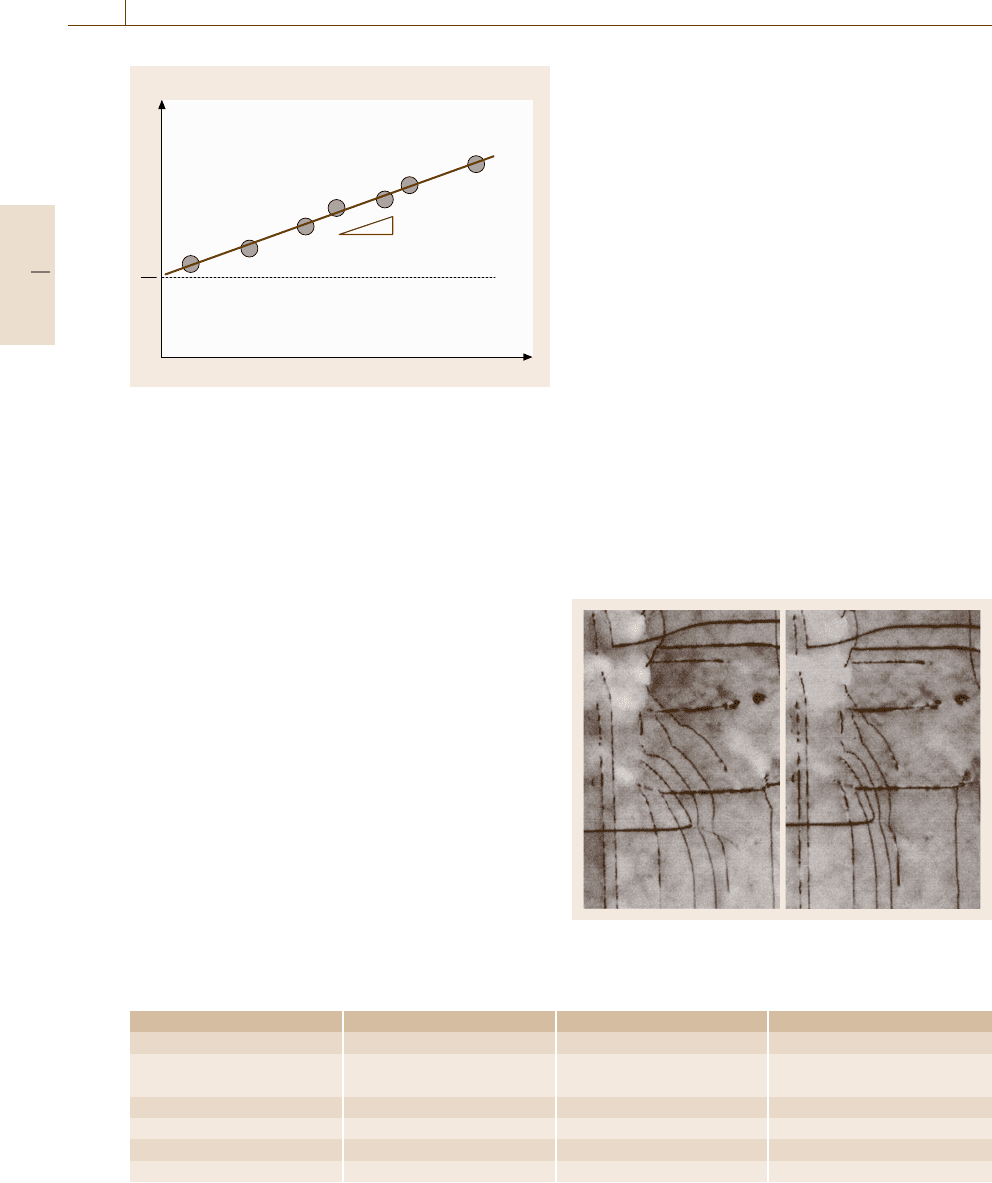
Fig. 5.107 A schematic Williamson–Hall plot for line
broadening analysis
Thus, by plotting β
s
(g) cos θ
g
versus sin θ
g
(the so-
called Williamson–Hall plot), as shown in Fig. 5.107,
we can evaluate the average size L of the crystallites
from the θ
g
-independent level.
Thus, the analysis of line broadening simultane-
ously enables evaluation of lattice strain ε or the internal
stress in the crystallites. With access to costly facilities
such as synchrotron radiation sources (for x-ray diffrac-
tion) or nuclear reactors (for neutron diffraction), one is
able to conduct experiments with great advantages for
probing the stress state deep into the sample [5.85].
Light Scattering Analysis of Particle Shape
The cloudy or milky appearance of latex and solid
polymer reflects a heterogeneous structure with two
coexisting phases [5.29] (colloid particles and liquid
in latex; crystalline and amorphous domains in solid
polymer) (Fig. 5.42). This is due to the high-angle scat-
tering of light over a wide range of wavelength, called
Mie scattering in contrast to the wavelength-dependent
Rayleigh scattering. When the particle size is smaller
than the light wavelength, we enter to a regime of
surface plasmon resonance. The resonance frequency
Table 5.5 Comparison of various stereology methods
Method 3-D reconstruction Resolution Test environment
3-D-SEM Stereogram < 10 nm Vacuum
LSCM+ Stacked tomographs lateral ≈ 1 μm Ambient, liquid, vacuum
nonlinear optical microscopy depth ≈ 1μm
3-DAP-FIM Stacked tomographs < 0.2nm UHV
X-CT Computed tomography ≈ 1 μm Ambient, vacuum
X-PCI Computed tomography afewμm Ambient
3-D-TEM Computed tomography < 10 nm Vacuum
depends on the dielectric function of the particle ma-
terial and the shape of the particle. As the particle is
elongated in one direction, the resonance is shifted to
longer wavelengths, so measuring the resonant wave-
length of the scattering, we can determine how the
particles are distorted from a sphere.
5.5.4 Quantitative Stereology
The great increase of computer power and resources
has made it possible to process the enormous amount
of data necessary for three-dimensional reconstruction
of a sample structure. There are various types of stereo-
logical techniques, as listed in Table 5.5, differing in the
reconstruction method.
Stereogram
Two micrographs are acquired for a sample that is
tilted by ±1−10
◦
in an eucentric way about a tilt-
ing axis at the center of the sample. If one views two
such images placed side by side with an appropriate
separation distance, three-dimensional stereovision of
the object is obtained, as demonstrated in Fig. 5.108.
Fig. 5.108 AsetofTEM stereograms showing disloca-
tions in Si
0.87
Ge
0.13
/Si(100)
Part B 5.5
Nanoscopic Architecture and Microstructure 5.5 Texture, Phase Distributions, and Finite Structures Analysis 275
The three-dimensional vision could also be provided
by an anaglyph image, an overlayed image of the two
micrographs that are superimposed with different col-
ors, when viewed with red–blue glasses. Quantitatively,
the surface profile is calculated from the disparity in
the stereo image pairs. The reconstruction of three-
dimensional structure is possible from such stereograms
acquired in TEM and SEM.
Stacked Tomographs
Laser Scanning Confocal Microscopy (LSCM). The high
depth resolution of ≈ 1 μm in laser scanning confo-
cal microscopy (LSCM) enables reconstruction of 3-D
images from successively acquired optical images of
optically sliced layers by changing the focal position.
The great advantage of LSCM is that there are no spe-
cial requirements for the test environment except that
the sample medium must be optically transparent.
Ordinary LSCM is based on one-photon processes
such as scattering or fluorescence of light. In thick sam-
ples, however, remaining light scattering deteriorates
the spatial resolution. Nonlinear fluorescent microscopy
uses an intense laser light whose photon energy is too
small for a one-photon excitation of a fluorochrome
but is large enough for a two-photon excitation. Due
to the quadratic dependence of the fluorescence in-
tensity on the intensity of excitation laser, the focal
point is effectively reduced so that the spatial resolu-
tion is enhanced whereas the background fluorescence
is removed. Two-photon microscopy has the merit of
avoiding photobleaching or optical damage that would
be caused when excitation lights of higher photon en-
ergy in the ultraviolet range is used for one-photon
excitation. Similar benefits are obtained in second-
harmonic generation microscopy in which the dielectric
response of the sample is nonlinear.
Three-Dimensional Atom Probe (3-DAP) Microanal-
ysis. As described in (Sect. 5.1.2), the field ion micro-
scope (FIM)[5.86] is an atom-resolving microscope in
which an imaging gas projects the positions of step-
edge atoms on a phosphor screen without destroying the
surface. If the applied voltage is high enough, the step-
edge atoms themselves evaporate due to the field. The
field-evaporating atoms (positively charged ions) are
conveyed by the bias field to the screen to form a direct
image of the atomic distribution on the tip, now destruc-
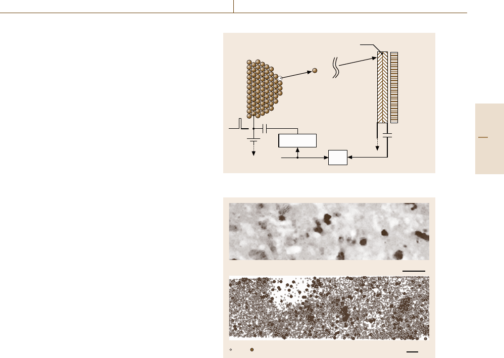
tively. As illustrated by Fig. 5.109, this field evaporation
can be induced by a pulse bias voltage applied to the
sample that is triggered as the starting signal for time
of flight (TOF) measurements of the evaporated ions.
Sample
tip
Position-
sensitive
detector
M
+
Ionized atom
Multichannel plate
(x, y, t)
HV
Pulsed HV
TOF
Stop signalTrigger pulse
Fig. 5.109 Experimental setup of 3-D atom-probe FIM
Nd Cu
20 nm
~10 nm
Fig. 5.110 Elemental mapping of Cu and Nd in Nd
4.5
Fe
75.8
B
18.5
Cu
0.2
Nb
1
alloy imaged by 3-D atom-probe FIM
(Courtesy of Dr. K. Hono)
The use of a multichannel plate in front of the phosphor
screen and a gated acquisition of the ion signal allows
one to detect specific element atoms sorted by the TOF.
Since the field evaporation takes place exclusively at
step edges, the evaporation processes are repeated as if
the atomic layers are being peeled back one by one. The
layer-by-layer acquisition of such atom probe signals
are used to reconstruct a three-dimensional map of ele-
ments in the sample tip. Figure 5.110 shows an example
of three-dimensional atom probe (3-DAP) images ob-
tained for a multicomponent alloy. The lateral and depth
resolution of 3-DAP microanalysis is ≈ 0.2nm,thebest
among the stereological methods developed so far.
Computed Tomography (CT)
Computed tomography is based on mathematical trans-
formations that enable reconstruction of the two-
Part B 5.5
276 Part B Chemical and Microstructural Analysis
Beam
source
Object
Detector
f (x,y)
y
x
r
p (r,θ)
θ
Fig. 5.111 Geometry in computed tomography
dimensional distribution function f (x, y) (e.g., of the
absorption coefficient) of an object from a set of
one-dimensional intensity profiles p(r,θ) detected by
the detector of a transmitted beam that is emitted
from a source in various directions θ, as illustrated in
Fig. 5.111. The computed tomographs obtained for suc-
cessive cross sections of the object are further stacked
to form a three-dimensional reconstruction of the whole
body.
X-Ray Computed Tomography (XCT). X-ray computed
tomography (XCT) using an x-ray for the source beam
is common equipment for clinical diagnosis, but can
also be used for materials analysis. In XCT, the beam
is fanned to scan an angle θ rather than scan position r.
The resolution of recent x-ray CT is ≈ 1 μm, compara-
ble with that of OM.
Magnetic Resonance Imaging (MRI). Magnetic reso-
nance imaging (MRI)orNMR-CT is also now common
for clinical diagnosis. MRI is based on the principle that
when the applied magnetic field in NMR hasaninten-
tional gradient (i.e., varies with position), the resonance
frequency of an NMR signal becomes dependent on
the position of the nucleus. The positional r-dependent
NMR signal frequency constitutes the profile function
p(r,θ) in which the angle θ is given by the direction of
the magnetic field gradient.
Three-Dimensional TEM (3-D-TEM). Although x-ray
crystallography can provide structural information at
atomic resolution, its application depends on whether
we can obtain crystalline arrays of the molecules
with good quality and analyze the diffraction data in
light of relevant phase information [5.87]. Further-
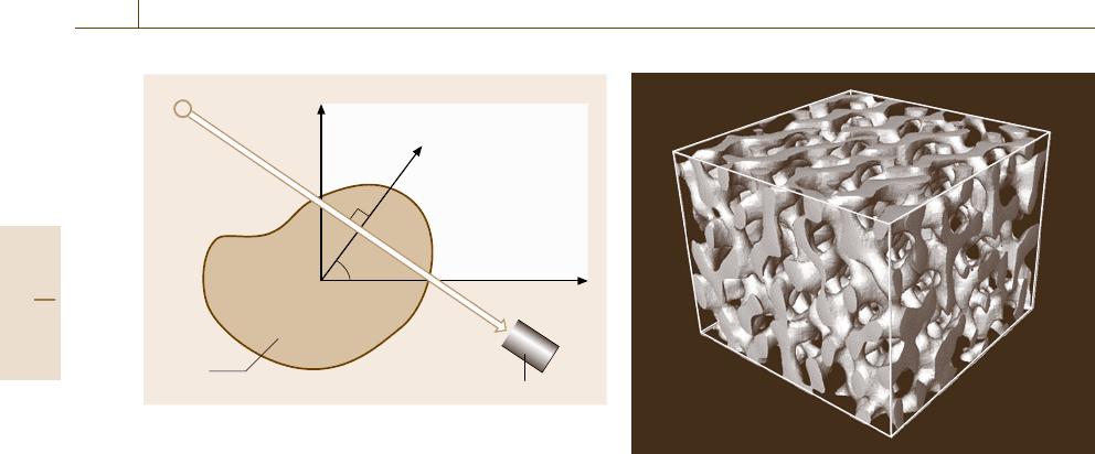
Fig. 5.112 A3-D-TEM image of a selfassembled tri-
block copolymer, poly(styrene-block-isoprene-block-styr-
ene). The box size is 270 nm ×270 nm×210 nm. 120 digital
images were acquired at tilt angles ranging from −60
◦
to
60
◦
with 1
◦
steps on a computer-controlled JEOL JEM-
2200FS operated at 200 kV (Courtesy of Dr. H. Jinnai)
more, a serious problem especially in biomolecules
is that crystallization may alter the conformation of
the molecules that could differ significantly from that
in which the molecules function in the natural en-
vironment. In single-particle electron microscopy, the
3-D structure of a single macromolecule (MW >
250 000 Da) is reconstructed at a resolution of 1–2.5nm
from a set of TEM images obtained in many differ-
ent sample orientations. In practice, sample damage
due to electron irradiation is reduced by the use of
cryo-microscopes and simultaneous observations of
many sample molecules (usually embedded in ice for
biomolecules) to overcome signal-to-noise problems.
Figure 5.112 shows a graphical rendering of 3-D-TEM
image obtained for a selfassembled polymer mater-
ial. 3-D-TEM is applicable to amorphous solids, but
not suitable for crystalline samples in which the TEM
images are too sensitive to diffraction conditions for
a simple three-dimensional reconstruction to be carried
out.
X-Ray Phase Contrast Imaging (PCI)
The contrast of conventional x-ray transmission images,
except for x-ray topography, is based on the positional
difference of the absorption coefficient [5.88]. For ma-
terials consisting of light elements such as biological
tissues and polymers, conventional x-ray transmission
microscopy, unless staining with heavy elements is
Part B 5.5
Nanoscopic Architecture and Microstructure References 277
possible, is not efficient enough. Phase contrast imag-
ing (PCI), as in optical microscopy (Sect. 5.1.2), is an
approach to overcome this problem also in x-ray re-
gions, though there is a difficulty due to the small
size of the phase shift in light elements. However,
in the soft x-ray region, the phase shift can be 10
3
times larger than in the hard x-ray region, so x-ray
PCI has been introduced. Among the four schemes
so far attempted, interferometric imaging, holography-
like imaging, diffraction enhancement imaging, and
Zernike-type microscopy, the interferometric method
is most sensitive for acquiring phase contrasts. The
method uses a Mach–Zehnder x-ray interferometer cut
out of a large Si single crystal ingot which is mounted
on a multiaxis goniometer set in the beam line of a syn-
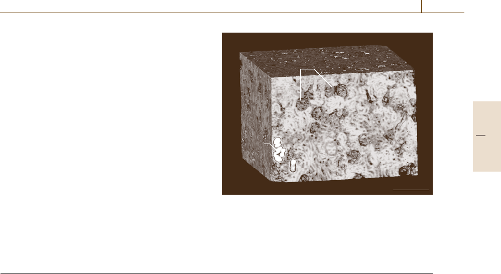
chrotron radiation source. X-ray phase shift computed
tomography [5.89] exploiting the ability of phase re-
trieval allows one to reconstruct 3-D images such as
those shown in Fig. 5.113.
G
T
500µm
Fig. 5.113 X-ray phase image of a tissue of a rat kidney.
Tubules in the tissue, a part of which was clogged by pro-
tein (T), were depicted. Glomeruli (G) were also revealed
(Courtesy of Drs. A. Momose, J. Wu, and T. Takeda)
References
5.1 A. Briggs, W. Arnold: Advances in Acoustic Microscopy
(Plenum, New York 1995)
5.2 K. Sakai, T. Ogawa: Fourier-transformed light scat-
tering tomography for determination of scatterer
shapes, Meas. Sci. Technol. 8, 1090 (1997)
5.3 S.V. Gupta: Practical Density Measurement and Hy-
drometry (IOP, Bristol 2002)
5.4 Z. Alfassi: Activation Analysis, Vol. I&II (CRC, Boca
Raton 1990)
5.5 A. Tonomura: Electron Holography (Springer, Berlin,
Heidelberg 1999)
5.6 J. Shah: Ultrafast Spectroscopy of Semiconductors
and Semiconductor Nanostructures (Springer, Berlin,
Heidelberg 1996)
5.7 T.Wimbauer,K.Ito,Y.Mochizuki,M.Horikawa,
T. Kitano, M.S. Brandt, M. Stutzmann: Defects in
planar Si pn junctions studied with electrically de-
tected magnetic resonance, Appl. Phys. Lett. 76,
2280 (2000)
5.8 L.V. Azároff: Elements of X-ray Crystallography
(McGraw-Hill, New York 1968)
5.9 J.M. Cowley: Diffraction Physics (North-Holland,
Amsterdam 1975)
5.10 B.D. Cullity: Elements of X-ray Diffraction (Addison-
Wesley, Reading 1977)
5.11 A. Guinier: X-ray Diffraction in Crystals and Imper-
fect Crystals, Amorphous Bodies (Dover, New York
1994)
5.12 G. Rhodes: Crystallography Made Crystal Clear (Aca-
demic, San Diego 2000)
5.13 S. Bradbury: An Introduction to the Optical Micro-
scope (Oxford Sci. Publ., Oxford 1989)
5.14 P.B.Hirsch,A.Howie,R.B.Nicholson,D.W.Pash-
ley, M.J. Whelan: Electron Microscopy of Thin Crystals
(Butterworths, London 1965)
5.15 D.B. Williams, C.B. Carter: Transmission Electron Mi-
croscopy (Plenum, New York 1996)
5.16 M.J. Whelan: Dynamical Theory of Electron Diffrac-
tion. In: Modern Diffraction and Imaging Techniques
in Materials Science, ed. by S. Amelinckx, R. Gevers,
G. Remaut, J. Van Landuyt (North-Holland, Amster-
dam 1970) p. 35
5.17 C.E. Lyman, D.E. Newbury, J.I. Goldstein, D.B. Wil-
liams, A.D. Romig Jr., J.T. Armstrong, P. Echlin,
C.E. Fiori, D.C. Joy, E. Lifshin, K. Peters: Scanning
Electron Microscopy, X-ray Microanalysis and Ana-
lytical Electron Microscopy (Plenum, New York 1990)
5.18 D. Bonnell (Ed.): Scanning Probe Microscopy and
Spectroscopy (Wiley, Weinheim 2001)
5.19 B. Bhushan, H. Huchs, S. Hosaka (Eds.): Applied
Scanning Probe Methods (Springer, Berlin, Heidel-
berg 2004)
5.20 K.S. Birdi: Scanning Probe Microscopes (CRC, Boca
Raton 2003)
5.21 V.J. Morris, A.R. Kirby, A.P. Gunning: Atomic Force
Microscopy for Biologists (Imperial College Press,
London 1999)
5.22 S. Morita, R. Wiesendanger, E. Meyer: Noncontact
Atomic Force Microscopy (Springer, Berlin, Heidel-
berg 2002)
Part B 5
