Czichos H., Saito T., Smith L.E. (Eds.) Handbook of Metrology and Testing
Подождите немного. Документ загружается.


288 Part B Chemical and Microstructural Analysis
Table 6.4 Kinetic energies, E
ref n
, for reference to the vacuum level. Values in parenthesis are referenced to the Fermi
level
Peak number n Assignment Kinetic energy, E
ref n
(eV)
Direct spectra Differential spectra
1 Cu M
2,3
VV 58 (62) 60 (64)
2 Cu L
3
VV 914 (919) 915
∗∗
(920
∗∗
)
3 Al KL
2,3
L
2,3
1388 (1393) 1390
∗∗
(1395
∗∗
)
4 Au M
5
N
6,7
N
6,7
2011
∗
(2016
∗
) 2021 (2026)
∗
For beam energies below 6 keV and for 0.25% < R ≤0.5% add 1 eV
∗∗
For 0.27% < R ≤ 0.5% add 1 eV
This table is derived from work in [6.50–52]
clean glass containers [6.49] until needed or analyzed
directly.
Details on how samples should be collected and
supplied to the analyst are given in ISO 18117 and
for mounting them for analysis in ISO 18116, listed
in Table 6.2. The guiding principle for mounting the
sample is to reduce the presence of any material that
causes gases in the vacuum system, contamination of
the surface to be analyzed or local charging of insulating
material.
We are now ready for analysis and need to consider
the spectrometer.
Calibrating the Spectrometer Energy Scale
Depending on the type of spectrometer and its intended
use, there are two ISO standards that provide pro-
cedures for calibrating the spectrometer energy scale.
ISO 17973 is for medium-resolution systems designed
for elemental analysis and is suitable for instruments
with a relative resolution R of ≤0.5%, used in either the
direct mode or the differential mode with a peak-to-peak
differentiation of 2 eV. ISO 17974 is for high-resolution
spectrometers intended for both elemental and chemical
state analysis. With both standards, high-purity metal
foils of the appropriate elements are used with their
surfaces cleaned by a light ion sputtering. The exact
peak energies are defined by a simple, accurate proto-
col [6.54], and those are then compared with tabulated
values obtained from traceable measurements.
For medium-resolution spectrometers, the peak
energies are given in Table 6.4, and for most spectrome-
ters, only Cu and Au foils are required. Note that values
are given referenced to both the vacuum level and, in
brackets, the Fermi level. In principle, energies can only
be accurately referenced to the Fermi level, since the
vacuum level – the level in the vacuum of the spec-
trometer at which a stationary electron exists – varies
from point to point in the spectrometer. This will change
after bake-out, and depends on local surface work func-
tions. This level generally exists at 4–5 eV above the
Fermi level, and for convenience, a value of 4.5eV is
used in these ISO standards and elsewhere. The vac-
uum level is used here, since all early work and the
handbooks [6.39–43] use the vacuum level reference.
A few spectrometers do not measure kinetic ener-
gies above 2 keV, and for these, an alternative energy
peak is provided in Table 6.4 using Al.
For high-resolution spectrometers, the vacuum level
is too vague, and data are Fermi level referenced. High-
resolution spectrometers are often also used for XPS,
where only Fermi level referencing is used, and this en-
hances consistency. For high-resolution spectrometers,
Cu and Au are again used, except in the exceptional cir-
cumstances where the spectrometer scale is limited to
2 keV, in which case Au must again be replaced by Al,
asshowninTable6.5.
To obtain the necessary level of accuracy, either
where the resolution R is poorer than 0.07% and when
Au is used, or where R is poorer than 0.04% and when
Al is used, a correction is required to the tabulated val-
ues such that the peaks are located at E
ref n
, where
E
ref n
= E
o
ref n
+cR+dR
2
. (6.4)
Table 6.5 Reference values for the peak positions on the
kinetic energy scale [6.52] E
o
ref n
for R < 0.04% if Al is
used, or R < 0.07% if Au is used
Peak number n Assignment E
o
ref n
(eV)
1 Cu M
2,3
VV 62.37
2 Cu L
3
VV 918.69
3 Al KL
2,3
L
2,3
1393.09
4 Au M
5
N
6,7
N
6,7
2015.80
These kinetic energies are referenced to the Fermi level.
This table is a refinement of earlier tables (after [6.50,51,53])
Part B 6.1

Surface and Interface Characterization 6.1 Surface Chemical Analysis 289
Table 6.6 Corrections to the reference kinetic energies for resolutions poorer than 0.07% when Au is used, or poorer
than 0.04% when Al is used
Peak number n Assignment c (eV) d (eV)
1 Cu M
2,3
VV 0.0 0.0
2 Cu L
3
VV 0.2 −2.0
3 Al KL
2,3
L
2,3
−0.3 −1.8
4 Au M
5
N
6,7
N
6,7
:5keVn(E) 0.0 0.0
5keV En(E) −0.3 4.4
10 keV n(E) −0.2 0.0
10 keV En(E) −0.1 0.0
This table is a simplification of a more complex table (after [6.51]) and is consistent with the more complex table for relative
resolutions in the range 0% < R < 0.2% to within 0.015 eV
The values of the coefficients c and d are given in Ta-
ble 6.6, where the resolution R,givenbyΔE/E,is
expressed in percent.
In these ISO standards, detail is provided of the
signal levels to use, the contributions leading to uncer-
tainties in the final calibration, and methods to ensure,
as far as is reasonable, that the instruments are kept in
calibration within their stated tolerance limits in order
to be fit for purpose.
Repeatability of the Intensity Scale
All electron spectrometers use electron multiplier detec-
tors, and these, unfortunately, age with use. Thus, even
though the analyst may use consistent spectrometer
settings each time, the absolute intensity of the meas-
ured spectrum will slowly reduce. This reduction may
be offset by increasing the detector multiplier voltage.
However, then the user may observe that the relative in-
tensities of peaks in the spectrum have changed, and
if we quantify a spectrum via equations such as (6.3),
the calculated value of X – the measured composition
– will appear to have changed. These effects mean that
the analyst needs to understand the behavior of the mul-
tiplier detector [6.55] in order to maintain long-term
repeatability of measurements. Indeed, a failure to un-
derstand detector behavior can lead to gross spectral
distortion [6.56].
With many pulse-counting systems, the electron
multiplier is designed to give a sufficiently large output
pulse that the detection electronics receives that pulse
well separated in magnitude from the ambient noise
in the system. However, the pulses from the multiplier
have a distribution of intensities, and so it is necessary
to increase the multiplier gain until all of the pulses are
clearly separated from the background noise. The gain
is set by the multiplier voltage, and the separation point
is defined by a discriminator in the detector electronics.
As the multiplier voltage is increased from a low level,
at a voltage usually in the range 1800–2400 V, the count
rate suddenly starts to rise, reaching 90% of its maxi-
mum over a range of about 250 V. The count rate then
rises more slowly to a saturation maximum value [6.57].
The transition from zero to the maximum count rate oc-
curs rapidly except at high count rates [6.57]. At high
count rates, the pulse height distribution broadens and
the transition occupies a wider voltage range. NPL has
adopted a procedure, for single-channel electron multi-
pliers, of setting the multiplier voltage at 500 V more
positive than the voltage required to observe 50% of
the saturation count rate when set to measure around
100 kc/s. These values are not critical but do lead to
precise setting of the multiplier voltage. This gives a re-
liable result and allows the user to track the multiplier
behavior as it ages in order to replace the multiplier
at a convenient time. If a significantly lower multiplier
voltage than this setting is used, the count rates are low-
ered and the system becomes very nonlinear. If higher
multiplier voltages are used, the linear counting range
extends to higher counting rates but this occurs at the
expense of the multiplier life [6.58].
In their normal use, all counting systems suffer
some loss of counts at high counting rates arising from
the counting electronics’ dead time. Information on
dead time may be found in references [6.57, 58]aswell
as ISO 21270. ISO 21270 also deals with the diagnoses
of the gross nonlinearities that have been seen in the
intensity scales of certain designs of detector [6.59,60].
If the detector is correctly set, it is important to
establish the constancy and repeatability of the instru-
ment’s intensity response. For AES, the ratio of the
intensity of the Cu L
3
VV peak to that of the M
2,3
VV
peak is a useful measure, particularly using the peak-
to-peak differential heights. Providing that sufficient
intensities are acquired to be statistically meaningful,
Part B 6.1

290 Part B Chemical and Microstructural Analysis
seven repeat measures of one peak followed by seven of
the other allows the trend in the ratio during acquisition
to be evaluated as well as the intensity ratio repeatability
standard deviation. With around 2 M counts per channel
at the peaks, ISO 24236 shows how repeatability stan-
dard deviation of better than 0.5% may be attained if the
data are recorded at 0.1 eV energy intervals and specific
Savitzky and Golay smoothing [6.37] is used. Any drift
in the absolute intensities or of the ratio between mea-
surements may indicate a source, analyzer or detector
instability that then needs to be investigated.
Calibrating the Intensity Scale
Interlaboratory studies to compare the shapes of spec-
tra obtained in different laboratories unfortunately show
that there are marked differences [6.61] that can lead to
variations of a factor of two in quantification if the same
relative sensitivity factors are to be used in all labora-
tories. These differences exist between similar models
of spectrometer from the same manufacturer and arise
mainly from the age dependence of the detector effi-
ciency D(E). D(E) exhibits a curve that rises with the
detected electron kinetic energy E from zero at E =0
to a maximum in the energy range 200–600 eV and then
to a slow decline at higher energies. In addition to the
detector efficiency, there are electron optical terms to
describe the spectrometer transmission function T(E).
These need to be combined to give the total instrumental
response.
Formally, one may write the intensity–energy re-
sponse function (IERF)as
IERF = T (E) D(E) , (6.5)
with additional terms, omitted here, that may arise from
stray electron or magnetic fields [6.62]. The term T (E)
is usually approximately proportional to E for spec-
trometers operated in the constant ΔE/E mode, and
proportional to E
−n
, where n ranges from 0 to 1, as
the energy increases in the constant ΔE mode. In the
constant ΔE/E mode, all voltages on electron optical
elements of the spectrometer are scanned so that they
remain in fixed proportion to each other. The resolu-
tion then deteriorates as the energy increases. This is the
mode generally used for AES, unless high-resolution
spectra are required, since very simple spectrometers
may then be used with high efficiency and with high in-
tensities at the high energies where the peaks are weak.
On the other hand, the constant ΔE mode is used for
high-resolution analysis so that ΔE, the spectrometer
energy resolution, is maintained at, say, 0.25 eV at all
energies. This is usually achieved by setting the pass
element of the spectrometer to detect, say, 25 eV elec-
trons, and then scanning this through the spectrum. If
we know the IERF, the true spectrum that we need,
n(E), is given by
n(E) =
I(E)
IERF
, (6.6)
where I(E) is the measured spectrum.
In order to calibrate spectrometers for their absolute
or relative IERFs, a series of studies were made using
different configurations of an instrumented spectrom-
eter with a Faraday cup detector to measure absolute
reference spectra [6.61–64]. These spectra were meas-
ured for Cu, Ag, and Au polycrystalline foil samples
using a 5 keV electron beam at 30
◦
to the surface
normal. Using these spectra, the absolute IERF may
be determined for any spectrometer. To facilitate this,
a software system has been designed for users to
self-calibrate their instruments based on their own mea-
surements for these foils [6.65]. The reason for using
three foils when, in principle, one would suffice, is
to evaluate the scatter between the three independent
IERF derivations in order to calculate the repeatability
of the average IERF derivation. These derivations can
be consistent to < 1%. In the calibration, certain other
diagnostics are important. For instance, internal scatter-
ing [6.66] may occur in some spectrometers, and if this
has any significant intensity, it leads to uncertainty in
the derived IERF. The above-mentioned software diag-
noses the extent of the internal scattering using the rules
established in [6.66] with the Cu and Ag samples.
The true spectral shape obtained in this way will
not change significantly with time provided the IERF
is determined at appropriate time intervals. Being ab-
solute, use may then be made of an extremely large
volume of theoretical knowledge as well as background
removal procedures based on physically meaningful al-
gorithms [6.67,68] in order to interpret different aspects
of the spectra.
As noted earlier, many analysts do not use any
significant theoretical evaluation of the spectra and sim-
ply use the peak-to-peak differential intensity. Relative
sensitivity factors for (6.1) are available from several
handbooks [6.39–43], but analysis shows that the lack
of control of the IERF leads to significant variability.
Additionally, different choices of modulation energy for
the differentiation increase that variability from source
to source [6.69], so that half of the published sensitiv-
ity factors for each element differ from the average by
more than a factor of 1.5. These issues are addressed
below.
Part B 6.1

Surface and Interface Characterization 6.1 Surface Chemical Analysis 291
Quantitative Analysis
of Locally Homogeneous Solids
It is useful to consider the basic derivation of sensitivity
factors so that the user appreciates why things are done
in certain ways and can link this text with older texts.
The Auger electron intensity per unit beam cur-
rent into a small solid angle dΩ for a sample of pure
element A involving the XYZ transition I
AXYZ
may
be calculated from the relation for homogeneous sys-
tems [6.70].
I
∞
AXYZ
=γ
AXYZ
n
AX
σ
AX
(E
0
)secα
× [1 +r
A
(E
AX
, E
0
,α)]N
A
Q
A
(E
AXYZ
)
× λ
A
(E
AXYZ
)cosθ
dΩ
4π
, (6.7)
where γ
AXYZ
is the probability that the ionized core
level X in element A is filled with the ejection of an
XYZ Auger electron, σ
AX
(E
0
) is the ionization cross
section of the core level X in element A for electrons
of energy E
0
, n
AX
is the population of the level X, α
is the angle of incidence of the electron beam from the
surface normal, r
A
(E
AX
, E
0
,α) is the additional ioniza-
tion of the core level X with binding energy E
AX
arising
from backscattered energetic electrons, Q
A
(E
AXYZ
)is
a term discussed later in this section, N
A
is the atomic
density of the A atoms, λ
A
(E
AXYZ
) is the inelastic mean
free path (IMFP) for the XYZ Auger electrons with en-
ergy E
AXYZ
in sample A, and θ is the angle of emission
of the detected electrons from the surface normal.
The inner shell ionization cross section is often
calculated using Gryzinski’s formula [6.71], but a de-
tailed analysis [6.72] shows that the formula of Casnati
et al. [6.73] is significantly more accurate. Plots of these
cross sections may be found in [6.72].
The parameter γ
AX
allows for the competing pro-
cess of x-ray emission, where
γ
AX
=1 −
Z
4
Z
4
+Z
4
0
, (6.8)
with Z
0
=32.4[6.74]forX= K,89.4[6.74]forX= L,
155.9[6.75]forX= M, and 300 for X = N shell [6.76].
The next term is the backscattering factor r
A
(E
AX
,
E
0
,α), and this is taken from the work of Shimizu [6.77].
General plots of this function may be found in [6.70].
Figure 4 in [6.78]showstheZ dependence of
[1 +r
A
(E
AX
, 5000, 30
◦
)] for various E
AX
i
, where the
backscattering enhancement may reach over a factor of
two. N
A
values are evaluated from published data for
elements [6.79, 80]. Figure 5 in [6.78]showsaplot
of N
A
versus Z. This is strongly periodic and spans
a range of values with a factor of eight between the max-
imum and minimum values. The weak correction factor
Q
A
(E
AXYZ
) is a term allowing for the reduction in over-
all escape probability of electrons from the solid arising
from elastic scattering [6.81]. This parameter ranges
from 0.9to1.0 and depends on the element and the elec-
tron energy. Values of Q may be taken from the plots of
Seah and Gilmore [6.82]. The inelastic mean free path,
λ
A
(E), can be taken from the TPP-2M formula [6.83]
given by
λ
A
(E) =
E
E
2
p
β ln(γ E)−(C/E) +
D/E
2
[Å],
(6.9)
where
E
p
=28.8
ρN
v
A
0.5
[eV ], (6.10)
β =−0.10+0.944
E
2
p
+E
2
g
−0.5
+0.069ρ
0.1
,
(6.11)
γ = 0.191ρ
−0.50
, (6.12)
C = 1.97−0.91W , (6.13)
D =53.4−20.8W , (6.14)
W =
ρN
v
A
. (6.15)
In these equations, ρ is the density (in g cm
−3
), N
v
is
the number of valence electrons per atom, and A is the
atomic weight. For metals, the value of E
g
, the band
gap, is zero. Recommended values for N
v
have recently
been published by Tanuma et al. [6.84]. Free software
is available to facilitate this process [6.5,85].
The above formulae allow us to calculate the inten-
sity for a pure element, and I
∞
A
(for simplicity, we now
omit to define the particular transition XYZ) may be
considered as a pure element relative sensitivity factor
(PERSF). These are what one would obtain by measur-
ing spectra in the reference handbooks [6.39–43], after
correcting for the IERF. To compute the composition,
one then needs to use not (6.2), but [6.70]
X
A
=
F
AM
I
AM
/I
∞
A
i
F
iM
I
iM
/I
∞
i
, (6.16)
where the I
iM
are the intensities for the elements i meas-
ured in the matrix M of the sample. The matrix elements
F
iM
are given by [6.70]
F
iM
=
N
i
N
M
Q
i
(E
i
)
Q
M
(E
i
)
[1+r
i
(E
i
)]
[1+r
M
(E
i
)]
λ
i
(E
i
)
λ
M
(E
i
)
. (6.17)
Part B 6.1

292 Part B Chemical and Microstructural Analysis
The difficulty of calculating the F
iM
when the matrix is
not known until the X
A
are calculated leads most an-
alysts to ignore the F
iM
and effectively replace them
by unity. The F
iM
vary from 0.1 to 7 in different sys-
tems [6.78], and so this is the error involved by ignoring
them.
Seah and Gilmore [6.78] show that (6.2) is in fact
valid if the PERSF, I
∞
A
, is replaced by the average ma-
trix relative sensitivity factor (AMRSF) I
Av
A
,givenby
I
Av
A
=γ
A
n
A
σ
A
sec α[1 +r
Av
(E
A
)]N
Av
Q
Av
(E
A
)
× λ
Av
(E
A
)cosθ
dΩ
4π
. (6.18)
In this equation, the items concerning effects inside
the atom retain their original element A specificity and
subscript (“A”), but those outside, such as the num-
ber density, become that for an average matrix (“Av”).
Appropriate equations for the average matrix terms are
given in references [6.78, 86] and may also be found in
ISO 18118.
Many of the above numbers are difficult to calculate,
and so experimental databases are often used. However,
we may now see why lack of calibration of the spec-
trometers and use of the wrong measures can lead to
significant errors. Tables of data for AMRSFsandtheir
constituent parts are available on the NPL website [6.4]
for the convenience of analysts.
Quantification of Inhomogeneous Samples
The general quantification of inhomogeneous layers
that vary over the outermost 8 nm is a complex issue
dealt with in detail elsewhere [6.87, 88]. However, for
AES there is a special case of particular interest to met-
allurgists and those studying catalysts: the case of the
segregated layer one atom thick with partial coverage.
Expressed as a fraction of a monolayer at the packing
density of the substrate s, the fraction of the monolayer
is given by θ
A
, where [6.70]
θ
A
= X
A
L
s
(E
A
)
a
s
cos θ, (6.19)
where a
3
s
is the atomic volume of the substrate atoms
and L
s
(E
A
) is the attenuation length of electrons of
energy E
A
in the overlayer. L
s
(E
A
) is related to
λ
s
(E
A
)[6.82], as discussed later at (6.21). In (6.19), θ
A
is unity at a
−2
s
atoms per unit area. Much early and some
recent AES work in this area ignores the difference in
concept between θ
A
and X
A
,leadingtoconfusionand
errors in the range 1–10.
Sputter Depth Profiling
The basic principle of sputter depth profiling is that
one removes the surface layer by layer using inert gas
ion sputtering, in situ, usually with 1–5 keV argon ions,
whilst monitoring the remaining surface by AES.For
samples with air-exposed surfaces there will be a level
of hydrocarbon contamination that is first removed, and
during this short period, the signals from the underly-
ing material rise rapidly, as seen in Fig. 6.4b. This effect
is not seen in Fig. 6.4a, as the surface there is not air
exposed. For elemental solids, the signal then remains
constant until the layer is removed. The signal then falls
to an appropriate level or zero for the next layer. If the
layer is a compound, one element may be preferentially
sputtered so that the quantified signal no longer reflects
the composition prior to sputtering. For the Ta
2
O
5
layer
in Fig. 6.4b, we see the composition fall from Ta
2
O
5
to approximately TaO as the oxygen is depleted. The
compound is not stoichiometric TaO but a distribution
of chemical states [6.89–91]overathinlayeroftheor-
der of the projected range [6.92] of the sputtering ion.
This range is typically slightly more than the analysis
depth [6.93].
The preferential sputtering of compounds has been
a long and rather frustrating area of research where the-
oretical models have been proposed [6.90, 94–97]but
predicting the effect in any quantitative way is currently
not possible. To quantify a profile involving a com-
pound, the best approach is to sputter a reference layer
of the compound under identical conditions to the sam-
ple in order to evaluate the spectral intensities expected.
Generally, those who conduct such profiles are less in-
terested in quantifying compound layers that they know
are there, and are more interested in changes in the lay-
ers as a result of, say, a heat treatment that leads to
changes in the interface shape. Thus there is already
a built-in reference layer. To measure changes at the
interfaces, good depth resolution is required.
In early studies of sputtered metallic layers, the
depth resolution Δz deteriorated roughly according to
Δz = kz
0.5
, (6.20)
where for Δz and z in units of nm, k is approx-
imately unity [6.90, 98]. This was caused by the
development of topography, which can be measured
by scanning electron microscopy (SEM; Sect. 6.2.4)
or atomic force microscopy (AFM; Sect. 6.2.3). For
single-crystal wafer studies, it was found that the depth
resolution, which starts as an exponential decay of one
monolayer (≈ 0.26 nm) for a submonolayer film, as
Part B 6.1

Surface and Interface Characterization 6.1 Surface Chemical Analysis 293
shown in Fig. 6.4a, degrades and saturates at approxi-
mately 1 nm [6.98,99] for thicker films.
A major development was made by Zalar [6.101],
who suggested rotating the sample whilst sputtering
in the same manner as when preparing samples for
transmission electron microscopy (TEM). With rotation
speeds of about 1 rpm, excellent results of 5 nm resolu-
tion are obtained, even for the difficult polycrystalline
metallic layers [6.102]. It is essential that the electron
and ion beams are properly aligned to the same point on
the sample surface, irrespective of the use of rotation or
not, in order to obtain the best depth resolution [6.103].
In the above, we have used the term depth reso-
lution without clearly defining it. In ISO 18115 it is
defined as the depth range over which a signal changes
by a specified quantity when reconstructing the pro-
file of an ideally sharp interface between two media
or a delta layer in one medium. In an attached note it
adds that, for routine analytical use, a convention for
the specified levels is 16–84% for layers such as those
shown in Fig. 6.4b. These levels arise from the standard
deviation points when the interface resolution is de-
scribed by a Gaussian function. For very high-resolution
profiles, the interface shape is described by exponen-
tials [6.45, 102] and the above convention, although
useful, then has no specific correlation with a physical
model.
Above, we have considered Δz, but also critical is
the measurement of the absolute depth z. Measurement
of the sputtered depth is covered in the ISO technical
report ISO/TR 15969. Usually, a stylus profilometer is
45
40
35
30
25
20
15
50
90
z
2
80706050403020100
10
0
5
5 keV
2 keV
1 keV
10 keV
500 eV
Y
Fig. 6.5 Calculated sputtering yields
of elements using argon ions at
45
◦
to the sample surface for
several energies as a function of the
atomic number of the sample, Z
2
(after [6.3,93,100])
used (Sect. 6.2.1), but certain laboratories prefer opti-
cal techniques (Sect. 6.2.2)orAFM (Sect. 6.2.3). AFM
can be particularly useful for small, shallower craters
where the roughness of the crater base is also of in-
terest. There are several issues that analysts need to be
aware of that are of increasing importance at shallower
depths. At the start of sputtering, some contamination
is removed. This takes a brief time. Next, the incident
ions are implanted, causing a slight swelling. As the
beam particles build up in the sample, the sputtering
yield changes until, after sputtering for approximately
1 nm, an equilibrium is established. After this, the sys-
tem remains constant if rotation is used; if not, a surface
topography may develop that slowly reduces the sput-
tering rate. For sputtering with argon ions, the build-up
of argon is typically 2.5% [6.104] and so these effects
are small and are generally ignored. A further effect,
seen for samples that react with air, is that the crater
base will swell as it oxidizes on air exposure prior to
the depth measurement. If a correlation of time and
depth is made for many craters, a straight line corre-
lation should be found, but it may not pass through the
origin. Typically, the offset may be up to 1 nm for Si
wafers.
Where a system comprises layers of different types,
the sputtering rate will change from layer to layer, and
an elapsed time to depth conversion cannot be made
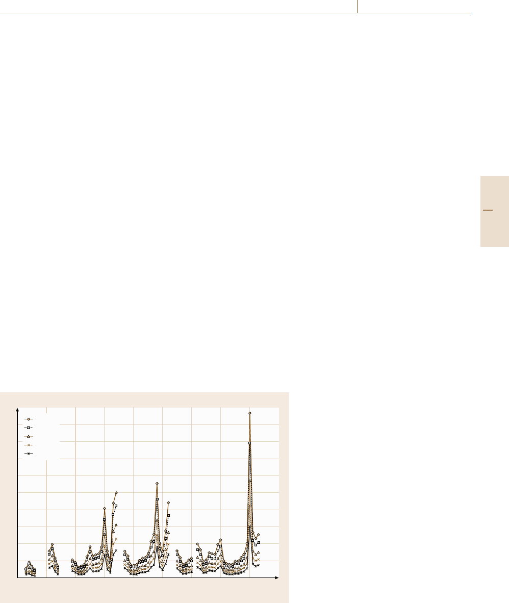
with one sputtering rate. Figure 6.5 shows the sputtering
yield for argon incident at 45
◦
for several energies and
many elements. The rates for different elements clearly
vary enormously. The rate then needs to be evaluated for
Part B 6.1

294 Part B Chemical and Microstructural Analysis
each layer separately or evaluated through calculation of
the relevant sputtering yields Y and a measurement of
the ion beam current density J at the AES measurement
point
d =
JtYa
3
e
, (6.21)
where t is the time for sputtering that layer, e is the
electronic charge, and a
3
is the atomic volume deduced
from
1000ρNa
3
= A . (6.22)
Ion beam currents generally need to be measured using
a screened Faraday cup, but focused ion beam currents
may be measured using an appropriately drilled hole in
a sample or the sample stage [6.105]. In (6.22), ρ is
the density of the element (kg/m
3
) of atomic weight A,
and N is Avogadro’s number. Thus, d may be deter-
mined if Y is known. Values of Y have been tabulated
for many elements and some compounds. Recent work
has led to significant improvements in the accuracy of
calculating Y for elements for Ne, Ar, and Xe bombard-
ing ions at 0
◦
and 45
◦
angles of incidence [6.93, 104],
with a typical uncertainty, for the calculations shown in
Fig. 6.5, of 10%. The equations are rather complicated,
and so plots of the yields may also be found as tables on
the NPL website [6.100]. The uncertainty from this con-
venient route, however, means that it is not as accurate
as a direct measurement of depth.
For sputter depth profiling, Ar is most popular. Oc-
casionally, if the argon AES peaks interfere with the
peaks to be measured, Ne or Xe may be used [6.104].
Some analysts prefer Xe, as the depth resolution is then
improved sometimes.
6.1.2 X-ray Photoelectron Spectroscopy
(XPS)
General Introduction
XPS has a considerable base in physics, in common
with AES, and is often conducted using the same instru-
ment. XPS uses characteristic x-rays to excite electrons
that are energy-analyzed by the same spectrometer that
is used for high-energy-resolution AES analysis. Thus,
XPS instruments often have an added electron gun for
AES. The x-ray source is generally of Mg or Al K
α
x-rays or, in many modern instruments, monochromated
Al K
α
x-rays. As shown in Fig. 6.2b, the x-rays of en-
ergy hν directly eject core electrons from the solid with
kinetic energy E given by
E = hν −E
1
, (6.23)
where it is these E
1
values, the core level binding en-
ergies, that are required in XPS. Thus, the E
1
values
are usually taken to be positive values, unlike (6.1),
and the binding energy scale is usually used directly
rather than the Fermi level referenced kinetic energy E.
The values of E
1
provide information about the chem-
ical state of the element analyzed. Tabulated binding
energies for the elements may be found in Bearden
and Burr [6.106] and for elements and compounds in
handbooks [6.107–109], textbooks [6.110], and web-
sites [6.111]. Note that, whilst Bearden and Burr use the
x-ray nomenclature for energy levels, as is common for
AES, this is rarely used in XPS. Here, the level number
and subshell letter with the spin–orbit coupling number
are given, so that M
V
or M
5
translates to 3d
5/2
. After
the initial excitation, the atom is left with a core hole
that can be filled by an Auger process ejecting an Auger
electron. This electron also appears in the measured
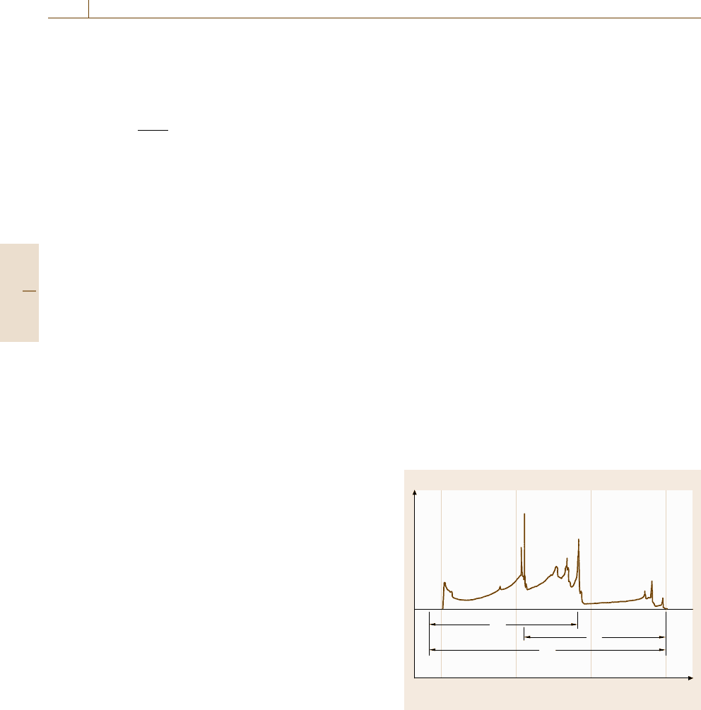
spectrum. Figure 6.6 shows a photoelectron spectrum
for copper with the photoelectron and Auger electron
peaks labeled.
The photoelectrons are mostly in the kinetic en-
ergy range 500–1500 eV, and so XPS is similar to AES
in its surface sensitivity. For a few (often important)
elements, the characteristic peaks may have kinetic en-
ergies as low as 200 eV.
Intensity
Electron kinetic energy (eV)
150010005000
FL XFL
X
X
X
X
A
A
A
A
A
E
A
E
B
hv
X
X
Fig. 6.6 X-ray photoelectron spectrum for Cu using an un-
monochromated Al x-ray source. The photoelectron peaks
are labeled “X” and the Auger electron peaks “A”. The po-
sitions of the Cu Fermi level (FL) and the photoemitted
Fermi level electrons (XFL) are indicated (after [6.53]).
The vacuum level, indicated by the start of the spectrum,
is 4.5 eV above the Fermi level (FL) and is shown exagger-
ated here for illustrative purposes
Part B 6.1

Surface and Interface Characterization 6.1 Surface Chemical Analysis 295
Handling of Samples
The handling of samples for XPS is generally the same
as that for AES, except that the types of sample tend
to be rather different. Environmental contaminants such
as poly(dimethyl siloxane) (PDMS) and similar mater-
ials are often analyzed, and so it is rare for samples to
be cleaned. More samples are insulating, and so greater
consideration needs to be given to charge control and
charge correction. Information on most of the impor-
tant methods for these is given in ISO 19318, but what
is achievable often depends on the specific instrumental
setup. Samples can be in the form of polymer films or
powders generally not studied by AES.
In some instruments, mounting the sample under
a grid or aperture works well for charge control, but
in others this leads to broader peaks. With monochro-
mated x-rays, an electron flood gun or very low-energy
ion flux may be required for charge control. Whatever
is used, the analyst should ensure that the sample is not
exposed to unnecessary levels of radiation, since most
of these samples are easily degraded by heat, electrons
or ions [6.115,116].
Calibrating the Spectrometer Energy Scale
As for AES, Cu and Au samples set with their angle of
emission ≤ 56
◦
are sputtered clean, and the measured
energies of the peaks listed in Table 6.7 are compared
with the energy values given there. As for AES,the
peak energy for the calibration is evaluated from the
top of the peak without background subtraction [6.54].
In Table 6.7, peak number 3 is an Auger electron
peak, and whilst this works well for unmonochromated
x-rays, it cannot be used accurately with a monochro-
mator. The lineshapes and energies of the K
α
1
and K
α
2
x-rays that characterize hν in (6.23) appear to be the
same in all unmonochromated instruments. However,
the lineshapes and energies of the K
α
x-rays, when
monochromated, vary significantly and depend on the
setup of the monochromator and its thermal stability.
By altering the monochromator settings, the measured
Table 6.7 Reference values for peak positions on the binding energy scale [6.113,114] E
ref n
E
ref n
(eV)
Peak number n Assignment Al K
α
Mg K
α
Monochromatic Al K
α
1 Au 4f
7/2
83.95 83.95 83.96
2 Ag 3d
5/2
− − 368.21
3 Cu L
3
VV 567.93 334.90 −
4 Cu 2p
3/2
932.63 932.62 932.62
This table is a refinement of earlier tables (after [6.53,112])
energies of the peak may be moved over a kinetic
energy range of 0.4 eV without too much loss of in-
tensity [6.113]. Thus, for monochromated systems, in
Table 6.7 the Cu Auger electron peak is replaced by the
3d
5/2
photoelectron peak from Ag. This action requires
the cleaning of an additional sample.
In ISO 15472, the calibration given in Table 6.7 is
included into a full protocol that includes methods of
conducting the calibration, assessing the uncertainties,
establishing tolerance limits, and evaluating a calibra-
tion schedule. For laboratories operating under a quality
system [6.117], and for analysts trying to ensure the
validity of their data, these are essential. Use of the
standard with a modern, well-maintained spectrometer
should result in calibration within tolerance limits of
±0.2 eV over 4 months or ±0.1 eV over 1 month before
recalibration is required. For many purposes ±0.2eVis
satisfactory.
Repeatability of the Intensity Scale
For XPS, the evaluation of the repeatability of the in-
tensity scale is similar to that for AES in Sect. 6.1.1.
Of critical importance are the comments made there in
relation to detectors and especially ISO 21270 on the
linearity of the intensity scale. For XPS, the intensity ra-
tio is determined from cleaned copper using the Cu 3p
and Cu 2p
3/2
peak areas after subtracting a Shirley
background [6.118]. Smoothing of the end-points for
establishing the Shirley background can improve the
precision and enable repeatabilities as good as 0.2% to
be achieved in a series of measurements. ISO 24237 de-
scribes the signal levels and procedures needed to get
the best quality data from a sample of copper, how to
build that into a monitoring protocol, and how to set up
tolerance limits for a control chart to try to ensure that
the intensity measurements remain fit for purpose.
Calibrating the Intensity Scale
Interlaboratory studies to compare the shapes of spec-
tra obtained in different laboratories unfortunately show
Part B 6.1

296 Part B Chemical and Microstructural Analysis
that there are marked differences [6.63]. These lead to
variations of a factor of two in quantification if the same
relative sensitivity factors were to be used in all labora-
tories.
The situation here and the rationale and protocol for
evaluating and using the IERF are precisely the same as
in Calibrating the Intensity Scale for AES, except that,
instead of measuring the spectra from the Cu, Ag, and
Au reference foils using 5 keV electrons, we use the Al
or Mg x-rays at incident angles in the range 0−50
◦
from
the surface normal.
Relative sensitivity factors for (6.3) are avail-
able from several handbooks [6.107–109], text-
books [6.119], and publications [6.69, 120]. Early
sensitivity factors varied significantly [6.69], and it is
not clear if these later values are consistent for the in-
struments intended or if significant uncertainties still
persist. An analysis has not been made since the assess-
ment in 1986 [6.69] showed that the sensitivity factor
datasets were very variable. This partly arose because
the IERFs of the instruments used were not measured
in these handbooks. In the next section we address the
basic concept of the peak intensities.
Quantitative Analysis
of Locally Homogeneous Solids
Following the procedure for AES, the x-ray photoelec-
tron intensity per photon of energy hν into a small solid
angle dΩ for a pure element A from the subshell X
i
is
0.45
200 eV
200 eV
1000 eV
1000 eV
0.4
0.35
0.3
0.25
0.2
0.15
0.1
0.05
0.5
ω
0
0
a) b)
100
Atomic number Z Atomic number Z
80604020
0.45
0.4
0.35
0.3
0.25
0.2
0.15
0.1
0.05
0.5
ω
0
0 10080604020
1000 eV
2600 eV
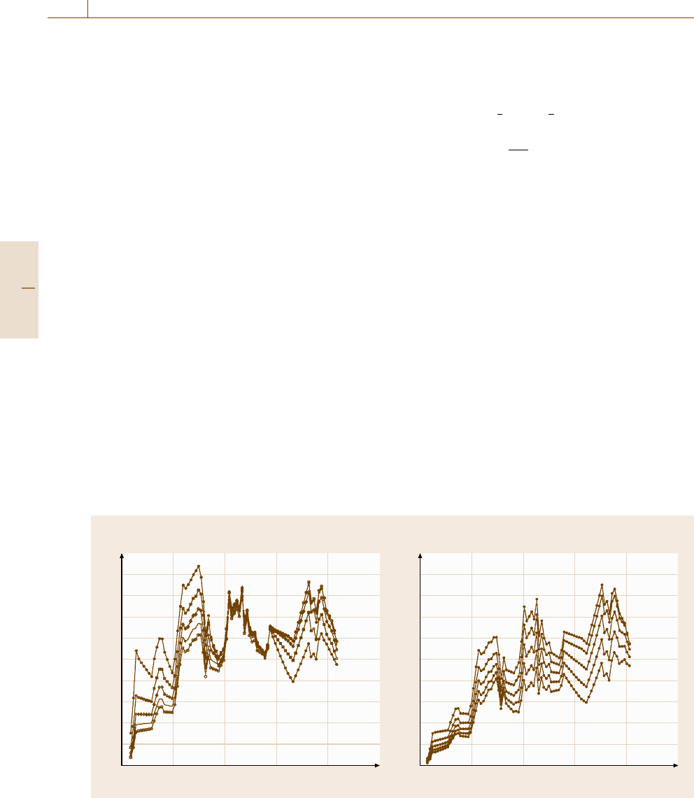
Fig. 6.7a,b Dependence of ω on Z for various electron kinetic energies: (a) at 200 eV intervals from 200 to 1000 eV and
(b) at 400 eV intervals from 1000 to 2600 eV (after [6.82])
given by
I
∞
AX
i
=n
AX
i
σ
AX
i
sec αN
A
Q
A
(E
AX
i
)λ
A
(E
AX
i
)
×
1 +
1
2
β
effAX
3
2
sin
2
γ −1
×cosθ
dΩ
4π
, (6.24)
where n
AX
i
is the population of electrons in the sub-
shell i of the core level shell X of element A, σ
AX
i
is the ionization cross section for that core level for
photons of energy hν, α is the angle of incidence of
the x-ray beam from the surface normal, γ is the an-
gle between the incident x-ray beam and the direction
of the photoemitted electrons, and the other terms are
as for AES. Values of the product of n
AX
i
and σ
AX
i
are taken from the data of Scofield [6.121]. Other cross
sections exist but have been shown to be less accu-
rate [6.122].
At the magic angle, where γ =54.7
◦
, the final term
in square brackets in (6.14) is unity. However, at other
angles this function is generally higher than unity for
γ>54.7
◦
. The values of β are tabled by Yeh and Lin-
dau [6.123] and by Reilman et al. [6.124]aswellas
others. The parameter β is valid for gas-phase work, but
in solids Jablonski [6.81]hasshownthatβ is reduced
to β
eff
by elastic scattering. Seah and Gilmore [6.82] re-
duce Jablonski’s Monte Carlo data to sets of equations
Part B 6.1

Surface and Interface Characterization 6.1 Surface Chemical Analysis 297
such as
β
eff
(θ) =β
eff
(0)(1.121 −0.208 cos θ
+0.0868 cos
2
θ) , (6.25)
where
β
eff
(θ)
β
eff
=0.876[1−ω(0.955 −0.0777 ln Z)],
(6.26)
where the value of ω may be read from graphs [6.82]or
Fig. 6.7.
The above calculation gives the PERSFsforXPS.
However, for quantification, as discussed above for
AES, we really need AMRSFsandsomayuse(6.3). If
we use PERSFsin(6.3) and effectively ignore the rele-
vant matrix factors, the errors involved range from 0.3
to 3 [6.86]. Using PERSFs we obtain (6.16)and(6.17),
exceptthatthe[1 +r(E)] term is replaced by the term
[1 +1/2β
eff
(3/2 sin
2
γ −1)]. Then [6.86,125]
I
Av
A
=n
AX
i
σ
AX
i
N
Av
Q
Av
(E
AX
i
)λ
Av
(E
AX
i
)
× G
Av
(E
AX
i
) , (6.27)
where [6.125]
G
Av
(E
AX
i
) =1 +
1
2
β
effAvAX
i
(θ)
3
2
sin
2
γ −1
.
(6.28)
Here, β
eff
is calculated via (6.25)and(6.26) with
Z
Av
=41 and ω
Av
deduced from Fig. 6.7 or the NIST
databases [6.5,126].
In the past, the differences between the PERSFsand
AMRSFs have not been recognized, and in general, the
experimental data have been for compounds and not ele-
ments and so relate more closely to AMRSFs. However,
these had no spectrometer calibration and furthermore
were often blended with PERSF calculations, leading to
parameters that were ill defined but which were adjusted
by manufacturers to give valid results on their equip-
ment when tested against certain reference compounds.
As noted in Sect. 6.1.1, tables of data for AMRSFsand
their constituent parts are on the NPL website [6.3] un-
der Reference data for the convenience of analysts.
Quantification
of Thin Homogeneous Overlayers
An important use of XPS is the measurement of over-
layer thicknesses of up to 8 nm. The intensities of a pure
layer of A of thickness d on a substrate of B are given
by
I
A
= I
∞
A
1−exp
−
d
L
A
(E
A
)cosθ
(6.29)
and
I
B
= I
∞
B
exp
−
d
L
A
(E
B
)cosθ
. (6.30)
In the approximation of no elastic scattering, the L
A
val-
ues would be the IMFPs λ
A
. However, in the presence
of elastic scattering, Cumpson and Seah [6.127]showed
that λ
A
should be replaced by the attenuation length L
A
and that (6.29)and(6.30) were valid for θ ≤ 58
◦
. Seah
and Gilmore [6.82] analyze these data to show, as an ana-
log to (6.16), that elastic scattering leads to
L
λ
=0.979[1 −ω(0.955 −0.0777 ln Z)]. (6.31)
More detailed calculations by Jablonski and Powell [6.128]
give similar results.
For general films, (6.29)and(6.30) are not easy to
solve for d from values of I
A
and I
B
, since if E
A
= E
B
,
the analysis must become iterative. For this reason,
Cumpson devised the Thickogram to help solve this
problem [6.129]. For metals and their oxides as over-
layers, (6.29)and(6.30) can be used for the oxygen
peak and the substrate in the metallic form. However,
any adsorbed moisture on the surface adds to the oxygen
peak [6.130], and a better method is to use the substrate
metal intensities in the oxide (o) and elemental (e) states
using XPS with peak synthesis. This has the advantage
that E
A
= E
B
sufficiently closely that their difference
may be ignored. Thus, in XPS,
d = L
o
cos θ ln(1 −R
expt
/R
o
) , (6.32)
where R
expt
= I
o
/I
e
and R
o
= I
∞
o
/I
∞
e
. The value of R
o
may be calculated, but for accurate measurements of d,it
is recommended that R
o
is measured experimentally us-
ing the same peak fitting as will be used for the analysis.
If the samples can be reasonably cleaned, and if there is
a significant range of thicknesses, a plot of I
o
versus I
e
gives the sought-after I
∞
o
and I
∞
e
as the intercepts of the
axes, since from (6.29)and(6.30),
I
o
I
∞
o
+
I
e
I
∞
e
= 1 . (6.33)
The use of (6.32) to quantify the thicknesses of thermal
SiO
2
layers on Si wafers has been evaluated in a major
international study [6.130, 131] involving comparison
with medium-energy ion scattering (MEIS), Rutherford
backscattering spectrometry (RBS), elastic backscat-
Part B 6.1
