Hawkes P.W., Spence J.C.H. (Eds.) Science of Microscopy. V.1 and 2
Подождите немного. Документ загружается.

868 M. Howells et al.
The beam then re-enters a vacuum environment where the objective
zone plate and the image detector are located. At energies below a few
keV, the most common detector is a backside-thinned CCD which is
directly illuminated by the X-ray beam; at higher energies, phosphor
screens imaged by a visible light lens onto a CCD detector are com-
monly used. Because of the desire to deliver 10–50 nm resolution using
detectors with 1–20 µm pixel size, the distance from the zone plate
objective to the detector is often in the range 1–2 m to give acceptably
high optical magnifi cation.
The approach described above is commonly used with bending
magnet and laboratory sources. Particular challenges arise when the
source phase space area is dramatically smaller than desired, which is
the case for undulator sources on low emittance storage rings. As an
example, Niemann has studied a variety of solutions for the condenser
of the current Berlin TXM which is illuminated by a BESSY II undulator
(Niemann, 1998). The adopted solution (Niemann et al., 2000) involves
a zone plate segment, and three fl at mirrors. The magnifi cation of the
zone plate is chosen to fully illuminate the object fi eld. The fi rst mirror
is fi xed but must be tilted for a change of wavelength. The other two
mirrors are mounted in a structure that rotates and delivers an incoher-
ent hollow-cone beam of which the inner-to-outer angular difference
(∆ϑ say) is determined by phase-space matching and the outer angle
(the NA) is determined by the last-mirror refl ection angle. Thus the
illuminated area and the NA can both be chosen, no wobbling is
required and Liouville’s theorem (which states that the phase space
area of an optical beam is a conserved quantity) is respected by allow-
ing ∆ϑ to fl oat. This system represents an elegant optical solution,
though it is mechanically quite complex. Other strategies to expand the
phase space of an XUV beam have been explored by the microfabrica-
tion community who are concerned about “fringing” in XUV lithogra-
phy (Murphy et al., 1993; White et al., 1995). One such approach is to
design a pseudorandom diffractive optic specifi cally to “spoil” the
phase space of a beam and match the object size and the NA (David
et al., 2003); such optics must meet the challenge of evenly fi lling both
the object plane and the back focal plane with light.
3.1.2 TXM Phase Contrast Layout
As noted above, phase contrast plays an important role in X-ray micros-
copy, particularly at higher photon energies. In order for phase varia-
tions at the specimen plane to produce intensity variations at the
detector, some method of mixing the wave diffracted by the specimen
with an undiffracted phase-reference wave must be employed. The
most common approach in X-ray microscopes is that of Zernike. In
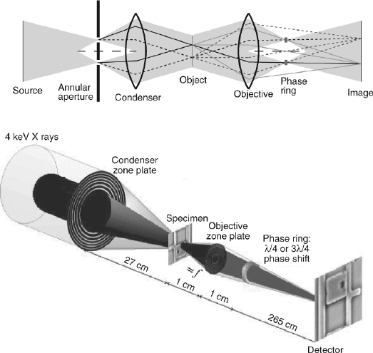
light microscopes, Köhler illumination is provided by using a relay
lens to image the source on to the front focal plane of the condenser.
Points at this front focal plane deliver parallel beams to the object plane
which (if undeviated by the object) are focused on to a phase ring at
the back focal plane of the objective where they are phase shifted
usually by ±π/2. Thus the ring aperture at the front focal plane of the
condenser provides a narrow, hollow cone of illumination of the speci-

Chapter 13 Principles and Applications of Zone Plate X-Ray Microscopes 869
men, and is conjugate to the phase ring. At the same time, light origi-
nating from a point scatterer in the object is focused to the detector,
where it interferes with the phase-shifted unscattered light (see Figure
13–17).
In X-ray microscopes, it is more diffi cult to use a relay optic to work
in the Köhler illumination condition; instead, the front focal plane ring
aperture is illuminated by nearly parallel light from the source (i.e.,
critical illumination) so that a much smaller area of the condenser is
illuminated. Because the light from the front aperture is much more
collimated than would have been the case with Köhler illumination,
the longitudinal location of the aperture and its corresponding phase
ring is much less critical than it is with visible light microscopes so the
Figure 13–17. Two illustrations of a Zernike phase contrast optical system. The upper one shows the
classical scheme used in light microscopes based on Köhler’s illumination (Born and wolf, 1999). Light
from each point of the annular aperture, placed in the front focal plane of the condenser, is delivered
to the object as a parallel beam. Two object points are shown receiving example rays from the source.
The rays may be undeviated by the object, in which case they are seen to pass through the phase ring
on their way to the detector. On the other hand the rays may be deviated (diffracted) by the object in
which case they reach the detector without passing through the phase ring. Interferences between
these two types of optical signal result in a mapping of the object phase variations into an intensity
pattern on the detector. The lower diagram shows a practical synchrotron radiation implementation
of a Zernike-phase-contrast TXM at the 4.1 keV beam line on ID21 at the European Synchrotron Radi-
ation Facility in Grenoble. An undulator X-ray source is followed by a crystal monochromator illu-
minating a condenser zone plate (which can be small since it does not have to act as a linear
monochromator). The condenser provides critical illuminated to the sample rather than Köhler and,
due to the good collimation of the incoming beam, the illuminated area of the condenser is projected
on to a phase ring in the back-focal plane of the objective zone plate so as to provide phase contrast.
The tradeoffs involved in choosing the illuminated area of the condense pupil are discussed in Section
3.3.6 (Courtesy J. Susini, ESRF.)
870 M. Howells et al.
primary alignment requirement is transverse to the X-ray beam direc-
tion. For computations it is convenient to consider the two rings as
built into the lens pupil functions. Evidently when the source is well-
collimated, as in the case of a synchrotron, there is no need for the two
rings to be exactly conjugate. The use of the ring aperture obviously
makes the illumination more coherent. The question of the best choice
of width and radius for the two rings or, equivalently, how much coher-
ence to have, we defer until later (Section 3.3.6). The phase ring itself
is constructed out of a material with a large phase shift per absorption
length f
1
/(2f
2
), such as any material used at energies just below an
absorption edge.
Zernike phase contrast X-ray microscopy was pioneered by Schmahl
and Rudolph (1987) and Ruldoph et al. (1990), and the Göttingen group
has shown impressive results in phase contrast for water-window
imaging of biological specimens. The ring aperture and phase ring
used in these experiments could be rapidly inserted or retracted
(Schmahl et al., 1994, 1995; Schneider, 1998). Phase contrast is arguably
even more important at higher X-ray energies where it is the dominant
contrast mechanism. For example a hard X-ray (4 keV) phase-contrast
microscope, illuminated by a system using a crystal monochromator
followed by a condenser zone plate, is operating at the ID21 beam line
at the European Synchrotron Radiation Facility in Grenoble, France
(see Figure 13–17). Imaging of functioning integrated circuits at 60 nm
resolution has been demonstrated (Neuhäusler, 2003); see Section 4 for
more information on this. In another example of this confi guration, the
National Synchrotron Radiation Research Center in Hsinchu, Taiwan
has installed a TXM built by Xradia. This instrument (alluded to in
Sections 1.4 and 2.4.4) has been used to demonstrate the long-existing
idea of using zone plate higher focal orders for imaging. In water-
window instruments which typically have focal lengths on the order
of 1–2 mm in fi rst order (and therefore 1/3–2/3 mm in third order), the
idea has not been readily adopted due to practical considerations of
working distance. However, in the Taiwan experiment (Tang et al.,
2006) a phase contrast image of a fabricated test object was made at
8 keV using a 50 nm outer-zone-width zone plate in third order. Lines
of minimum width 30 nm were imaged clearly and the authors esti-
mate a resolution below 25 nm. This is evidently a most important
development (see Section 5).
3.1.3 Scanning Ttransmission X-Ray Microscope (STXM) Layout
Scanning transmission X-ray microscopes (STXMs) typically use a
zone plate to demagnify a pinhole source to a small focus spot through
which the specimen is scanned. While initial demonstrations using
synchrotron radiation used pinhole optics (Horowitz and Howell, 1972;
Rarback et al., 1980), the use of zone plate optics in scanning micro-
scopes was pioneered by Rarback et al. (1984) and later by (Niemann,
1987; Niemann et al., 1988). (Normal incidence optics with synthetic
multilayer refl ective coatings have also been used in the 50–120 eV
range (Haelbich, 1980a; Haelbich et al., 1980b; Ng et al., 1990)). Since
scanning microscopes require coherent illumination to reach their
Chapter 13 Principles and Applications of Zone Plate X-Ray Microscopes 871
maximum resolution, they have often used undulators as high bright-
ness sources (Rarback et al., 1988; Kenney et al., 1989; Morrison et al.,
1989b) though excellent performance has also been obtained using
bending magnet sources on low emittance storage rings (Kilcoyne
et al., 2003). While a large number of STXMs are now in operation, we
describe here the characteristics of the most recent in a series (Rarback
et al., 1988; Jacobsen et al., 1991; Feser et al., 1998, 2000) of undulator-
based scanning microscopes built at Stony Brook University for opera-
tion at the National Synchrotron Light Source at Brookhaven National
Laboratory in New York. A soft X-ray undulator plus spherical grating
monochromator with an energy resolution that can be as good as
0.06 eV at 290 eV (Winn et al., 2000) is used to deliver soft X-rays to a
2D exit slit which can limit the beam size in the range 25–120 µm in
both x and y. This slit then serves as a secondary radiation source for
zone plates of either 80 or 160 µm diameter and zone widths of 30–
45 nm (Spector et al., 1997; Tennant et al., 2000), producing a focal spot
of 36–54 nm Rayleigh resolution. The beam emerges from the ultra high
vacuum synchrotron beam line into an atmospheric pressure environ-
ment by passing through a 100 nm thick Si
3
N
4
window. The zone plate
includes a central stop of about half the zone plate diameter; this stop
must be made quite thick (0.3 µm gold is common for soft X-ray applica-
tions) so that the undiffracted light transmitted through the large
central stop is kept to a very small level compared to the fl ux in the
focused X-ray beam. The zone plate is then followed by an order sorting
or selecting aperture (OSA) so that a pure fi rst-order focal spot is
obtained.
While steering mirrors are used to scan the beam in visible light
scanning microscopes, it is easier to maintain signal uniformity by
keeping the beam and zone plate fi xed and scanning the specimen
through the focal spot. This is accomplished using an X-Y-Z stack of
stepping motor stages for large motion with 1 µm precision, and a piezo
scanning stage for 50–100 µm range and nanometer precision. Because
piezos have nonlinearities and hysteresis in their response to scan volt-
ages, some form of closed-loop feedback is generally used, based on
position signals such those provided by linear voltage differential
transformers (Kenney et al., 1985), capacitance micrometers (Jacobsen
et al., 1991), or laser interferometers (Shu et al., 1988; Kilcoyne et al.,
2003); the latest Stony Brook STXM allows the user to choose between
capacitive or laser interferometer feedback. The specimen is then fol-
lowed by a high effi ciency X-ray detector; common choices include the
use of gas-based proportional counters which offer extremely high
effi ciency of detection for those X-rays that make it through a thin
entrance window (Rarback et al., 1980; Kenney et al., 1985; Feser et al.,
2000) but which suffer from a count-rate limit of about 1 MHz. Alterna-
tives are phosphor-coated screens followed by photomultipliers to
detect the resulting visible light (Maser et al., 2000), and solid state
detectors which are capable of signifi cantly higher signal rates (Barrett
et al., 1998; Wiesemann et al., 2000; Feser et al., 2001, 2003; Guttmann
et al., 2001). In the Stony Brook STXM, the user can choose between
proportional counter and segmented silicon detectors, and a visible
872 M. Howells et al.
light microscope is also placed on the detector stage with X-Y-Z motor-
ized motion so as to pre-locate desired regions of the specimen. Another
approach, which works for either a TXM or a STXM, is co-indexing of
off-line light microscopes with the X-ray microscope (Meyer-Ilse et al.,
1994, 2001; Kilcoyne et al., 2003).
Scanning microscopes offer different characteristics than full-fi eld
imaging systems do. These include the ability to quickly change from
scanning very large areas at low resolution to taking high resolution,
small fi eld scans, and reduced radiation dose because the 5–20% effi -
cient zone plate is located upstream of the specimen rather than down-
stream. Because of the need to mechanically scan the specimen in most
present microscopes, and the need for coherent illumination, imaging
times are generally longer (in the range of one or a few minutes, rather
than seconds in the case of many TXMs). At the same time, the require-
ment for coherent illumination means that the etendue or phase space
that the monochromator must accept is greatly reduced, so that aber-
rations are reduced and it is relatively easy to obtain very high spectral
resolution. These characteristics make scanning transmission X-ray
microscopes especially well suited to low-dose spectromicroscopy
applications, as will be described below.
Phase contrast has historically seen less use in scanning transmis-
sion X-ray microscopes. However, refractive and diffractive effects by
the specimen lead to a redistribution of signal on the detector which
can be interpreted to give phase contrast images (as will be discussed
below). The ultimate approach is to use a 2D detector (such as a CCD
camera) to detect the entire intensity distribution at each pixel of a
scanned image; Chapman has used this in an impressive demonstra-
tion of Wigner deconvolution microscopy to recover the phase and
magnitude distribution of the specimen as well as the zone plate objec-
tive (Chapman, 1996a), while Morrison et al. have used this to obtain
fi rst moment images which reveal the dominant phase gradient at each
pixel location (Eaton et al., 2000; Morrison et al., 2002). Coupled with
the potential power of these approaches are signifi cant challenges: the
readout time of large pixel detectors is often not in the few or sub
millisecond pixel timescale required for fast scanning, and the result-
ing 4D data fi les are quite large. More fundamental is the question of
statistical signifi cance in each pixel of a large array detector when
radiation dose to the specimen must be considered; in some cases it
may be preferable to divide a weaker signal into fewer detector seg-
ments. This approach has been used by Feser et al. (Figure 13–18) who
have used a detector with only 8 segments to obtain quantitative phase
contrast images while operating at per-pixel acquisition times of a few
milliseconds and producing data fi les of manageable size (Feser et al.,
2003) (see Figure 13–18). Additional approaches to obtaining phase
contrast in scanning microscopes include the use of zone plate dou-
blets (Kaulich et al., 2002) or phase modifi ers (Polack et al., 2000) to
produce differential interference contrast. Undoubtedly different
experiments will involve different choices in the tradeoff of the fi ne-
ness of segmentation of scanning microscope detectors, but in any case

Chapter 13 Principles and Applications of Zone Plate X-Ray Microscopes 873
it is clear that phase contrast plays an interesting role in scanning
transmission X-ray microscopy as well as in microprobes as will be
noted below.
3.1.4 Scanning Fluorescence X-Ray Microprobe (SFXM) Layout
Scanning fl uorescence X-ray microprobes (SFXM) use a focused X-ray
beam to stimulate the emission of characteristic fl uorescence X-rays
from specifi c elements in the specimen. When linearly polarized radia-
tion (such as is usually obtained from synchrotron sources) is used, a
fl uorescence detector placed 90º to the beam in the polarization plane
will detect a minimum of coherent scattering signal; this detector must
then have some means of discriminating between different X-ray emis-
sion energies. Energy-dispersive detectors accomplish this by measur-
ing the number of electron-hole pairs created by each X-ray in a
semiconductor material, while wavelength-dispersive detectors use a
crystal optic or a grating to separate the X-ray energies. Energy disper-
sive detectors generally have large solid angle collection, and multi-
element detectors can be used to overcome the ∼50 kHz count rate limit
determined by charge readout time, while wavelength dispersive
detectors offer better separation between nearby spectral lines and
larger dynamic range for detecting low concentration elements amongst
other fl uorescing elements of higher concentration. There is a long and
rich history of synchrotron-based microprobes (Horowitz and Howell,
1972; Sparks, 1980; Rivers et al., 1988; Thompson et al., 1988; Hayakawa
et al., 1989), and a variety of optical approaches including the use of
compound refractive lenses and Kirkpatrick-Baez mirror optics are
now achieving submicron resolution. We outline here some of the
characteristics of microprobes using zone plate optics (Barrett et al.,
1998; Yun et al., 1998a; Suzuki et al., 2001; Kamijo et al., 2003) by
Figure 13–18. Amplitude (left) and phase (right) contrast images of a germanium test pattern imaged
using a scanning transmission X-ray microscope with a segmented detector. Also shown is a schematic
view of the detector. The undefl ected beam from the fi rst order focus is directed into bright fi eld seg-
ments 1, 2 and 3 (these segments also allow differential interference contrast). The defl ected beam is
detected in the angular segments 4, 5, 6, and 7 for dark fi eld imaging and differential phase contrast.
(Reprinted from Feser et al., © 2003, with permission of EDP Sciences.)
Zero order
OSA
First
order
focus
1
1
2
3
4
5
6
7
1 µm
874 M. Howells et al.
considering the example of the 2-ID-E microprobe at the Advanced
Photon Source at Argonne National Laboratory near Chicago.
This microprobe operates using a side-defl ecting crystal monochro-
mator to transfer an off-axis part of the central cone produced by a
hard X-ray undulator. In the vertical direction, the objective zone plate
images the source directly onto the specimen, while in the horizontal
direction the variable width monochromator exit slit is imaged. Astig-
matism effects are avoided in the resulting focused beam by the fact
that the depth of focus is much larger than the difference between
the positions of the horizontal and vertical foci of the zone plate (in
addition, the zone plate can be tilted to compensate for more severe
source astigmatism). Zone plates of diameter 160–320 µm and outer-
most zone width of 100 nm are typically used, giving focal lengths of
12–25 cm at 10 keV. While the probe size can be as small as 150 nm, a
larger horizontal source size is often chosen to give more fl ux at the
cost of resolution. The specimen is mounted at 15º to the incident
beam to provide access to both the incident X-ray beam and the fl uo-
rescence detector, and it is scanned by motor-driven stages with 0.1 µm
step size. A multi-element germanium fl uorescence detector is used
to collect the fl uorescent signal; one can either record the signal in a
limited number of pre-defi ned energy windows for rapid analysis
with modest data fi le size, or record the full fl uorescence spectrum
per pixel for improved quantitation of elements with closely spaced
fl uorescence energies. The region consisting of the specimen and
detector is located inside a glovebox which can be purged with helium
to eliminate fl uorescence from argon in air which would otherwise
obscure a number of low-Z elements, and to reduce the absorption of
low-Z fl uorescence signals by air. Per-pixel dwell times are on the
order of one second, so that the experimenter must be judicious in the
choice of scan area (the use of common position indexing between a
visible light microscope and the microprobe aids in rapid specimen
location).
Zone plates of diameter 50–100 µm and outermost zone width of
100–300 nm are typically used, giving focal lengths of 5–30 mm. While
the probe size can be as small as the Rayleigh resolution of 1.22 times
the outermost zone width, a larger virtual source size is often chosen
to give more fl ux at the cost of resolution. The specimen is mounted at
45º to the incident beam to provide access to both the incident X-ray
beam and the fl uorescence detector, and it is scanned by motor-driven
stages with 0.1 µm step size. A multi-element germanium fl uorescence
detector is used to collect the fl uorescent signal; one can either record
the signal in a limited number of pre-defi ned energy windows for
rapid analysis with modest data fi le size, or record the full fl uorescence
spectrum per pixel for improved quantitation of elements with closely
spaced fl uorescence energies. The region consisting of the zone plate,
specimen, and detector is all located inside a glovebox which can be
purged with helium to eliminate fl uorescence from argon in air which
would otherwise obscure a number of low-Z elements. Per-pixel dwell
times are on the order of one second, so that the experimenter must be
judicious in the choice of scan area (the use of common position index-

Chapter 13 Principles and Applications of Zone Plate X-Ray Microscopes 875
ing between a visible light microscope and the microprobe aids in
rapid specimen location).
Trace element mapping by fl uorescence detection with sensitivities
down to about 100 parts per billion, or about 10
−17
grams of iron within
a (200 nm)
2
spot, represents the majority of microprobe applications.
However, X-ray microprobes can be used in a number of other ways
as well, including measurements of crystal strain in small regions
(Rebonato et al., 1989; Cai et al., 1999; Soh et al., 2002) and differential-
aperture measurements of microstructure and strain (Larson et al.,
2002). The phase contrast methods described above for STXM are
equally applicable in SFXM, and offer a much-needed way to image
the overall mass and ultrastructure of specimens while simultaneously
forming trace element or strain maps.
3.2 Fundamentals of Contrast in the TXM
It is useful to have an analytical treatment that provides insight into
the way a microscope produces contrast and at the same time allows
simple calculations to assess experimental plans. This was provided
by Rudolph and coworkers (1990) in a form that allows amplitude-
contrast, Zernike-phase-contrast and dark-fi eld imaging, to be included
in a unifi ed description, that is largely independent of the microscope
design. Assuming only that we have an imaging microscope, we con-
sider fi rst the Zernike phase-contrast TXM.
We are interested in the contrast C or the contrast parameter Q (see
Section 1) between an interesting feature F and a background feature
B generated via the phase shifter S. F and B are defi ned to have the same
thickness but in reality the background material (water for example)
may be thicker than the feature so we allow for that by adding a layer
L. If we defi ne the complex transmission factors of F, B, S and L as a
F
⋅ p
F
≡ [exp{−2πβ
F
t
F
/l}] ⋅ [exp{2πiδ
F
t
F
/l}] etc where 1–δ–iβ is the refractive
index and t is the thickness, then following Rudolph et al. (1990) we can
obtain the image intensities (I
F
and I
B
) and thence C and Q
C
II
II
II
II
Iaaaaa ppp
FB
FB
FB
FB
FBSFBS FBS
=
−
(
)
+
(
)
=
−
(
)
+
=+
,
**
Θ
22
2Re
−
{
+−
+
}
=
2
2
2
222
2
aa p
aaa ppaa
Ia
BS S
FFB FBBL
BB
Re
Re .
*
*
aaa
SL
22
The dose D (the energy deposited per unit mass of sample) needed to
detect a feature of area d
2
, thickness t
F
= d and density ρ with signal-
to-noise ratio S/N can now be calculated (Rudolph et al., 1990) as
follows
D
S
N
hc
d
aa
FL
=
(
)
−
2
3
22
2
1
λρ Θ
where hc = 1240 eV-nm represents the product of Planck’s constant and
the velocity of light. The above relations are convenient because, in

876 M. Howells et al.
addition to phase contrast, they also describe amplitude-contrast (t
s
=
0) and dark-fi eld (t
s
= large) experiments. The formula for D yields dose
plots like Figure 13–5 and also tells us that the number of X-rays (of
energy E) per unit area required to make the measurement with the
given resolution and signal-to-noise ratio is Dρ/(µE), where µ is the X-
ray absorption coeffi cient.
In the multi-keV X-ray energy range, the phase contrast is substan-
tially larger than the absorption contrast for suitable choices of the
thickness of the phase shifter. The best result is typically achieved by
attenuating the direct beam by the phase plate so that its amplitude
is comparable to that of the scattered signal, resulting in an interfer-
ence of two beams of similar amplitude. The available choices of
phase plate thickness to optimize this are positive phase contrast
(phase shift = π/2, 5π/2, . . .) or negative phase contrast (phase shift =
3π/2, 7π/2, . . .). Figure 13–19 shows contrast plots of some of these
possibilities. Although these plots are useful for providing compara-
tive information, they represent a considerable idealization; the phase
shift is assumed to be applied to 100% of the undiffracted light and
0% of the diffracted light, the thickness of the phase shifter is chosen,
at each energy, to give the stated phase shift and the optical system
is assumed 100% effi cient. Under these assumptions, the dark-fi eld
contrast is identically equal to one. This suggests that dark-fi eld has
a dose advantage that will be dependent on the practical value of the
nominally zero signal due to the undiffracted light and on the strength
of the dark-fi eld signal (Chapman et al., 1996c; Vogt et al., 2001a).
3.3 Partial Coherence
3.3.1 History
The resolution of microscopes, including X-ray microscopes, depends
on the angular widths of the light beams delivered to, and collected
100 1,000 10,000
Energy (eV)
Phase 7π/2
Amplitude
1
10
-1
10
-2
10
-3
10
-4
Fractional contrast
100 1,000
10,000 100,000
Energy (eV)
Amplitude
Phase 3π/2
Phase 7π/2
1
10
-1
10
-2
10
-3
Fractional contrast
Protein in water
Glass
Figure 13–19. Intrinsic amplitude and Zernike phase contrast for two types of sample of thickness
30 nm relative to a background material of the same thickness: protein in water (left) and vacuum in
glass, representing a crack (right). The protein is modeled assuming a density of 1.35 g/cm
3
, composi-
tion of H
50
C
30
N
9
O
10
S and a phase ring made of copper. Glass is modeled assuming a density of 2.5 g/cm
3
,
composition Si
16
Na
12
K
1
Ca
7
Mg
6
P
1
O
57
and a phase ring made of gold. It is noteworthy that the phase
contrast can be much greater than the amplitude contrast even in the 290–540 eV water window.

Chapter 13 Principles and Applications of Zone Plate X-Ray Microscopes 877
from, the sample . The analysis of this effect was pioneered in the 1950s
by Hopkins, Wolf and others and was part of a movement to apply the
linear-systems ideas, widely used by the engineering community, in
the optical arena. This work has been reviewed by Hopkins (1957),
Thompson (1969), and in various texts (Wilson and Sheppard, 1984;
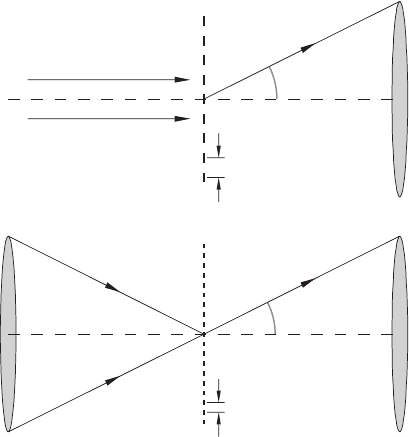
Goodman, 1985; Born and Wolf, 1999). The main point is that the fi nest
features (highest spatial frequencies) in the sample diffract the illumi-
nating beam by the largest angles θ. The best geometry to include such
large defl ection angles is therefore one that has a wide-angle beam
both inward to, and outward from, the sample. This implies broadly
that in a TXM, a large-area source providing spatially incoherent illumi-
nation gives better resolution than a point source giving coherent illu-
mination (Figure 13–20), although such a comparison is not as simple
as it sounds (Goodman, 1968). It would be wrong to conclude from this
that STXMs which use coherent illumination have intrinsically worse
resolution than TXMs. In fact, scanning microscopes with large area
detectors and transmission microscopes with large illumination angles
both deliver incoherent bright-fi eld images with the same resolution
and transfer function provided only that they use objective lenses of
equal resolution.
The fi rst application of linear-systems concepts in X-ray microscopy
was in the analysis of STXM images (Jacobsen et al., 1991; Zhang et al.,
θ
θ
d
d/2
Figure 13–20. Schematic showing why the transfer function for incoherent
imaging extends to twice the spatial frequency of coherent imaging for a given
optic numerical aperture. For coherent imaging (top) the marginal ray is
deviated by an angle theta due to diffraction by the sample periodicity d. For
incoherent imaging (bottom) some light is deviated by 2θ due to the sample
periodicity d/2. Both TXM and STXM with large area detector deliver incoher-
ent bright-fi eld images. (After Jacobsen et al., 1992b.)
