Hawkes P.W., Spence J.C.H. (Eds.) Science of Microscopy. V.1 and 2
Подождите немного. Документ загружается.

898 M. Howells et al.
Cluster 1 Cluster 2 Cluster 3 Cluster 4 Cluster 5
525 530 535 540 545 550
Photon energy (eV)
2 µm
0.0
0.5
1.0
1.5
2.0
Optical Density
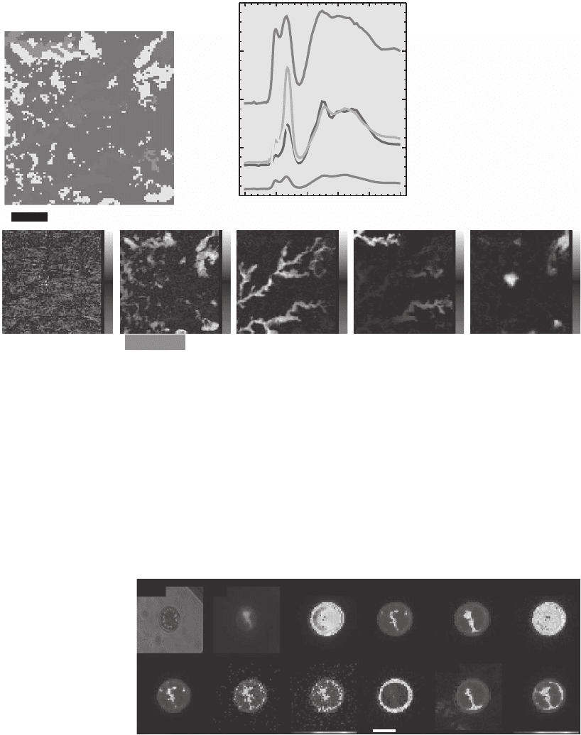
Figure 13–34. Cluster analysis in a spectromicroscopy study of lutetium in hematite. Lutetium is
serving as a homologue to americium in an investigation of the uptake and transport of nuclear waste
products in groundwater colloids. By using a pattern recognition algorithm to search for pixels with
spectroscopic similarities, a set of signature spectra is automatically recovered from the data (shown
here in a color-coded classifi cation map) and thickness maps can be formed based on these signature
spectra. Analysis at the oxygen edge reveals two different phases of reactivity for lutetium with hema-
tite. Analysis by Lerotic (2004), from a study by T. Schäfer, INE Karlsruhe. (See color plate.)
light epi
Si
Ca Mn
Fe
Ni
PS
Cu
K
Zn
10 µm
Figure 13–35. Visible light and epifl uorescence micrographs, and false color
X-ray fl uorescence element maps of a centric diatom collected from the south-
ern Pacifi c. In this region of the ocean, iron availability is a biolimiter with an
impact on oceanic uptake of carbon dioxide from the atmosphere. X-ray micro-
probes allow one to study iron content on a protist-specifi c basis. (Reprinted
from Twining et al., © 2003, with permission from American Chemical Society.)
(See color plate.)
using spectromicroscopy, imaging of the structure and electromigra-
tion failure of integrated circuits, measurements of strain in crystalline
materials using microdiffraction, and studies of surface properties
using photoelectrons. Studies of polymer systems represent one of the
Chapter 13 Principles and Applications of Zone Plate X-Ray Microscopes 899
fi rst uses of zone plate spectromicroscopy (Ade, 1992) (see Figure
13–36), and subsequent work has ranged from exploring fundamental
questions such as confi nement-induced miscibility (Zhu et al., 1999) to
studies of specifi c industrially useful materials using both absorption
contrast (Smith et al., 2001; Rightor et al., 2002; Hitchcock et al., 2003;
Croll et al., 2005) and linear dichroism (Ade an dHsiao, 1993). Other
studies have measured the degree to which polymers can seep into
wood at the cellular level in particleboard (Buckley et al., 2002). These
represent only a few examples; a much wider survey is given in recent
reviews (Ade, 1998; Urquhart et al., 1999).
Modern integrated circuits are incredibly intricate, with oxidation
layers sometimes only a few molecular layers thick, and metallization
planes and vias which connect them, having dimensions in the 100 nm
range. The ability of X-ray microscopes to image thick specimens (espe-
cially using phase contrast at higher energies; see Figure 13–37) is well
suited to studies of the properties and failure modes of such circuits.
As one example, Schneider et al. (2002b, 2002c, 2003) have studied
electromigration failures as they take place, leading to observations of
the propagation of voids from the point of their original formation (see
Figures 13–38 and 13–39), while Levine et al. have done tomographic
imaging of electromigration voids using a STXM (Levine et al., 2000).
For industrial applications of chip inspection, a very signifi cant devel-
opment has been the recent commercial availability (Xradia, Inc.) of
PMMA
6 µm
Pre C edge
Transmission x-ray micrographs
Polystyrene
Energy (eV)
280 290 300 310 320
C 1s π
*
C=C
C 1s π
*
C=O
Continuum
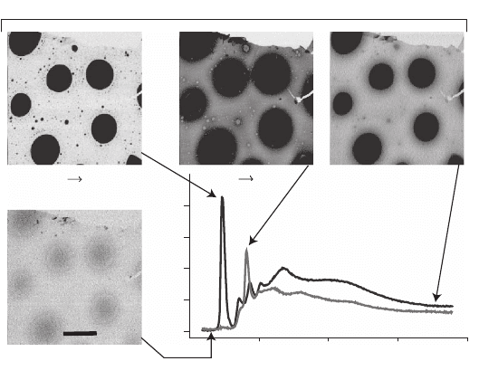
Figure 13–36. One of the fi rst applications of X-ray transmission spectromi-
croscopy was to the study of polymers, where the chemical selectivity of near-
edge absorption resonances allows one to make maps based on XANES
spectral signatures. In this example, polymethylmethacrylate (PMMA) was
spun cast with polystyrene (PS) before annealing, giving rise to phase segrega-
tion. Images acquired at specifi c absorption resonances show very different
contrast and can be used to form compositional maps of the polymers. (Cour-
tesy of D.A. Winest, NCSU.)
900 M. Howells et al.
laboratory-based tomography systems using zone plate optics and
operating at a suffi ciently high energy (5.4 keV) so as to allow tomo-
graphic data sets to be acquired and reconstructed; this allows one
to study various metallization layers in intact, working chips (Wang
et al., 2002) (see Figure 13–40).
Another way in which zone plate X-ray microscopes are used to
study material properties is through microdiffraction, where one
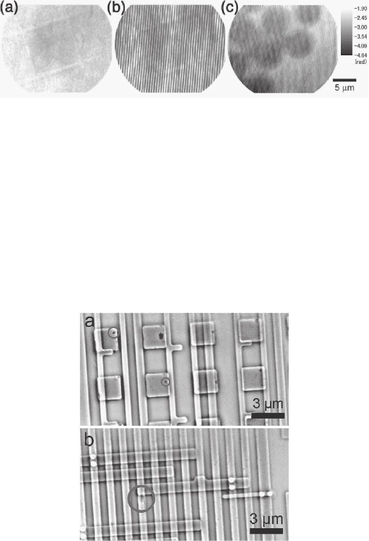
Figure 13–37. Interferometric TXM imaging of polystyrenes at 9 KeV. In these experiments, a hard X-
ray micro-interferometer has been constructed by using two overlying objective zone plates with a
slight transverse offset to produce an interferometric fringe pattern as shown in (b). Compared to the
single-objective image (a), interference fringes with a visibility of as high as 60% can clearly be seen.
Analysis of the interferometric image (b) is then used to obtain the quantitative contrast image of the
polystyrene spheres shown in (c). This example shows how low absorption contrast objects can be
imaged in hard X-ray microscopes. (Courtesy of T. Koyama, Himeji Institute of Technology.)
Figure 13–38. Zernike phase contrast provides one means to image the metallic layers of integrated
circuits in regions where the underlying silicon wafer has been thinned. A common failure mode in
integrated circuits is electromigration in which voids in a conducting layer or via are formed. These
images obtained using a TXM operating at 4 keV at the European Synchrotron Radiation Research
Facility or ESRF, show what appear to be such voids (circles) within test structures for advanced
microprocessors. (Reprinted from Schneider et al., © 2003, with permission from Elsevier.)
Chapter 13 Principles and Applications of Zone Plate X-Ray Microscopes 901
SEM
SEM
SiC low-k
barrier
TXM, E
1
=524.5 eV TXM, E
2
=700.5 eV
1 µm
2 µm
1 µm
X rays
Cu
SiOCH
low-k film
SiO
2
Si
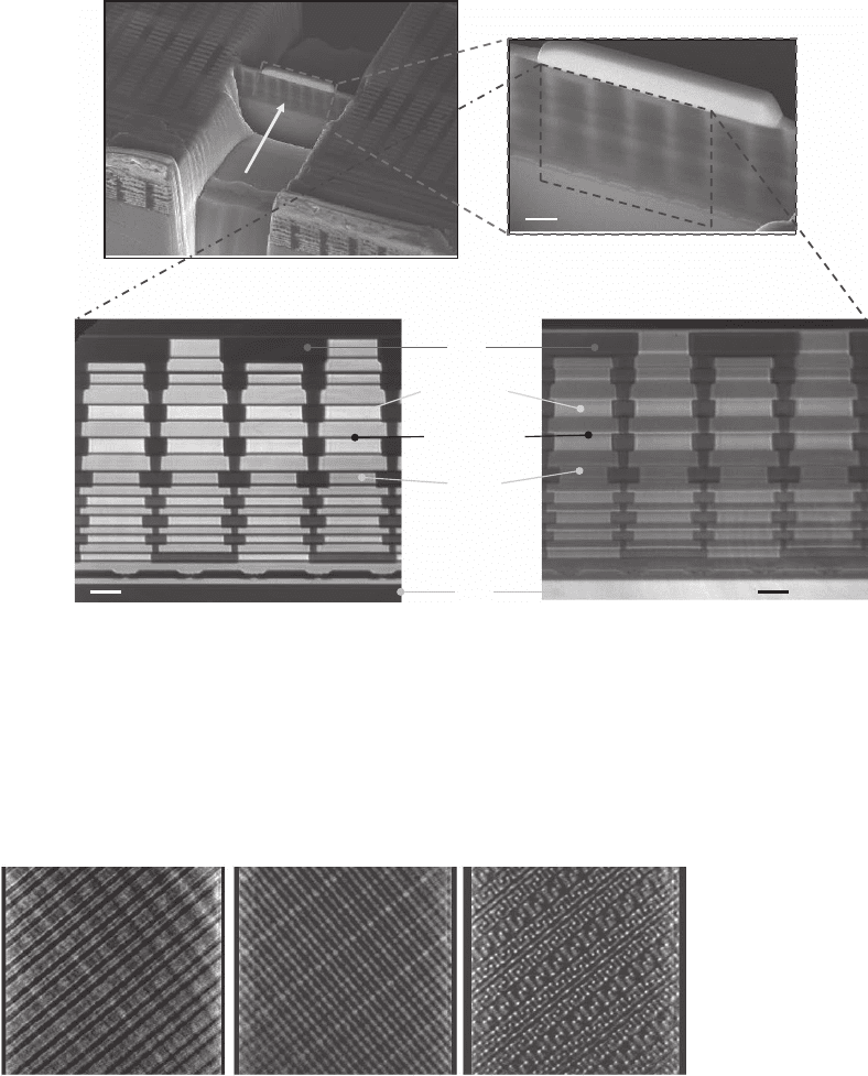
Figure 13–39. In studies of integrated circuits, it is often important to study changes in metallization
from layer-to-layer by imaging in cross section. In this example, a focused ion beam (FIB) system was
used to prepare a thinned, fully passivated cross section of copper interconnect structures within an
electrically functional test structure as shown in the two scanning electron micrographs at top. A soft
X-ray TXM at the BESSY II synchrotron facility in Berlin was then used to image this cross section at
525.5 eV (left) and 700.5 eV (right) to highlight different metal and dielectric layers in the chip, with
features as fi ne as 20 nm visible. (Courtesy of G. Schneider, BESSY.)
Figure 13–40. Tomographic imaging of an integrated circuit done with a com-
mercial laboratory X-ray microscope (Xradia). An integrated circuit had the
silicon wafer underneath a region of interest thinned to about 15 µm, after
which a tilt series of TXM images was acquired over 8 hours using a rotating
anode source operation at 5.4 keV. The fi gure shows slices extracted at depths
corresponding to the center of three Cu interconnect layers in the tomographic
reconstruction with an estimated resolution of 60 nm in the transverse dimen-
sion and 90 nm in depth. This system can be used for chip inspection at a chip
fab plant, among other applications. (Courtesy W. Yun, Xradia.)
902 M. Howells et al.
examines not the undeviated transmission image through the speci-
men, but the signal that is Bragg diffracted (usually in the Laue geom-
etry) by specifi c crystalline regions within the specimen (see for
example (Engström et al., 1995)). Measurement of the position of the
Bragg peaks can give values of the local lattice constants so that repeti-
tion of the measurement over a grid of points provides a strain map of
the sample. This has been applied to optoelectronic devices (Cai et al.,
1999), magnetic domain evolution (Evans et al., 2002) as well as for
examination of the strain at the midpoint and edges of mesoscopic
structures (Murray et al., 2005) (see Figure 13–41).
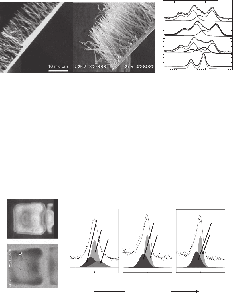
Since photoelectrons emerge only from within the top 100 nm or so
of a bulk specimen, methods that use photoelectron detection are ideal
for studies of surface phenomena. Photoelectron emission microscopes
using X-ray illumination of a broad area and sub-30 nm resolution
electron optics are beyond the scope of our concentration on zone plate
microscopes, though we note that they are used with great success and
at very high spatial resolution (see Figure 13–42). Another type of
photoelectron microscope is a Scanning PhotoEmission Microscope or
SPEM using a zone plate to produce a fi ne focus and an electron spec-
trometer for signal detection (Ade et al., 1990b; Ko et al., 1998; Yi et al.,
2005) (see Fig. 43); activities in this area were recently reviewed by
Günther et al. (2002).
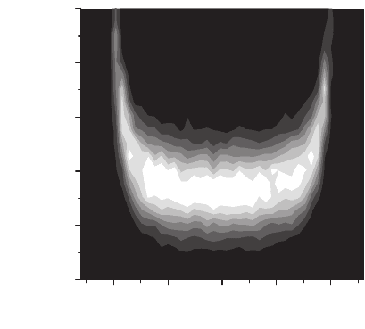
-10 -5 0 5 10
Distance from feature center (µm)
SiGe [008] Bragg angle (degrees)
53.78
53.80
53.82
53.84
53.86
53.88
Figure 13–41. In X-ray microdiffraction, a detector is set to collect diffraction
from small, crystalline features of the specimen that can be selectively illumi-
nated by the microfocus beam. Local variations from perfect crystal order are
seen as changes in the width or angle of the diffraction peaks. In this example,
a 20 µm wide, 0.24 µm thick Si
0.86
Ge
0.14
pseudomorphically strained fi lm is
located on a Si [001] surface. A determination of the angle of the SiGe [008]
diffraction peak as a function of position on the sample reveals elastic relax-
ation at the free edges of the SiGe feature, and demonstrates the ability of a
zone-plate STXM to study the strain distribution of patterned microstructures.
(Reprinted from Murray et al., © 2005, with permission from American Insti-
tute of Physics.) (See color plate.)
Chapter 13 Principles and Applications of Zone Plate X-Ray Microscopes 903
4.4 Magnetic Materials
X-ray magnetic circular dichroism (XMCD) exploits changes in absorp-
tion due to the relative orientation of magnetic domains and incident
circularly polarized radiation. It draws upon the fact that in magnetic
Intensity (a.u)
166 165 164 163 162 161 160
Binding Energy (eV)
S 2p
MoS
2
tips
sidewalls
base
Figure 13–42. In Electron Spectroscopy for Chemical Analysis or ESCA microscopy, a monochromatic
beam is used to illuminate a region several micrometers across; electron optics are then used to image
a tunable electron ejection energy to reveal surface chemistry. Though this does not involve zone plate
imaging, we include it here due to its widespread use with tunable X rays. In this case a 90-nm resolu-
tion ESCA microscope was used to locate aligned MoS
2
nanotube bundles and select certain areas
along the axes of the tubes for detailed examination. The image at left was acquired using Mo 3d
electrons, while S 2p, Mo 3d, and valence band spectra taken at the tips and sidewalls and the growth
base from the Si wafer appear strongly affected by the low dimensionality of the nanotubes and
differ signifi cantly from the corresponding spectra taken on a reference MoS
2
crystal. (Reprinted from
Kiskinova et al., © 2003, with permission of EDP Sciences.)
SEM Image
SPEM image
Relative B.E. (eV) Relative B.E. (eV)
Degree of ageing
2
1
3
Relative B.E. (eV)
MgCO
3
MgO
Mg 2p, 2
MgCO
3
MgO
Mg(OH)
2
Mg 2p,1
Intensity (a.u.)
MgCO
3
MgO
Mg 2p, 3
50-550-5 5 0 -5
Figure 13–43. Scanning photoemission microscope (SPEM) study of a plasma display cell. In this micro-
scope the specimen is scanned through the zone plate focus while photoelectrons are collected by an elec-
tron spectrometer. This fi gure shows a SPEM image, a scanning electron micrograph, and photoelectron
spectra from several regions of the sample. In a plasma display cell, light of the appropriate color emerges
through a front glass window which is protected from plasma damage by a composite insulating layer
including MgO. The photoelectron spectra show aging in the Mg(OH)
2
component of the layer over the
life of the display cell. (Reprinted from Yi et al., © 2005, with permission from the Institute of Pure and
Applied Physics.) (See color plate.)
904 M. Howells et al.
materials the density of certain electronic states is different for elec-
trons of spin parallel to the magnetization, compared to electrons of
spin anti-parallel. The absorption of circularly polarized photons
selects between electron spins, and depends on the component of spin
parallel to the helicity of the photon (the direction of the photon beam).
Images taken with a particular polarization of the illumination beam,
at saturated magnetization states, or at L
2
versus L
3
absorption edges
can by themselves show magnetic contrast effects, while difference
images between two polarization states at an absorption edge can be
used to obtain element-specifi c images of magnetic contrast only. While
much work has been done using photoemission microscopes (Stöhr
et al., 1993) and there are recent exciting results using X-ray holography
(Eisebitt et al., 2004), zone plate microscopy provides two primary
approaches. One method is to use a TXM with a large-angle-collection
condenser zone plate and exploit the fact that the radiation from syn-
chrotron bending magnet sources is circularly polarized above and
below the synchrotron plane; this was the fi rst method demonstrated
(Fischer et al., 1996) and it has led to considerable success for the study
of out-of-specimen-plane magnetism (Fischer et al., 2001a) (see Figure
13–44) and has recently been extended to the study of in-specimen-
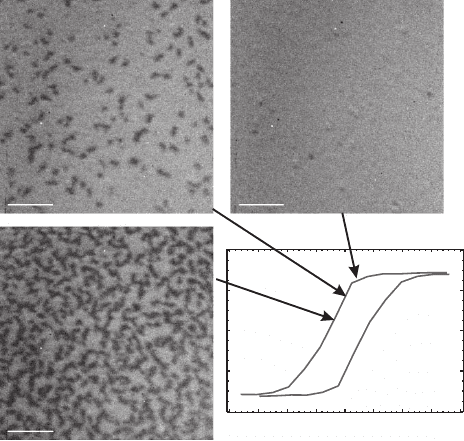
+400 Oe
-400 Oe
0 Oe
(b)
(c)
1 µm
-4 -3 -2 -1 0 1 2 3 4
-400
-200
0
200
400
Field (kOe)
M (emu/cc)
Figure 13–44. X-ray magnetic circular dichroism (XMCD) images of the magnetic domain structure
of a 50-nm thick (Co
83
Cr
17
)
87
Pt
13
alloy fi lm recorded at the Co L
3
absorption edge (777 eV) and in an
external fi eld of (a) +400 Oe, (b) 0 Oe, and (c) −400 Oe. (d) M vs. H hysteresis loop obtained via VSM
measurement. The arrows indicate the point in the reversal cycle at which each image is recorded.
Domain structure is apparent as the magnetization of the fi lm is driven around the hysteresis loop
and the net magnetization reversal can be seen to be the average of the reversal of individual domains,
with the number of reversed domains increasing as the strength of the applied fi eld is increased.
(Reprinted from Im et al., © 2003, with permission from American Institute of Physics.)
Chapter 13 Principles and Applications of Zone Plate X-Ray Microscopes 905
plane magnetic structure as well (Fischer et al., 2001b). Another more
recent approach is to use a STXM with a variable polarization undula-
tor source. In either case, the pulsed nature of synchrotron radiation
from electron bunches means that one can cycle an applied magnetic
fi eld in synchrony with the arrival of short (∼100 psec) pulses of X-rays,
and thereby accumulate images corresponding to controlled time
delays before and after application of the pulsed fi eld (Stoll et al., 2004)
(see Figure 13–45). A more extended discussion of magnetic contrast
X-ray microscopy is provided in a recent review by Fischer (Fischer,
2003).
5 Conclusion
In this chapter, we have outlined some of the principles and character-
istics of X-ray microscopes using zone plate optics, and have attempted
to convey an incomplete but representative survey of their applications
in scientifi c studies. We have seen that the resolution and effi ciency of
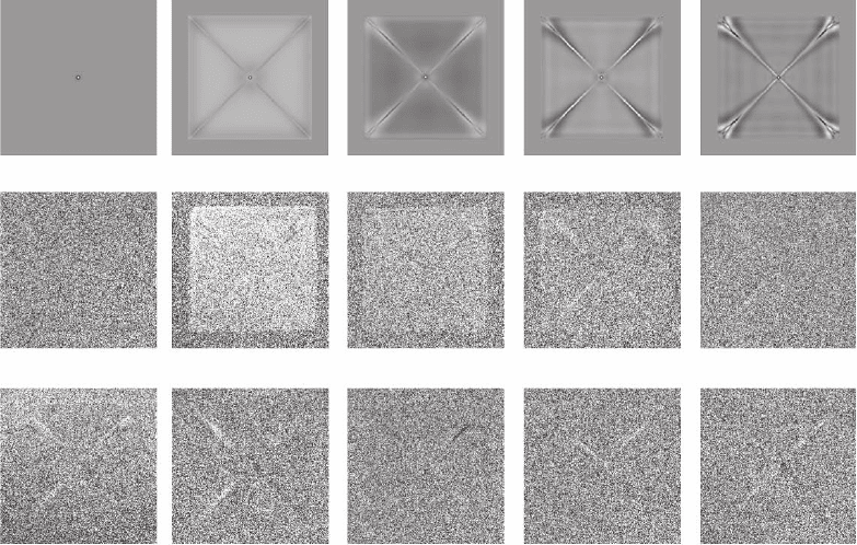
(a)
(b)
∆t=-400 ps ∆t=+400 ps ∆t=+500 ps ∆t=+600 ps
∆t=+800 ps
∆t=+900 ps ∆t=+1000 ps ∆t=+1200 ps ∆t=+2000 ps ∆t=+2400 ps
Figure 13–45. Time-resolved XMCD imaging of a magnetized Ni-Fe fi lm patch as the magnetization
is reversed in an applied magnetic fi eld. a) The z-component of the dynamic magnetization at selected
time delays obtained from micro-magnetic simulations (OOMMF). b) XMCD images from the XM-1
TXM taken with various time delays between the application of the pulsed magnetic fi eld and the
arrival of radiation from electron bunches in the storage ring. By integrating over many bunches with
a particular time delay, one can study the temporal evolution of the z-component of the magnetization
at delay times varying from probe pulse 400 ps before the pump, up to 2400 ps after the pump.
(Reprinted from Stoll et al., © 2004, with permission from American Institute of Physics.)
906 M. Howells et al.
zone plates has improved considerably over the lifetime of the fi eld,
although, in spite of constant efforts and the application of the best
technology, the rate of improvement has been slow. For some time the
“Moore’s Law” graph for zone plate resolution has had a slope of about
a factor of two per decade. However, as we have seen, this area of
development has been especially active in recent times. There is now
some optimism that the 10 nm barrier may be broken and the present
art is nowhere close to hitting fundamental limits. Resolution is not
the whole story, however; many applications are combining imaging
with tilt of the specimen for tomography, with energy tunability for
spectromicroscopy, and with fl uorescence detection for elemental iden-
tifi cation. These represent the application of zone plate optics to extend
the boundaries of previously existing techniques with active communi-
ties, so these areas are likely to expand. Another general trend of the
last few years has been the growth in hard X-ray applications of zone
plate imaging. This has been especially benefi cial for tomography and
microanalysis and, as recent experiments have shown, the use of hard
X-ray zone plates in high order may soon approach the best resolution
of soft X-ray zone plates in fi rst order. At the time of this writing
it seems that technical developments in X-ray microscopy and its
marriage with promising application areas is happening at an ever-
increasing pace and we can now forecast that these activities have a
bright future with more confi dence than ever before.
Acknowledgments. Naturally an enterprise like writing this review
depends greatly on the willingness of our colleagues around the X-ray
microscopy community to provide us with advice information and
images and we thank the many people who have done that. We espe-
cially thank Janos Kirz and Henry Chapman for reading the manu-
script and our immediate colleagues at Stony Brook, Brookhaven and
Berkeley for many helpful discussions. Work by MH and TW was sup-
ported by the Director, Offi ce of Energy Research, Offi ce of Basics
Energy Sciences, Materials Sciences Division of the U. S. Department
of Energy, under Contract No. DE-AC03-76SF00098. Work by CJ was
supported by the National Institutes of Health under grant R01
EB00479-01A1, and the National Science Foundation under grants
DBI-9986819, ECS-0099893, and CHE-0221934.
References
Abraham-Peskir, J. (1998). Structural changes in fully hydrated Chilomonas
paramecium exposed to copper. Eur. J. Protisto. 34, 51–57.
Abraham-Peskir, J. (2000). X-ray microscopy with synchrotron radiation:
Applications to cellular biology. Cel. Molec. Biol. 46(6), 1045–1052.
Ade, H. (1998). X-ray spectromicroscopy. Experimental Methods in the Physical
Sciences. 32, 225–262 (R. Celotta and T. Lucatorto, Eds.). (Academic Press
New York).
Ade, H. and Hsiao, B. (1993). X-ray linear dichroism microscopy. Science 262,
1427–1429.
Chapter 13 Principles and Applications of Zone Plate X-Ray Microscopes 907
Ade, H., Kirz, J., Hulbert, S., Johnson, E., Anderson, E. and Kern, D. (1990a).
Scanning photoelectron microscope with a zone plate generated micro-
probe. Nucl. Inst. Methods Phys. Res. A 291, 126–131.
Ade, H., Kirz, J., Hulbert, S.L., Johnson, E., Anderson, E. and Kern, D. (1990b).
X-ray spectromicroscopy with a zone plate generated microprobe. Appl.
Phys. Lett. 56, 1841–1843.
Ade, H., Zhang, X., Cameron, S., Costello, C., Kirz, J. and Williams, S. (1992).
Chemical contrast in X-ray microscopy and spatially resolved XANES spec-
troscopy of organic specimens. Science 258, 972–975.
Agard, D. and Sedat, J. (1983). Three-dimensional architecture of a polytene
chromosome. Nature 302, 676–681.
Anderson, E.H., Bögli, V. and Muray, L.P. (1995). Electron beam lithography
digital pattern generator and electronics for generalized curvilinear struc-
tures. J. Vacuum Sci. Tech. B 13, 2529–2534.
Anderson, E.H., Olynick, D.L., Harteneck, B., Veklerov, E., Denbeaux, G., Chao,
W., Lucero, A., Johnson, L. and Attwood, D. (2000). Nanofabrication and
diffractive optics for high-resolution X-ray applications. J. Vacuum Sci. Tech.
B 18(6), 2970–2975.
Aoki, S. (1994). Grazing incidence X-ray microscope with a Wolter type mirror.
In Radiation in the Biosciences (B. Chance, D. Deisenhofer, S. Ebashi, D.T.
Goodhead, J.R. Helliwell, H.E. Huxley, T. Iizuka, J. Kirz, T. Mitsui, E. Ruben-
stein, N. Sakabe, T. Sasaki, G. Schmahl, H. Sturhmann, K. Wüthrich and G.
Zaccai, Eds.) (Oxford University Press, New York).
Aoki, S. and Kikuta, S. (1974). X-ray holographic microscopy. Japan. J. Appl.
Phys. 13, 1385–1392.
Aristov, V.V. and Erko, A.I., Eds. (1994). X-ray Microscopy IV. (Bogorodskii
Pechatnik, Chernogolovka, Russia).
Atlwood, D. (1999). Soft X-rays and Extreme Ultraviolet Radiation. (Cambridge
Univ. Press, Cambridge).
Baez, A.V. (1960). A self-supporting metal Fresnel zone-plate to focus extreme
ultra-violet and soft X-rays. Nature 186, 958.
Baez, A.V. (1961). Fresnel zone plate for optical image formation using extreme
ultraviolet and soft X radiation. J. Opt. Soc. Amer. 51, 405–412.
Baez, A.V. (1989). The early days of X-ray optics: A personal memoir. J. X-ray
Sci. Tech. 1, 3–6.
Baez, A.V. (1997). Anecdotes about the early days of X-ray optics. J. X-ray Sci.
Tech. 7(2), 90–97.
Barbee, T.W., Jr. (1981). Sputtered layered synthetic microstructure (LSM) dis-
persion elements. In Proceedings of the Conference on Low Energy X-ray Diag-
nostics (American Institute of Physics, Monterey, CA).
Barrett, R., Kaulich, B., Oestreich, S. and Susini, J. (1998). Scanning microscopy
end station at the ESRF X-ray microscopy beamline. In X-ray Microfocusing:
Applications and Techniques (I. McNulty, Ed.). Proceedings of the SPIE, Vol.
3449, 80–90 (SPIE, Bellingham, WA).
Batterman, B. and Cole, H. (1964). Dynamical diffraction of X-rays by perfect
crystals. Rev. Mod. Phys. 36, 681–717.
Beetz, T. and Jacobsen, C. (2003). Soft X-ray radiation-damage studies in PMMA
using a cryo-STXM. J. Synchrotron Rad. 10(3), 280–283.
Behets, G.J., Verberckmoes, S.C., Oste, L., Bervoets, A.R., Salome, M., Cox,
A.G., Denton, J., De Broe, M.E. and D’Haese, P.C. (2005). Localization of
lanthanum in bone of chronic renal failure rats after oral dosing with lan-
thanum carbonate. Kidney Int. 67(5), 1830–1836.
Bennett, P.M., Foster, G.F., Buckley, C.J. and Burge, R.E. (1993). The effect of
soft X-radiation on myofi brils. J. Microsc. 172, 109–119.
