Hawkes P.W., Spence J.C.H. (Eds.) Science of Microscopy. V.1 and 2
Подождите немного. Документ загружается.


878 M. Howells et al.
1992) in which the intensity point spread function and its Fourier
transform, the optical transfer function (OTF) were calculated. In fact,
the magnitude of the OTF, known as the modulation transfer function
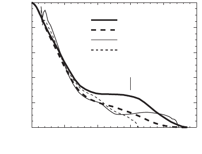
(MTF), was both calculated and measured for the Stony Brook STXM
and good agreement was obtained (Figure 13–21). Similar analysis has
been provided for TXMs (Jochum and Meyer-Ilse, 1995; Niemann et al.,
2000). Jochum and Meyer-Ilse provided a fairly general treatment of
the application of coherence theory to X-ray microscopy including
imaging of two-point and step objects by a realistic TXM in bright-fi eld
amplitude contrast. Other discussions of coherence issues have been
provided by (Heck et al., 1998; Chao et al., 2003).
3.3.2 Fourier Optics Treatment
Partially coherent imaging by a microscope can be described generally
by the methods of Fourier optics (Wilson et al., 1984; Goodman 1985;
Born and Wolf, 1999). This method uses a real-space and a frequency-
space description of waves in which frequencies (u) are closely
related to directions (θ) according to the general (1D) relation u =
sin θ/l. Following Chapman et al. (1996c), we consider fi rst a STXM
with amplitude point spread function h(x) imaging a sample of ampli-
tude transparency t(x). Using capital letters to represent Fourier trans-
forms, the pupil function of the lens is H(u) where u is the general
frequency coordinate, that is conjugate to the object-plane spatial coor-
dinate x and has a maximum value of NA/l where NA refers to the
beam-limiting lens. Any point in the lens pupil or the detection plane
can be represented by a u value. Since the detector in a STXM is placed
in the far fi eld of the X-ray focal spot, the diffraction pattern formed in
the detection plane in the absence of a sample will be H*(−u). When
0.0
0.2
0.4
0.6
0.8
1.0
MTF
05101520 25
Spatial frequency (µm)
-1
Horiz. (theory)
Vert. (theory)
Horiz. (exp.)
Vert. (exp.)
33 nm feature
Figure 13–21. Measured and calculated modulation-transfer function for
STXM imaging with a nickel zone plate of outer zone width 45 nm and diam-
eter 100 nm. The calculated curve was derived from the known zone plate
aperture function and the size and distance of the source pinhole. (Reprinted
from Jacobsen, © 1991, with permission from Elsevier.)

Chapter 13 Principles and Applications of Zone Plate X-Ray Microscopes 879
the sample is present and the spot is at x
s
, the wave fi eld immediately
behind the sample is h(x)t(x − x
s
) and the fi eld in the far-fi eld detection
plane is given by the Fourier transform of that. The detected intensity
is therefore
F(u,x
s
) = |H(u)⊗
u
T(u)e
2πix
s
.u
|
2
(33)
where ⊗ represents convolution and the convolution and shift theo-
rems have been used. The same quantity F(u,x) can also be represented
in another useful way. By inserting the representations of H(u) and
T(u) as Fourier integrals into the convolution integral Eq. (33) and using
the Fourier-integral defi nition of the delta function (Born and Wolf,
1999) and then its sifting property, we obtain (Chapman et al., 1996c)
F(u,x) = |h(x)⊗
x
t(x)e
−2πix.u
|
2
, (34)
The fi rst of the above two equations represents the diffraction pattern
formed in the detection plane by a STXM at each scan position as
F(u,x
s
), regarded as a function of u for a given x
s
. The second equation
represents a coherent image in a TXM, for illumination direction u, as
F(u,x), regarded as a function of x for a given u. In the fi rst case the
exponential represents the scan shift x
s
and in the second case it rep-
resents the incoming plane wave at direction u. This optical equiva-
lence of the STXM and TXM is known as “reciprocity” and is discussed
further in Section 3.3.4. To get the delivered intensity image I(x) in
either case one has to integrate the signal in the detection plane over
the particular distribution of u values that are used. That is in STXM
we integrate over the intensity response function of the detector |D(u)|
2
while in TXM we similarly integrate over the intensity distribution in
u delivered by the source |S(u)|
2
.
IFDd
IFSd
STXM
DET
TXM
SOURCE
and
xuxuu
xuxuu
()
=
()()
()
=
()()
∫
∫
,
,.
2
2
(35)
If the condenser is approximately incoherently illuminated (as speci-
fi ed in §10.5.1 Eq. 13 of (Born and Wolf, 1999) for example), which is
often the case for TXMs, (Schneider, 1998; Vogt et al., 2001a), then the
effective source (Hopkins, 1957), |S(u)|
2
, will be the condenser lens
aperture function. The expression for the fully incoherent bright fi eld
image (|D(u)|
2
= 1 or |S(u)|
2
= 1) is the same in both TXM and STXM and
is obtained by inserting Eq. (33) into Eq. (35) and applying Parseval’s
theorem (Chapman et al., 1996c)
I
BF
(x) = |h(x)|
2
⊗
x
|t(x)|
2
(36)
The coherent bright fi eld image is available from a STXM by using an
axial point detector and from a TXM by using an axial point source.
Both are given by F(0,x) although neither is widely used in X-ray
microscopy. The process of integrating over S or D, which is carried
out automatically by the hardware of the TXM or STXM, is generally
convenient but it destroys potentially useful information about the

880 M. Howells et al.
sample. A procedure for capturing this information, by storing the
full detection-plane pattern at every pixel position of the STXM image,
has been described and implemented to obtain phase- and amplitude-
contrast images by Chapman etal. (1996a). The speed of the procedure
was limited by the speed of early 1990s computers but, given the im -
provement of computers since then, it may well be time to re-examine
this approach (see Section 3.1.3). Eq. (34) and Eq. (36) show respectively
that coherent imaging is linear in the amplitude and incoherent imaging
is linear in the intensity. On the other hand, as we see below, partially
coherent imaging is not linear in either.
3.3.3 Contrast Transfer
In the case that we do not have |D(u)|
2
= 1 or |S(u)|
2
= 1, the above pro-
cedure used to obtain Eq. (36) does not lead to such a simple result but
rather to the following expression representing partially coherent
imaging in a STXM (Kintner et al., 1978; Wilson and Sheppard, 1984;
Born and Wolf, 1999).
ICTTedd
i
xmpmp mp
mpx
()
=
(
)
()
(
)
∫∫∫∫
−∞
+∞
−−
()
⋅
[]
;*
2π
(37)
CDHHdmp u u m u p u;
(
)
=
()
−
(
)
−
(
)
−∞
+∞
∫∫
2
*
(38)
For a TXM S replaces D in the last equation. The integration variables
m and p in Eq. (37) are frequencies similar to u but m represents a ray
incident on the sample while p represents a ray emerging from it. The
ranges of frequencies included in these beams by the form of S or D
determine the range of periodicities (m–p) in the sample that contrib-
ute to the image and thus determine the extent of the MTF in frequency
space. The function C(m; p) is known in optics as the transmission
cross coeffi cient (Born and Wolf, 1999) or the partially coherent trans-
fer function (Wilson and Sheppard, 1984) and provides a sample-
independent description of the effect of both the illumination and the
optical system on the transfer of information from object to image. It
is not a true transfer function, since the transfer is not linear, but is a
member of a wider class of “bilinear transfer functions.” Such func-
tions are described, for example, by (Saleh, 1979) and have been applied
to partially coherent X-ray imaging by Vogt et al. (2001a).
C(m; p) is widely used in the optical and electron microscopy com-
munities and its properties have been worked out in detail; see for
example (Sheppard and Wilson, 1980; Wilson and Sheppard, 1984). It
is normally a 4D function but in the case of a 1D object it becomes the
2D function C(m;p). The value of C(m;p) is then equal to the overlap
integral of the three appropriately shifted aperture functions in the
integrand of (38) (Kintner et al., 1978; Wilson and Sheppard, 1984; Born
and Wolf, 1999). For many cases of interest in both TXM and STXM,
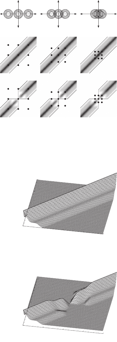
all three are circular disks or annuli (Figures 13–22 and 13–23). For the
ideally incoherent bright-fi eld image the value of D or S is taken to be
unity for all frequencies and the overlap then depends only on the

Chapter 13 Principles and Applications of Zone Plate X-Ray Microscopes 881
abc
a
b
-4
-2
0
2
4
m
-4
-2
0
2
4
p
-4
-2
0
2
4
m
-4
-2
0
2
4
p
Figure 13–22. Imaging of a one-dimensional
cosine amplitude grating object using a STXM.
The top row shows the signal in the detection
plane. The center row shows gray-level plots of
C(m, p) in m–p space overlaid with the support
boundary of C(m, p) (solid lines) and the spectra
T(m)T*(p) as in Eq. (37) (spots). The middle row
shows bright fi eld and the bottom row dark
fi eld. The three columns correspond to grating
frequencies that are (a) >2, (b) between 1 and 2
and (c) <1 expressed in units of the maximum
values of m and p which are both equal to NA/l.
(Reprinted from Chapman et al., © 1996c, with
permission from Elsevier.)
Figure 13–23. Bright-fi eld (a) and dark-fi eld (b)
partially coherent transfer functions for a 1D
object and an annular lens with inner radius
equal to 0.44 times the outer (after Chapman
et al., 1996c). The function C(m, p) is plotted
against m and p, expressed as multiples of their
maximum value NA/l. (Reprinted from
Chapman et al., © 1996c, with permission from
Elsevier.)
882 M. Howells et al.
difference m–p of the shifts of H and H*. That is, there is only one
response to the sample frequency s = m − p irrespective of m, which
indicates a linear system with MTF equal to C(m–p;0)/C(0;0). For forms
of D or S corresponding to partial coherence, the system is not linear.
On the other hand a dark-fi eld confi guration must have detector (or
source) and lens aperture functions which have zero overlap at m = p
= 0. For example D or S could be the Babinet inverse of H. For circular
functions of the latter type C(m;p) = 0 if sign(m) ≠ sign(p). Examples of
both bright- and dark-fi eld transfer functions for aperture geometries
that are representative of a STXM and that show the above character-
istics are given by Chapman et al. (1996c) (Figure 13–23). The response
of the same systems to a grating-like object are also given (Figure 13–
22). Dark-fi eld STXM is particularly well suited to imaging samples
with small features such as gold labels that scatter by large angles
(Chapman et al., 1996b). The procedures outlined above allow the cal-
culation of the MTF and the resolution behavior of both types of X-ray
microscope based on a knowledge of the resolution-determining lens
and the geometry of the source or detector. It is noteworthy that, as in
other types of microscope, the resolution does not depend on aberra-
tions of the condenser if there is one. As noted in Section 3.1.2 and
illustrated in Figure 13–17, the placement of a ring aperture and phase
ring to get Zernike phase contrast in a TXM may be modeled as modi-
fi cations of the source and lens aperture functions. By this means the
above method of analysis may be applied to this case as well (Mondal
and Slansky, 1970; Sheppard and Wilson, 1980; Morrison, 1989a).
3.3.4 Reciprocity
The general conclusion of the above analysis is that the optical systems
of the TXM and STXM are the same with the position of the lens, before
or after the sample, interchanged and the role of the source and detec-
tor interchanged. This is the “reciprocity” relationship (Zeitler and
Thomson, 1970) that has long been recognized in the visible-light and
electron imaging communities and has been explained in the context
of X-ray imaging by Morrison (1989a; Morrison et al., 2002). Thus we
might expect that, given identical resolution-determining lenses, a
TXM and a STXM (both operating in incoherent bright-fi eld mode)
could equally well utilize wide-angle beams and get good resolution.
For TXM the requirement would be that the condenser should deliver
a wide angle to the sample and for STXM that the detector should
collect a wide angle from the sample. However, in the past, the practical
realization of a wide-angle condenser for a TXM has been much harder
than a wide-angle detector for a STXM as we discussed in the con-
denser zone plate section above.
In practice the TXM/STXM relationship is not quite as symmetrical
as the above account suggests because of the general use of objective
zone plates with a central stop for STXM (Section 3.1.3) but not for
TXM. The stop produces a point-spread function, which has a narrower
central peak but larger side lobes. As a consequence the frequency
response (the MTF, see Figure 13–21) is increased in the high-frequency
and decreased in the low-frequency region.

Chapter 13 Principles and Applications of Zone Plate X-Ray Microscopes 883
3.3.5 The Infl uence of Coherence on Resolution
Calculations of the transfer function as an overlap area of three aper-
ture functions in the integrand of Eq. (38) were discussed in Section
3.3.3. For standard TXM, these functions are circular and two of them
are the same. One can therefore follow (Hopkins, 1957) and characterize
the illumination by a coherence parameter s defi ned by the ratio of the
condenser and objective numerical apertures or s = NA
c
/NA
o
. Full
coherence is represented by σ = 0 and full incoherence by σ = ∞,
although s = 1 is usually suffi cient to get close to fully incoherent
behavior. We start by considering the modulation (the percent dip in
the valley between the peaks) for a two-point object with separation
0.61l/NA
o
. The modulation is 26.5% for incoherent illumination (Born
and Wolf, 1999) and according to the Rayleigh criterion, the two points
are just resolved. It is common practice (Born and Wolf, 1999) to extend
the Rayleigh criterion to other pairs of objects and defi ne them as
resolved if the modulation is at least 26.5%. An example is the two-point
object with in-phase coherent illumination for which the just-resolvable
separation is 0.82l/NA
o
. Further detail of this can be seen from the plots
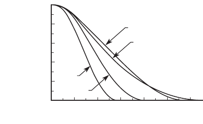
in Figure 13–24 (Jochum and Meyer-Ilse, 1995). An important example
for X-ray imaging is the modulation due to a periodic object, in particu-
lar a square-wave transmission object. Such an object can either be
prepared by standard lithography methods (Jacobsen et al., 1991) or, for
fi ner line widths, by preparing thin cross-sections of synthetic multilay-
ers (Chao et al., 2003), to yield resolution test patterns for an X-ray
microscope. If the resolution is defi ned as the half period of the fi nest
square wave that can be i mage d w it h 26. 5% modu lation and is expressed
as k
1
l/NA
o
, then according to (Chao et al., 2005), the diffraction-limited
value of k
1
is 0.5 for a coherent system (s = 0) and 0.4 for s = 0.38 (the
actual value for XM-1 illuminating the 15 nm zone plate). Thus the dif-
fraction-limited resolution of their experiment was 0.8∆r
n
while the
0.8 1 1.2 1.4
1/d
1.6 1.8 2
1
0.8
0.6
0.4
0.2
0
Contrast
σ = 1
σ = 0
σ = 0.5
σ→
∞
Figure 13–24. Image contrast as a function of 1/d where d is the point separa-
tion of a two-point object imaged by a lens with a circular pupil and coherence
parameter σ equal to 0 (coherent case), 0.5, 1 and infi nity (incoherent case). d
is expressed in units of l/NA. Note that the Rayleigh resolution corresponds
to 15.3% intensity contrast (defi ned as (I
max
− I
min
)/(I
max
+ I
min
)), which is the
same as 26.5% modulation. It occurs at d = 0.61l/NA for both s = 1 and s →
infi nity. (Reprinted from Jochum and Meyer-Ilse, © 1995, with permission
from Optical Society of America.)
884 M. Howells et al.
achieved value was about 1.0∆r
n
(<15 nm). The data demonstrating the
achieved resolution are shown in Figure 13–13. By reciprocity, the same
arguments about the value of the diffraction-limited resolution would
apply for a STXM in incoherent bright fi eld using the same zone plate.
With a large enough detector one would achieve s = 1 and the same
diffraction-limited resolution as a TXM with s = 1.
3.3.6 Coherence in Zernike Phase Contrast
We return now to the question of the choice of width and radius for
the two rings in a Zernike phase-contrast confi guration of the TXM,
or, equivalently, how much coherence is desirable in this case. This
choice has been discussed by (Mondal and Slansky, 1970) and is essen-
tially a trade off between light collection and the distorting effects of
the fact that the phase ring must have fi nite area, which applies an
unintended phase change to a certain portion of the diffracted light
causing the so-called halo effect (Wilson and Sheppard, 1981). It is
generally thought that one needs very little coherence, that is, the ring
aperture can leave a large fraction of the condenser area open. This is
true if the requirement is merely to make otherwise invisible phase
features, especially phase jumps, become visible. However, with low
coherence, a phase step is rendered as a double-peaked zero-crossing
function and a phase rect function is rendered as two such double
peaks. How much coherence do we need to get anything resembling a
faithful rendition of the object? We have not found much attention to
this point in the literature but the treatment by Martin (1966) provides
an answer, which is confi rmed by our own computer modeling. To get
a rendition of a rect function that looks like the original function one
needs to have the coherence width w
c
= lf
cond
/∆r
ring
of light arriving at
the sample at least equal to the width of the rect function.
3.3.7 Propagation-Based Phase Contrast
Another way to achieve phase contrast is to exploit the exp[iπ(x
2
+ y
2
)/
lz] phase shifts that occur in the (x, y) plane as a result of the propaga-
tion of a coherent wavefi eld through a distance z. This is exploited in
X-ray holographic microscopy which has had many successes (Aoki
and Kikuta, 1974; Joyeux, et al. 1988; Jacobsen et al., 1990; McNulty et
al., 1992; Snigirev et al., 1995; Lindaas et al., 1996; Eisebitt et al., 2004)
but which is so far not used for routine X-ray imaging. The exception
is in the use of holography for phase contrast tomography at higher X-
ray energies, where Cloetens and coworkers have achieved consider-
able success in routine micrometer-resolution tomography using a
phosphor/lens/CCD detector system (Cloetens et al., 1999). While it
lies beyond the scope of the present article’s emphasis on zone plate
X-ray microscopy, this unique approach is providing impressive 3D
reconstructions of diffi cult specimens including foams.
3.4 Tomography in X-Ray Microscopes
3.4.1 Principle of Operation
As was noted in Section 2.4.5 on hard X-ray zone plates, the transverse
resolution of a zone plate operated in fi rst diffraction order is given by

Chapter 13 Principles and Applications of Zone Plate X-Ray Microscopes 885
0.61l/NA = 1.22∆r
n
, and the depth of focus is 2l/(NA)
2
= 8(∆r
n
)
2
/l. Since
present zone plates have outermost zone widths ∆r
n
that are much
larger than the wavelength λ, this means that the depth of focus is nec-
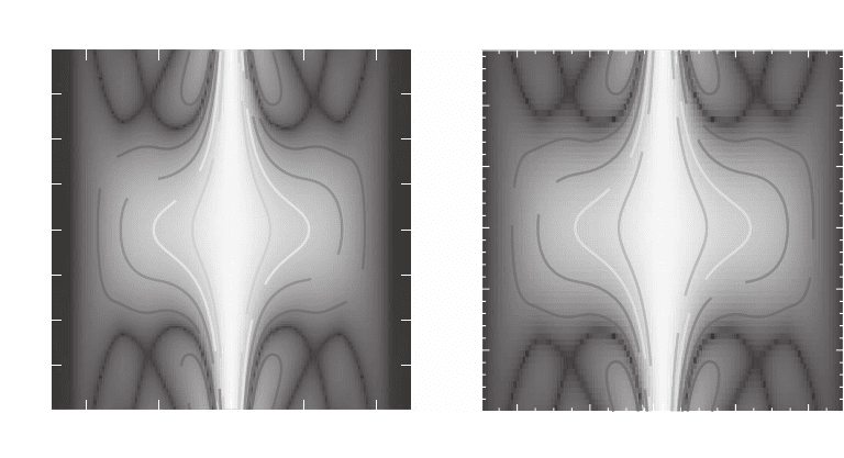
essarily large compared to the resolution as can be seen by the illustra-
tion of the 3D modulation transfer function in Figure 13–25. This
provides an opportunity for 3D imaging if the object is smaller than the
depth of focus because a 2D image can then be interpreted as providing
a simple projection through the specimen, which is precisely what con-
ventional tomography requires at each viewing angle. Tomography
with electron microscopes is long established, and following earlier
demonstrations by Haddad et al. (1994) using the Stony Brook STXM
and Lehr (1997) using the Göttingen TXM, a number of groups are now
using X-ray microscopes for tomographic studies of frozen hydrated
biological specimens and integrated circuits, among other applications
that will be described below in Section 4. However, although the tech-
nique used in these studies is improving, they have still not reached the
resolution achieved by the same microscopes in 2D.
3.4.2 The Depth-of-Focus Limit
Given the successes achieved and the amount of current interest, what
are the issues to be faced in improving the resolution of tomography
10%
10%
30%
50%
70%
10%
50%
70%
Contrast versus defocus:
δ
rN
=45 nm,
λ
=2.5 nm
-20 -10 0 10 20
Spatial frequency (
µm
-1
)
-8
-6
-4
-2
0
2
4
6
8
Defocus (
µm)
10%
30%
50%
70%
10%
30
%
50%
70%
-40 -20 0 20 40
Spatial frequency (
µm
-1
)
-1
0
1
Defocus (
µ
m)
Contrast versus defocus:
δ
rN
=20 nm, λ=2.5 nm
30%
Figure 13–25. Properties of soft X-ray tomography using zone plate optics. At the left is shown the
3D modulation transfer function for monochromatic, spatially incoherent bright-fi eld imaging with a
45-nm-outer-zone-width zone plate with a half-diameter central stop, as a function of depth. One can
see that the good in-focus frequency response is preserved over a total depth of about 8 µm. This is
useful for many tomography experiments that rely on the fact that the delivered image is a projection
of the object. At the right is the same information for a 20-nm-zone plate; as can be seen, the fi gure
scales quite well from the fi gure at the left according to the square of the ratio of the fi nest zone widths.
With a 20-nm-zone plate and monochromatic illumination, good frequency response is only preserved
over a depth of about 0.5–1 µm which is much more restrictive, illustration the challenges of improved
resolution in TXM zone-plate tomography. (Reprinted from Wang et al., © 2000, with permission from
Blackwell Publishing.)
886 M. Howells et al.
in X-ray microscopes? One of them concerns the same depth of focus
that makes such tomography straightforward. To our knowledge all
demonstrations of soft X-ray tomography reconstructions have used
zone plates with outermost zone width ∆r
n
no fi ner than 35 nm, so that
the depth of focus in the water window region is at least 4 µm which
has been comparable to the specimen size. As higher resolution zone
plates are employed, the depth of focus will decrease as the square of
improvements in transverse resolution, so that a 15 nm outermost-
zone-width zone plate would have a depth of focus of about 0.8 µm.
This approaches the ∼0.5 µm thickness accessible to cryo electron
tomography of frozen hydrated cell regions at 6–8 nm resolution
(Grimm et al., 1998; Medalia et al., 2002) and thus would seem to leave
a much-reduced “window of opportunity” for soft X-ray tomographers
and apparently deny them the important goal of high-resolution, 3D
imaging of intact eucaryotic cells.
The depth-of-focus limit evidently poses a considerable challenge to
those who would exploit zone plate X-ray microscopes for tomography.
Accordingly we discuss in the sections that follow several different
approaches to dealing with the challenge.
3.4.3 Avoiding the Depth-of-Focus Limit by Reconstructing a
Partial Volume
Zone-plate-microscope X-ray tomography as described above is based
on geometrical optics and is mathematically identical to the technique;
Computed Axial Tomography (CAT). Therefore, following an estab-
lished CAT procedure (see for example Ritman et al., 1997), it is possi-
ble to reconstruct partial volumes that are embedded in a larger overall
object. This has been done in the X-ray microscope context by Xradia
Inc. In this case the depth-of-focus limit applies to the partial volume,
rather than the whole volume, and the signals generated by the out-of
focus parts of the sample reduce, after combining all members of the
tilt series, to a smooth background. The size of the region that is always
in focus will be roughly a sphere of diameter equal to the depth of
focus.
3.4.4 Avoiding the Depth-of-Focus Limit by
Wide-Band Illumination
In fact, the depth of focus calculation given above is for monochromatic
illumination, which is applicable to demonstrations of tomography of
frozen hydrated cells in a STXM with a high-energy-resolution mono-
chromator (Wang et al., 2000). In some existing soft X-ray TXMs using
the zone plate condenser as a linear monochromator, an energy resolv-
ing power of E/∆E ≈ 200 has been used for objective zone plates with
N = 375 zones (the recommended bandwidth for such an objective
would usually have been E/∆E > N, (Thieme, 1988). However, Weiss et
al.(2002) have shown that use of E/∆E ≈ 200 leads to signifi cant changes
of the modulation transfer function (MTF) as a function of defocus
(similar results for different cases have been given by Schneider et al.
(2002a)). In their calculations for a zone plate with 40 nm outermost
zone width, the MTF at a spatial frequency of 15 µm
−1
(corresponding
to a spatial half-period of 33 nm) declines from about 0.28 in focus to
Chapter 13 Principles and Applications of Zone Plate X-Ray Microscopes 887
about 0.015 at a defocus of 4 µm in the monochromatic case. When the
bandwidth is increased to E/∆E ≈ 200, the MTF at 15 µm
−1
is reduced
in focus to only about 0.11 but at the same 4 µm defocus it degrades
much less to 0.08. Therefore these calculations show that, although the
transverse resolution and effi ciency for the collection of structural
information, are both made worse by the use of high-bandwidth radia-
tion, the depth of fi eld is improved. (To our knowledge no calculations
have yet addressed the possibly interesting question of how this trad-
eoff with nonmonochromatic illumination compares with the tradeoff
resulting from using a lower or higher resolution zone plate). In
summary, it will require further investigation to determine whether
manipulation of the bandwidth can enable signifi cant improvements
to the tomographic resolution without reduction of the reconstructible
sample volume.
3.4.5 Avoiding the Depth-of-Focus Limit by
Through-Focus Deconvolution
One possible approach to beat the depth of focus limit is to use through-
focus deconvolution as is done in light and electron microscopy. In
electron microscopy, the recording of defocus image sequences is
routine; each defocus provides positive and negative phase contrast at
various bands of spatial frequencies along with zeroes in the transfer
function, and the combination of several images can provide a com-
plete image of the specimen (Reimer, 1984). In fl uorescence light
microscopy, through-focus image sequences can yield a high quality
3D image through the use of deconvolution of the 3D point spread
function (Agard and Sedat, 1983; Carrington et al., 1995). However,
there are important differences between these examples and the situ-
ation present in X-ray microscopy. In electron microscopy, this approach
is usually applied to thin samples for which phase contrast dominates
(indeed the specimen focus can be quickly estimated by looking for a
minimum in image contrast). In light microscopy, the use of fl uores-
cence means that the object is a sparse, pure-real function (incoherent
emission from independent fl uorescence emitters with no sensitivity
to the relative phase of the illumination) so that the deconvolution can
be done based on the intensity point spread function. While some form
of generalized through-focus deconvolution may provide the needed
breakthrough, quantitatively reliable results such as are needed for
assembly into a tomographic reconstruction will have to account for
the fact that biological specimens imaged at water-window energies
produce both absorption and phase contrast so one will require exact
knowledge of the complex bilinear transfer function of the zone plate
optic and illumination system. In other words, the problem of 3D
deconvolution of a strongly absorbing, optically thick, complex object
with partial coherence is much more diffi cult than the cases in which
optical sectioning is typically used at present.
3.4.6 Avoiding the Depth-of-Focus Limit by Use of Higher
Energy X-Rays
The use of shorter wavelengths (higher X-ray energies) to increase the
monochromatic depth of fi eld 8(∆r
n
)
2
/l is a guaranteed way to extend
