Hawkes P.W., Spence J.C.H. (Eds.) Science of Microscopy. V.1 and 2
Подождите немного. Документ загружается.


858 M. Howells et al.
The list of issues posed by condenser zone plates does not end here.
Another serious limitation is that to change the X-ray energy one has
to change the condenser zone plate or at least change its object and
image distances. This is so diffi cult that systems fed by a condenser
zone plate are effectively not energy tunable. Evidently there are plenty
of reasons to seek alternatives to condenser zone plates and to this we
now turn.
2.4.4 Alternatives to Condenser Zone Plates
Zone plates defl ect the outermost ray by an angle equal to their NA (Eq.
(7)). As explained in the previous section, manufacturable NAs of con-
denser zone plates are too small to match those of the best objectives
by a factor of two or more. Now we know that a mirror can defl ect an
X-ray roughly by
22δ or twice its critical angle. Thus we can character-
ize a mirror by an effective outer zone width ∆r
n
=
()
λδ/4 2 , that a
zone plate would need to have to produce a defl ection equal to twice
the critical angle of the mirror. Since δ is proportional to λ
2
(Section 1.1),
one can see that the effective outer zone width will vary rather slowly
with both energy and electron density. For example, for a platinum,
mirror it varies from 6 nm at 0.5 keV to 4 nm at 5 keV. While for a silicon
dioxide mirror it varies from 12 nm to 10 nm over the same energy
range. This shows that if a suitable geometry can be arranged a single-
refl ection mirror system could be a very effective condenser. However
it is important to note that a refl ective condenser will not provide a
monochromatic beam and that either a line source or a monochromator
will be required. As we shall see, a separate monochromator coupled
with a refl ective condenser will have major advantages.
To produce the desired hollow-cone illumination, a grazing inci-
dence ellipsoid of revolution in the form of a hollow tube would be
suitable. The input angle should match the angle of the beam from the
source or monochromator and the output angle should match or exceed
the objective NA. For a synchrotron beam this design will normally
lead to under fi lling of the sample which can be overcome by wobbling.
The fabrication accuracy should be such that the point spread function
of the mirror is small compared to the size of the object fi eld. Mirrors
of this general type have been made for some time (often starting from
glass capillaries) and are quite widely used in the hard X-ray research
community as reviewed for example by Bilderback (2003).
Single-refl ection monocapillary X-ray mirrors have now been in use
for microscopes at both laboratory and synchrotron sources (Section
1.4) for the last year or two and have enjoyed considerable success. For
example, documentation of the performance of the X-ray microscope
at the Taiwan synchrotron, including details of its condenser mirror,
is provided by Tang et al. (2006) and Yin et al. (2006). These condenser
mirrors have many advantages compared to condenser zone plates.
Specifi cally, capillary-mirror condensers
1. Are more readily available
2. When fed by a separate monochromator allow the TXM to be truly
energy-tunable
3. Are able to match the NA of any currently available objective zone
plate

Chapter 13 Principles and Applications of Zone Plate X-Ray Microscopes 859
4. Are a factor 3–15 times more effi cient with no unwanted orders
5. Are more robust, longer lived and easier to clean
6. Are less fragile with respect to thermal or mechanical damage
7. Do not requi re a band-width-selecting pinhole close to the focus
and thus do not limit the size of the sample holder nor its ability
to rotate.
As indicated earlier (Section 1.4) we believe that the advent of an
energy-tunable TXM is important and may enable the TXM to become
competitive for spectromicroscopy.
2.4.5 Zone Plates with Shaped Grooves
Until now we have talked about square-wave zone plates with a gap
to period ratio of 0.5 that behaved according to the theory of a thin
zone plate, even if the thickness was greater than ∆r
n
. Just as a blazed
refl ection grating with a saw-tooth profi le has much better effi ciency
than a square-wave grating (even if the latter is a perfect phase grating
with π phase shifts), so one can get higher effi ciency from zone plates
with shaped groove profi les. Considering that a zone plate is intended
to synthesize a smooth spherical wave front from a succession of ring-
shaped parts, we might expect that the optimum groove shape will be
a parabola that increases the phase shift smoothly across the zone-plate
period. In fact the mathematical treatment (Tatchyn et al., 1982; Michette,
1986) shows that the thickness function is
tr f r fr r rrd
rd rr
iiiii
ii i
()
=+−+
{}
≤< −
=−≤≤
−−
22 2
1
2
1
0
/()
()
δ
(32)
where d
i
/r
i
can be calculated (Tatchyn et al., 1984) and is the outer frac-
tion of the ith period which is to be left open. The fi rst-order effi ciency
of a nickel zone plate made according to this specifi cation would be
about 80% at 7 keV.
It is hard to microfabricate a smooth curve but one can still get much
of the advantage of this scheme by approximating the parabolic profi le
by a stepped structure (Di Fabrizio et al., 1994; Yun et al., 1999). For
example Di Fabrizio et al. (1999) have made a nickel zone plate with
four equal width steps of optical delay 0, 0.25, 0.5 and 0.75 wavelengths.
The measured fi rst order effi ciency of this zone plate was 55% at 7 keV,
which represents a substantial improvement in effi ciency and suppres-
sion of unwanted orders compared to traditional soft X-ray perfor-
mance and shows the benefi ts of both groove-shaping and phase-plates.
Evidently the use of several thickness steps implies that the outermost
zone must be several times wider than the fi nest line width that can
be achieved with the particular fabrication process, so that this approach
involves a tradeoff between spatial resolution and effi ciency.
2.4.6 Hard X-Ray Zone Plates
While much of the effort of the last three decades has gone into devel-
oping zone plate microscopy in the 290–540 eV “water window” region
for studies of 0.1–10 µm thick specimens, there is increasing activity in
hard-X-ray zone-plate imaging at energies of roughly 2–15 keV. Scan-
ning fl uorescence X-ray microprobes (SFXM) using zone plate optics
are providing new capabilities for trace element mapping, and hard

860 M. Howells et al.
X-ray transmission X-ray microscopes (TXMs) using absorption or es -
pecially phase contrast are able to image much thicker objects than
their soft-X-ray counterparts. Zone plates for these energies must be
much thicker to achieve good effi ciency (see Figure 13–11) which places
increasing demands on zone aspect ratio in lithographically patterned
zone plates and means that the minimum zone width (and thus fi rst
order spatial resolution) were initially in the 50–100 nm range. However,
as noted in Section 1.4, several 45 and 50 nm hard X-ray zone plates are
now operational at synchrotrons. At the same time, because the ratio
of phase shifting to absorption f
1
/f
2
improves as the energy is increased,
the achievable effi ciency becomes much higher and the depth of focus
increases considerably (Jacobsen, 1992), which is helpful for applica-
tions such as tomography. Quantitatively, the transverse resolution of
a zone plate is given by 0.61λ/NA = 1.22∆r
n
(Eq. (8)) and the depth of
focus by 2λ/(NA)
2
= 8(∆r
n
)
2
/λ. Therefore, a zone plate with ∆r
n
= 50 nm
has a depth of focus of about 160 µm at 10 keV as opposed to about 8 µm
at 500 eV. In addition, the focal length f = 2r
n
(∆r
n
)/λ (Eq. (6)) for such a
zone plate with 100 µm diameter increases from 2 to 40 mm, which
considerably eases some of the challenges of mechanical design for
specimen temperature control, insertion of fl uorescence detectors, and
so on.
In spite of the challenges of fabricating thicker zone plates using
lithographic techniques, much success has been achieved. In some
cases the initial electron beam lithography write has been transferred
into a thicker plating mold using reactive ion etching as described
above; this has led to the commercial availability (Xradia, Inc.) of a
variety of high-aspect-ratio zone plates including one with outer-zone
width 50 nm and thickness 700 nm, or an aspect ratio of 14 : 1 (an
example of an earlier Xradia zone plate is shown in Figure 13–14).
Additional parameters of zone plates given at the latest X-ray micros-
copy conference (Himeji, Japan, June 2005) are given in Section 1.4.
Other approaches have involved using an electron-beam-written zone
2 µm1 µm
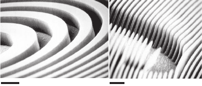
Figure 13–14. A hard X-ray zone plate with 100 nm outermost zone width and
1.6 µm thickness of gold for use at 5.4 keV in a commercial X-ray microscope
system (Xradia, Inc.). The simultaneous achievement of narrow zone widths
for high spatial resolution, and signifi cant zone thickness so as to achieve a π
phase shift, means that the achievement of high aspect ratio nanostructures
is important. This zone plate has an aspect ratio of 16 : 1 and a theoretical
focusing effi ciency of 31%. (Figure Courtesy W. Yun, Xradia.)
Chapter 13 Principles and Applications of Zone Plate X-Ray Microscopes 861
plate as a mask for the subsequent processing of a thicker zone plate
using X-ray lithography (Shaver et al., 1980; Lai et al., 1992), including
the fabrication of 2.5 µm thick zone plates with a fi nest zone width of
0.25 µm (Krasnoperova et al., 1993). Another approach is to stack two
or more zone plates together (Shastri et al., 2001).
Sputter-sliced or “jelly roll” zone plates (Schmahl et al., 1980) repre-
sent a completely different approach in fabrication. The goal of this
approach is to start with a rotating wire and then build up alternating
layers of weakly and strongly refractive material by sputtering or evap-
oration. The resulting structure is then sliced to yield zone plates of
the appropriate thickness. In this case the achievement of high aspect
ratios is not at all challenging; instead, the challenges include avoiding
error and roughness accumulation in realizing the proper zone radii,
the diffi culties of maintaining perfect cylindrical symmetry, and the
challenges involved in slicing the structure.
Recent results from the groups involved (Bionta et al., 1994; Tamura
et al., 2002; Duvel et al., 2003) show that the technique is making steady
progress to the point where a zone plate consisting of 70 Cu/Al layer
pairs with outer zone width of 0.16 µm and aspect ratio of more that a
thousand has been used to focus a 100 keV beam from Spring 8 to
0.5 µm FWHM (Kamijo et al., 2003). Similarly a sputter-slice soft X-ray
zone plate with an aspect ratio of 200, made at Göttingen Germany,
had 188 layer pairs of the alloy Ni80-Cr20 and silica. It showed a mea-
sured effi ciency of 3.8% at 4.1 keV but had a focal spot size considerably
greater than the 17-nm outer zone width. These are intriguing results,
though for the moment, the sputter-sliced approach has not yet pro-
duced optics with an optical performance consistent with the intended
geometrical parameters.
2.4.7 Thick Zone Plates
Up until now we have used kinematical diffraction theory to under-
stand the properties of zone plates. In this theory, the incident wave
and the diffracted signals from each volume element are all treated as
independent. However, in reality, the “incident” wave and the “dif-
fracted” waves in the solid structure are coherently coupled and if the
zone plate is thick enough, the effect of this coupling will become
evident at the output. Such coupling is known in perfect-crystal dif-
fraction where it leads to anomalous transmission in Laue-geometry
experiments (the Borrmann effect), while on a larger size scale, “Bragg-
effect” holograms show similar behavior. Zone plates can be designed
to exploit coupled-wave effects and these devices offer the possibility
of very high effi ciency and resolution in high diffraction orders, thus
exceeding the resolution limit of the outermost zone width which
applies when operating in fi rst order. Theoretical treatments of diffrac-
tion by thick periodic structures have been developed in the hard-X-ray
community (dynamical diffraction by crystals) (Batterman and Cole,
1964) and the optical-holography community (coupled-wave theory)
(Kogelnik, 1969; Solymar and Cooke, 1981). The coupled-wave method
has been applied to X-ray gratings and zone plates by Maser (1994) and
more recently by Schneider (1997) who has given a solution that includes
862 M. Howells et al.
the case of high orders and gap-to-period ratios other than 0.5.
Schneider’s solution predicts that, in the soft X-ray region, absolute
effi ciencies of 30–50% in a single high order are indeed possible with
line-to-space ratios of 0.1–0.5 and aspect ratios greater than 30 : 1.
Hambach et al. (2001) have followed up these calculations with a series
of experiments, mostly with copolymer gratings with aspect ratio 10 : 1.
The predictions of the theory were broadly confi rmed and a maximum
effi ciency of 15.3% was achieved at 13 nm wavelength. This value was
75% of the prediction of the coupled wave theory and 25 times greater
than the prediction of thin-grating (kinematic) theory. The authors
suggested that zone plates based on this principle may fi nd application
as condensers for table-top microscopes using isotropically emitting
sources. A similar verifi cation of dynamical diffraction theory for the
case of sectioned multilayers illuminated with 19.5 keV X-rays in Laue
geometry has been published recently (Kang et al., 2005). A refl ecting
effi ciency of 70% was observed. Both of these experiments used grat-
ings as being representative of the diffraction-effi ciency behavior of a
conventional zone plate (Hambach) and a sputter-sliced zone plate
(Kang) respectively. However, as pointed out by Maser (1994), thick
zone plates, like volume holograms, have a directional selectivity based
on Bragg’s law. That is, the zones must be oriented so that the incoming
wave is locally Bragg-refl ected by the zones and there will be a rocking
curve outside of which the high effi ciency is lost or moved to another
order. Eq. (6) can be regarded as showing that Braggs law is obeyed at
every zone of a conventional zone plate operating at magnifi cation
unity. On the other hand, for high magnifi cation or demagnifi cation
applications, the zones must be tilted by an angle that varies with
radius. The diffi culty of doing this in practice is currently delaying the
application of these ideas to practical zone plates.
A promising recent alternative to tilt angles that vary continuously
with angle has been to apply the concepts of the “sputter-sliced” zone
plate (see previous section) to produce linear half-zone-plates called
“multilayer Laue lenses.” One starts with an atomically smooth fl at and
deposits alternating zone materials, starting with the highest-order
zones, so that error accumulation mainly affects the coarser, low-order
zones. Two multilayer Laue lenses together can achieve two dimen-
sional focusing in the manner of a Kirkparick-Baez mirror pair.
Although tilting by a continuously variable angle is not exactly achieved
by this approach it can be approximated by tilting the whole lens to a
compromise Bragg angle. One dimensional line foci as narrow as 19 nm
have already been reported by this method (Maser et al., 2004; Kang
et al., 2006) and these authors believe that 5 nm may be possible in
future work.
3 X-Ray Microscopes
In the previous sections we have described some of the characteristics
of X-ray interactions and focusing optics. We now turn our attention
to a discussion of X-ray microscopes currently in operation. They fall

Chapter 13 Principles and Applications of Zone Plate X-Ray Microscopes 863
into two classes: full-fi eld imaging and scanning, which are both illus-
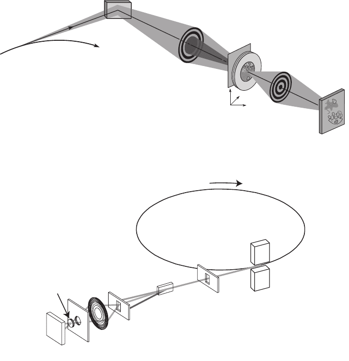
trated in Figure 13–15. A large number of microscopes are listed in
Table 13–3. We also describe three specifi c microscopes as examples: a
transmission X-ray microscope (TXM) operated at Lawrence Berkeley
National Laboratory, a scanning transmission X-ray microscope (STXM)
operated at Brookhaven National Laboratory, and a scanning fl uores-
cence X-ray microprobe (SFXM) operated at Argonne National
Laboratory.
A key difference between TXM, STXM, and SFXM concerns the
illumination phase space that can be accepted. In STXM and in SFXM,
the size of the spot delivered by the zone plate objective is a convolu-
tion of the geometric image of the source and the point spread function
Plane
mirror
Bending
Magnet
Condenser
zone plate
Objective
zone plate
Pinhole
X-ray
sensitive
CCD
Sample
stage
National Synchrotron Light Source
X-ray Ring
X1 undulator
Monochromator
Zone plate
Order sorting
aperture
Detector
Specimen
So
ft
x
ra
ys
2.8 GeV electrons
Figure 13–15. Schematic of the main components of a transmission X-ray microscope or TXM (top:
courtesy of D. Attwood, Lawrence Berkeley National Laboratory) and a scanning transmission X-ray
microscope or STXM (bottom: courtesy of Y. Wang, then of Stony Brook.) (See color plate.)

864 M. Howells et al.
Table 13 –3. Zone plate microscopes.
Microscope/ Illumination/ Focusing, Contrast Techniques,
location Light source monochromator imaging mechanisms X-ray energy Citation
MES STXM ALS grating STXM absorption, NEXAFS, MCD (Tyliszczak, 2004;
undulator magnetization 100 to 2000 eV Warwick, 2004)
Polymer STXM ALS bend grating STXM absorption NEXAFS (Warwick, 2002;
magnet 250 to 750 eV Kilcoyne, 2003)
XM-1 TXM ALS bend zone plate TXM absorption, magn- Tomog, MCD (Meyer-Ilse, 2000a)
magnet condenser/mono etization, phase 200 to 1800 eV
XM-2 TXM ALS bend zone plate TXM absorption, Tomog
magnet condenser/mono phase 200 to 7000 eV
2-ID-B APS multilayer-coated STXM abs, fl uor, phase tomography (McNulty, 2003a;
undulator grating XANES, tomog 600 to 4000 eV McNulty, 2003b)
2-ID-D APS Crystal/multilayer STXM diffraction strain mapping (Cai, 2003, McNulty,
undulator 6 to 20 keV 2003a)
2-ID-E APS Crystal/multilayer STXM fl uor, XANES 5 to 35 keV (McNulty, 2003a)
undulator diff, microdiff
26-ID APS Crystal/multilayer STXM abs, fl uor, diff 3–30 keV (McNulty, 2003a)
undulator TXM XANES
BL20B2 Spring8 crystal STXM absorption 4 to 113 keV opt (Suzuki, 2003,
bend magnet testing, tomog Takano, 2003)
BL47XU Spring8 crystal TXM absorption 5–37.7 keV (Suzuki, 2003, Uesugi,
undulator tomography 2003)
BL20XU Spring8 crystal—250 m STXM absorption 8–37.3, 24–113 keV, (Suzuki, 2003)
undulator beam line mbeams
BL24XU Spring8 crystal TXM phase contrast 8.77–12.85, (Tsusaka, 2001,
undulator 12.4–18.17 keV Kagoshima, 2003)
BL12 Ritsumeikan zone plate TXM absorption water window, (Takemoto, 2003)
bend magnet condenser/mono

Chapter 13 Principles and Applications of Zone Plate X-Ray Microscopes 865
8A1 U7 SPEM Pohang grating STXM photoemission nanoXPS (Shin, 2003, Yi, 2005)
undulator 100 to 1000 eV
1B2 hard xray Pohang crystal TXM absorption 6.95 keV (Youn, 2005)
b. magnet
U41TXM BESSYII zone plate TXM absorption, phase water window, 2D, (Guttman, 2003,
undulator condenser/mono 3D imaging Wiesemann, 2003)
UE46TXM BESSYII zone plate TXM absorption, MCD (Eimüller, 2003)
undulator condenser magnetization 0.2 to 2 keV
TWINMIC ELETTRA grating STXM & absorption NEXAFS (Kaulich, 2003)
Undulator TXM phase contrast 250 to 2000 eV
BL2.2 ESCA ELETTRA grating STXM absorption nanoXPS (Casalis, 1995;
undulator photoemission 200 to 1400 eV Kiskinova, 2003)
KINGS STXM laser plasma gas fi ltered STXM absorption water window (Michette, 2000)
spectrum
X1A STXMs NSLS grating STXM absorption NEXAFS, (Jacobsen, 2000a)
undulator diffraction cryomicroscopy
phase contrast 250 to 1000 eV
ID21 ESRF grating, STXM & absorption NEXAFS (Susini, 2000)
microscopes undulator
crystal TXM fl uorescence 200 to 7000 eV
diffraction
phase contrast
ID22 imaging ESRF crystal STXM absorption 5 to 70 keV (Weitkamp, 2000)
undulator fl uorescence
phase contrast
Aarhus TXM ASTRID zone plate TXM absorption typically 517 eV (Uggerhøj, 2000)
bend magnet condenser/mono
XRADIA Chromium refl ective TXM absorption, phase tomography (Scott, 2004)
anode condenser 5.4 keV

866 M. Howells et al.
of the optic. As Figure 13–16 shows, for an objective with diffraction-
limited (as opposed to aberration-limited) resolution, the effect of the
geometric source size becomes negligible if the product p = wθ of
source width w times the full angle θ accepted by the optic is less than
the wavelength λ in each dimension. This is commonly summarized
by saying that scanning microscopes require single-mode illumina-
tion, although it is understood that a spatially fi ltered, incoherent
source is not the exact equivalent of a single-mode optical cavity. The
situation in TXM is much different; for incoherent bright fi eld imaging,
each pixel in the object can be imaged independently of its neighbors
(within good approximation), so one can illuminate all object pixels
simultaneously and with nominally incoherent light. If object resolu-
tion elements are imaged in 1 : 1 correspondence to detector pixels in a
TXM, the number of “modes” of phase space p/λ that can be accepted
in the x direction is approximately equal to the number of detector
pixels in that direction and the same holds for y. As a result, TXMs are
often operated with bending magnet synchrotron radiation sources or
laboratory sources which deliver high fl ux (photons per solid angle),
while STXMs and SFXMs are often operated with undulator sources
which deliver high brightness (photons per solid angle per source
area). The issues of microscope illumination and its effects on image
formation will be discussed in more detail in Section 3.1.
0.0
0.5
1.0
1.5
2.0
2.5
3.0
3.5
4.0
-1.00 -0.75 -0.50 -0.25 0.00 0.25 0.50 0.75 1.00
Normalized spatial frequency
p/λ
30%
5%
5%
5%
10%
10%
10%
20%
20%
20%
30%
30%
50%
50%
50%
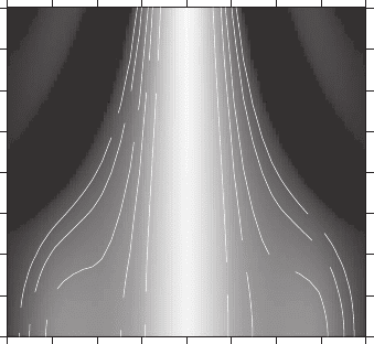
Figure 13–16. The aim in operating a scanning microscope or microprobe is
to have a diffraction-limited focus. Therefore the source must be suffi ciently
demagnifi ed so that it contributes negligibly to the focal width. This contour
plot shows how the modulation transfer function (MTF) of an optic with a
half-diameter central stop is affected by increasing the phase space parameter
p of the source. This parameter p = wθ (source full width w times the full angle
θ accepted by the optic) should be less than about the wavelength λ in both
the x and y directions to achieve maximum spatial resolution. The normalized
spatial frequency is defi ned to be unity at the MTF cutoff of 1/∆r
n
. (From Winn
et al., 2000.)
Chapter 13 Principles and Applications of Zone Plate X-Ray Microscopes 867
3.1 Microscope Layouts and Illumination Schemes
3.1.1 Transmission X-Ray Microscope (TXM) Layout
Full-fi eld transmission X-ray microscopes (TXMs) typically use a zone
plate to produce a magnifi ed image of the specimen on a 2D detector.
This approach was pioneered by the group of G. Schmahl at the Uni-
versität Göttingen, who, after initial experiments including refl ection-
grating monochromators (Niemann et al., 1976) switched to using a
condenser zone plate as the sole monochromator (Rudolph et al., 1984).
This latter approach is now used by a number of TXMs, including the
XM-1 at Lawrence Berkeley Lab (Meyer-Ilse et al., 1994, 2001) for which
we provide some example numbers. As shown in Figure 13–15a, the
beam from the synchrotron bending magnet source is defl ected by a
grazing-incidence mirror which fi lters out the power due to high-
energy X-rays, passes through a thin metal fi lter to remove visible and
ultraviolet radiation, and is then imaged by the condenser zone plate
onto a pinhole located just upstream of the specimen. As noted in
Section 2.4.3 on condenser zone plates, the condenser zone plate (of
diameter D = 9 mm) and the pinhole (of diameter d ≈ 10–20 µm), together
are equivalent to a monochromator of resolving power equal to D/(2d)
(Niemann, 1974). Because the light transmitted by the objective zone
plate includes a signifi cant undiffracted (zero order) component which
must not reach the detector, the illumination of the sample needs to be
hollow-cone and this is achieved by means of a stop built into the
condenser, blocking a central circle of radius about one third to one
half of the condenser radius. The objective zone plate used by XM-1 in
the resolution test described above had the following characteristics:
outer zone width ∆r
n
= 15 nm, diameter d = 30 µm, 500 zones of 80 nm
thick gold (giving a maximum aspect ratio of 5 : 1), and focal length
f = 0.3 mm at 815 eV. This is the highest resolution zone plate used to
date and slightly larger outer zone widths (25–30) are used for routine
user operations. The vertical phase space area of the synchrotron
source is generally smaller than its horizontal phase-space area and
smaller than that of the microscope (which equals object full-width d
times twice the objective NA). Since the condenser zone plate cannot
expand the phase space, both the object width and the numerical aper-
ture of the objective of a TXM will generally be underfi lled. To counter
the under fi lling of the object fi eld, the condenser is usually “wobbled”
up and down during the course of an exposure. This type of micro-
scope layout, in which the source is imaged on to the sample, is known
as “critical illumination” (Born and Wolf, 1999) and is widely used for
amplitude contrast.
Traditionally, the specimen has been placed in an atmospheric pres-
sure environment and to accomplish this, thin vacuum windows
(100 nm Si
3
N
4
or Si are common) can be used between the condenser
and the specimen, and also between the specimen and the objective
zone plate. Because the focal length of the objective zone plate is quite
small (for example, in the case of a 25-nm-outermost-zone-width, 60-
µm-diameter zone plate operating at 530 eV it would be 1.3 mm.), the
specimen region lying between these two windows is quite constrained.
