Hawkes P.W., Spence J.C.H. (Eds.) Science of Microscopy. V.1 and 2
Подождите немного. Документ загружается.


838 M. Howells et al.
Much of modern X-ray microscopy centers on the exploitation of X-
ray absorption edges. X-ray absorption edges arise when the X-ray
photon reaches the threshold energy needed to completely remove an
electron from an inner-shell orbital. The energy at which this occurs is
approximately given by the Bohr model as E
n
= (13.6 eV)(Z-z
shield
)
2
/n
2
,
where Z is the atomic number, z
shield
approximates the partial screening
of the nucleus’ charge by other inner-shell electrons (z
shield
≈ 1 for K
edges), and n is the principal quantum number (n = 1 for K edges, 2
for L edges, and so on). This produces the step-like rise in the cross-
section for photoelectric absorption that can be seen in the plots of f
2
in Figure 13–2. If one takes one image I
1
at an energy E
1
just below an
element’s absorption edge where the incident fl ux is I
01
, and a similar
image I
2
at an energy just above an absorption edge, one can recover
the mass per area m
x
/A of the element x from (Engström, 1946)
m
A
EE II I I
EE
x
=
(
)
(
)
−
(
)
−
(
)
ρ
µµ
12
3
101 202
2112
3
ln ln
(1)
This approach works well for mass concentrations greater than about
1%. Another way in which X-ray absorption edges are exploited is by
means of the “water window.” At X-ray energies between the carbon
and oxygen absorption edges at 290 and 540 eV, respectively, organic
materials show strong absorption contrast while water layers up to
several µm thick are reasonably transmissive (Wolter, 1952); this is
particularly valuable for imaging hydrated biological and environmen-
tal science specimens.
For those elements which have absorption edges below the energy
of incident X-rays so that inner-shell ionization occurs, the aftermath
of absorption involves the emission of either a fl uorescent photon or
an Auger electron of characteristic energy. The energy of these fl uores-
cent photons, and the fl uorescence yield (Krause, 1979) (the fraction of
events which result in fl uorescence rather than Auger electron emis-
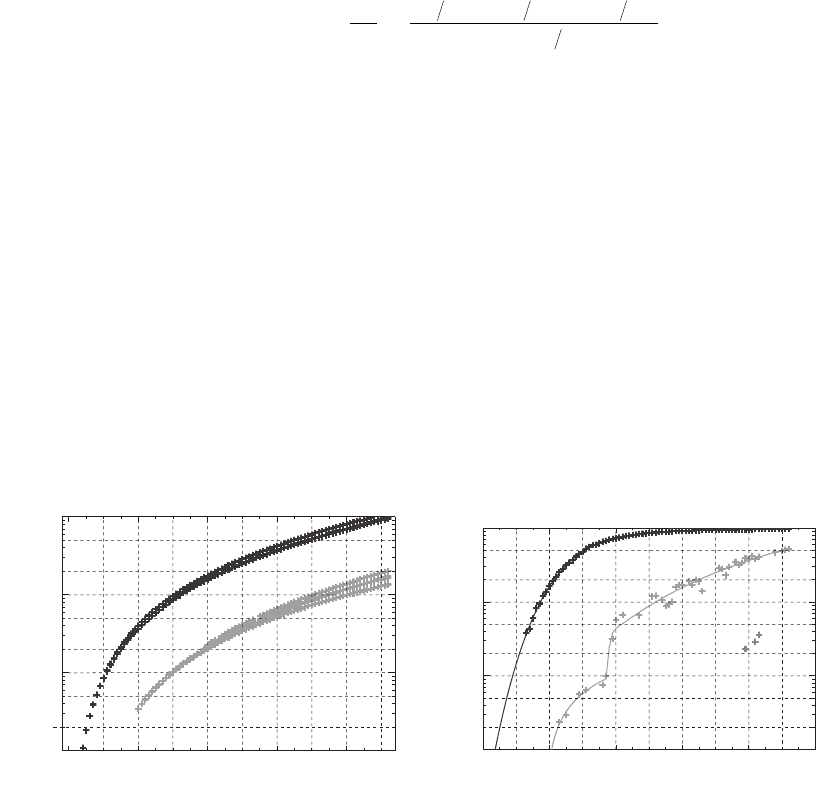
sion), are both shown in Figure 13–3. At X-ray energies below 1 keV,
L
L
M
K
K
0.20
020406080
Atomic number Z
0.001
0.01
0.10
1.00
Fluorescence yield Y
2
20
5
50
1
10
100
Fluorescence energy (keV)
0.1
0.2
0.5
0.50
0.05
0.02
0.005
0.002
10 30 50 70 90
020406080
Atomic number Z
10 30 50 70 90
Figure 13–3. Energies (left) and fl uorescence yields (right) for K and L edge emission. (Krause,
1979.)

Chapter 13 Principles and Applications of Zone Plate X-Ray Microscopes 839
Auger emission dominates, and scanning photoemission microscopes
(SPEM) use electron spectrometers to exploit these electrons for surface
studies (Ade et al., 1990a; Günther et al., 1997; Warwick et al., 1997; Ko
et al., 1998). At higher energies, the fl uorescence signal dominates and
detection of these characteristic X-rays provides information on the
concentration of various elements in the specimen. Most scanning
fl uorescence X-ray microprobes (SFXM) (Horowitz and Howell, 1972;
Sparks, 1980) use energy dispersive detectors where the number of
electron-hole pairs created by each fl uorescent photon is used to
measure its energy, though crystal-based wavelength dispersive spec-
trometers can also be used. Exact quantitation of the elemental concen-
tration requires accurate knowledge of a number of factors, including
the solid angle acceptance of the detector and its quantum effi ciency,
the degree to which fl uorescent photons are reabsorbed in the speci-
men, and other factors, so that in most cases comparison is made with
standards with known elemental concentration and matrix concentra-
tion similar to that of the specimen under study. When compared with
electron microprobes, X-ray microprobes do not suffer from expansion
of the probe beam due to electron scattering, or a large continuum
background, so that the sensitivity to trace elements is often in the 100
parts per billion range.
Because X-ray interactions are well understood and do not involve
signifi cant complications due to multiple scattering at energies below
about 10 keV, reliable predictions of image contrast can be made. If we
have a normalized signal I
f
from a feature-containing pixel and I
b
from
a background region, the signal to noise ratio obtained with N illumi-
nating photons is (Glaeser, 1971; Sayre et al., 1977a)
SNR
Signal
Noise
==
−
+
=
N
II
II
N
fb
fb
Θ
(2)
where we have used the Gaussian approximation to Poisson statistics
(which is quite good for NI greater than about 10) and the assumption
that there are no other noise sources with signifi cant fl uctuations. The
contrast parameter Θ is different from the usual defi nition of contrast
due to the square root in the denominator. With this defi nition, the
number of photons required to see a feature with a desired signal to
noise ratio SNR is given by N = (SNR)
2
/Θ
2
, and a common choice for
the minimum detectable signal to noise ratio is the Rose criterion of
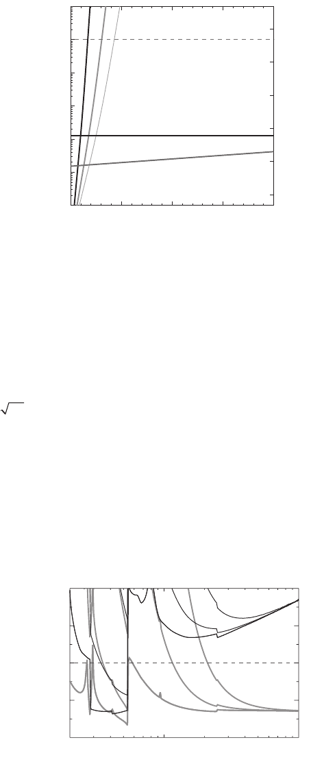
SNR = 5 (Rose, 1946). Using this approach, Sayre et al. showed that
“water window” X-ray microscopes are able to image organic speci-
mens in micrometer-thick water layers with greatly reduced radiation
dose compared to electron microscopy (Sayre, 1977b; Sayre, 1977a).
This conclusion remains true even when modern energy-fi ltered elec-
tron microscopes are considered (Grimm et al., 1998; Jacobsen et al.,
1998) (see Figure 13–4). Other investigators have extended the same
approach to include the effects of phase contrast (Rudolph et al., 1990;
Gölz, 1992) (see Figure 13–5) and the reduction of modulation transfer
at high spatial frequencies (Schneider, 1998), while Kirz et al. have used
this approach to compare elemental mapping using both differential
absorption and X-ray fl uorescence (Kirz et al., 1978, 1980a, 1980b).

840 M. Howells et al.
1.3 Focusing Optics
Microscopes require focusing optics, or some other means to provide
a magnifi ed view of the object. X-rays refl ect well from single refractive
interfaces only at grazing angles of incidence less than a critical angle
of
θδ
c
≈
2
which is typically in the range of 1–5º for soft X-rays. (Once
a particular angle has been selected, this same relationship gives a
critical energy above which the refl ectivity becomes low; this can be
used to low-pass-fi lter the energy spectrum from a radiation source).
While a number of labs have explored the use of axially symmetric
paraboloid or hyperboloid optics (Wolter, 1952; Aoki, 1994), most present
efforts center on the use of two orthogonal cylindrical grazing mirrors
in the Kirkpatrick-Baez geometry (Kirkpatrick and Baez, 1948). Advan-
tageous characteristics of these optics include their relatively long focal
Protein in ice: 30 nm resolution
(100% efficient optics, detectors)
Electron microscopy
(zero loss)
100 kV
300 kV
phase contrast
X-ray microscopy
at 520 eV
3000 kV
absorption contrast
10
0
10
-1
10
1
10
2
10
3
10
4
300 kV electrons: e
-
/Å
2
051015 20
Ice thickness (µm)
Dose in Gray
10
11
10
10
10
9
10
8
10
7
10
6
10
5
(x-ray cryo mass loss)
Energy (eV)
500200 2000 50001000 10,000
10
14
10
12
10
10
10
8
10
6
Skin dose (Gray)
5 µm
ice
25 µm
ice
125 µm
ice
Phase
contrast
Absorption
contrast
5 µm
25 µm
125 µm
5 µm
25 µm
125 µm
Feature: 20 nm protein
(x-ray cryo mass loss)
Figure 13–4. Estimated radiation dose required for imaging 30-nm-protein
features as a function of ice thickness for 200 keV electron microscopes and
520 eV soft X-ray microscopes. (Reprinted from Jacobsen et al., 1998, with per-
mission from Springer Science+Business Media.)
Figure 13–5. Estimated radiation dose required for imaging 20-nm-thick
protein features in various ice thicknesses as a function of X-ray energy; 100%
effi cient optics and detectors are assumed.
Chapter 13 Principles and Applications of Zone Plate X-Ray Microscopes 841
length (several centimeters is typical) and their low chromaticity, so
that the incident beam energy can be tuned for spectroscopy without
any need to adjust the focus on the specimen. Optics of this sort have
recently achieved better than 100 nm resolution probe sizes using 12 keV
X-rays (Hignette et al., 2003; Yamamura et al., 2003; Mimura
et al., 2004), although the profi le of the focus always has some degree
of “tail” outside of the geometrical image of the source due to scattering
from the residual surface roughness of even the best available mirrors.
Synthetic multilayer X-ray mirrors (Spiller, 1972; Barbee et al., 1981) can
increase the incidence angle well beyond θ
c
for narrow-bandwidth
radiation, and can achieve good refl ection effi ciencies for normal inci-
dence refl ection at photon energies below about 200 eV. This approach
has seen rapid improvements due to the development of EUV projec-
tion lithography at 95 eV. However, notwithstanding recent progress in
mirror manufacture, it is important to recognize that even a perfectly
made Kirkpatrick-Baez mirror system still suffers from aberrations,
especially obliquity of fi eld, which severely restrict its fi eld of view and
therefore its performance as a microscope. On the other hand, it is still
well-able to focus points on or near the optical axis, which has led to a
resurgence in its popularity for microprobes and relay mirrors that are
imaging small sources such as synchrotrons.
When Röntgen discovered X-rays, he immediately tried to focus
them using refractive lenses but without success. The reason for this
is now well known: the focal length for a planoconvex lens with radius
of curvature R
c
is given by f
R
= −R
c
/δ, so that at 10 keV a glass lens with
R
c
= 1 cm would have a focal length of about 2 km. This does not pre-
clude the usefulness of refractive optics, however; a series of lenses
with small R
c
can be placed together to produce a signifi cant net focus-
ing effect. One simple way to achieve this result in 1D is to drill a series
of holes in a solid block (Snigirev et al., 1996), and more recent work
using parabolic optics has demonstrated a resolution of about 100 nm
for hard X-ray imaging (Lengeler et al., 2002) with theoretical promise
for sub-10 nm resolution imaging (Schroer and Lengeler, 2005). Because
the ratio of phase shift to absorption increases with increasing X-ray
energy, these optics work primarily at energies above about 5 keV, and
at higher energies one will ultimately need to consider the contribu-
tions of inelastic scattering to the image due to the overall thickness of
the optic. Still, this approach is of interest especially since these optics
can be easily water cooled for high power applications.
The third way to focus X-rays is to use diffraction. While bent crys-
tals can provide focused beams of Bragg or Laue diffracted X-rays,
most work in X-ray microscopy centers on the use of microfabricated
diffractive optics in the form of Fresnel zone plates. Efforts in X-ray
microscopy using zone plate optics date back nearly a half century
(Baez, 1960, 1961), and X-ray Fresnel zone plates are now benefi ting
from a high degree of development. Apart from detailed literature that
we will cite in Section 2. general reviews can be found in the books by
(Michette, 1986; Attwood, 1999). Due to their popularity as high resolu-
tion optics for X-ray microscopy, the properties of Fresnel zone plates
are described in some detail below.
842 M. Howells et al.
1.4 Overview and Recent Trends
We now review some of the general trends of X-ray microscopy tech-
nology and how they might affect the planning of a new X-ray micros-
copy program today. In this section, the number of relevant references
is essentially unlimited so we cite instead the subsections of this article
where many of the references can be found.
Modern X-ray microscopy was based on the twin ideas of the water
window (Section 1.1) and microfabricated zone-plate lenses (Section 2).
These led initially to life-science experiments consisting of 2D imaging
of wet samples in room-temperature air with “naturalness” as the
unique capability of the technique. Early versions of both the TXM
(Section 3.1.1) and STXM (Section 3.1.3) were capable of such imaging
and this style of X-ray microscopy has proved to be good fi t to
many of the needs of the polymer (Section 4.3) and environmental
research communities (Section 4.2) among others. However, the room-
temperature approach could not be easily adapted to 3D imaging of
radiation-sensitive samples and this was something for which there
was, and still is, considerable demand. On the other hand the STXM,
even in its earliest realizations, was ready to begin the development of
spectromicroscopy (Sections 3.5 and 4). This development has contin-
ued over the last two decades or so and spectromicroscopy is now quite
a mature fi eld that spans a wide range of application areas.
The step which has brought 3D imaging of biological samples within
reach has been the recent move toward X-ray microscopy of hydrated
biological samples frozen to cryogenic temperatures. For this type of
sample, X-ray microscopy fi lls an important gap in the range of resolu-
tion values and sample thicknesses which can be covered by other
methods. The use of cryogenic temperatures is the key step in provid-
ing enough radiation-damage protection (see Figure 13–5) to enable
tomographic (three-dimensional) imaging (see Section 3.4). However,
3D cryomicroscopy requires signifi cant technical additions to the X-ray
microscope including either the use of a vacuum sample chamber or a
gas-stream approach to keeping the sample cold plus the mechanical
devices required for recording a tilt series.
The interest in 3D imaging is also high in materials science and
engineering where there is a similar gap in the coverage by other
methods and where generally the samples have higher atomic num-
ber and have nonaqueous background materials (microcircuits for
example). For these samples the water window has no advantage and
X-ray microscopy at higher X-ray energies allows examination of bigger
samples and has in fact been going on for some time. Even in biology
there are factors pushing in the same direction. Ice is equally transpar-
ent at 1.5 keV as in the water window. Moreover, the 1.5–3.0 keV region
gives less absorption and more phase contrast so there is only a moder-
ate increase of the required radiation dose compared to the water
window (see Figure 13–5). In this energy range zone plates also have
longer focal lengths (important for sample tilting) and greater depth
of focus (important for 3D reconstruction (Section 3.4.2)). Overall,
multi-keV 3D X-ray microscopy has great promise but it poses some-
Chapter 13 Principles and Applications of Zone Plate X-Ray Microscopes 843
what different challenges with respect to the resolution/effi ciency
capabilities of the zone plates and the provision of a suitable
condenser.
When possible we have cited publications in widely available jour-
nals rather than conference proceedings. However, good snapshots of
the fi eld are provided at three-year intervals by the series of conference
proceedings dating back to 1984 (Section 1.1). As of the time of writing
of this chapter the most recent one (of which the proceedings
(Kagoshima et al., 2006) have not yet been published) was in Himeji,
Japan, in 2005. We have in some cases cited papers in the proceedings
of this conference because they have not appeared yet elsewhere. This
is especially true for papers on multi-keV X-ray microscopy which was
a growth subject at Himeji. Reports on the high-aspect-ratio zone plates
needed for multi-keV X-ray microscopy (Section 2.4.6) were particu-
larly promising. Zone plates of 50 nm outer zone width for 3–10 keV
and 30 nm for 1–3 keV were reported with effi ciencies of at least 10%
(Section 2.4.6). Several of the former are operational in synchrotrons.
Equally signifi cantly, images were shown using a 50 nm zone plate in
3rd order to get sub-30-nm resolution (Section 2.4.6).
The resolution values of the best water-window and multi-keV three-
dimensional images are both currently around 60 nm. The underlying
reasons for this value probably include the depth of focus effect (3.4b)
for water-window images and the zone plates for multi-keV ones. If
this is true, then the above reports suggest that the tools are now in
place for improvements to the 60 nm limit.
On condensers the news was also good. It was announced publicly
for the fi rst time that single-refl ection monocapillary mirrors have been
in use for some time as condensers for multi-keV TXMs and that exten-
sive 3D imaging has already been done using them. The condenser
system (Section 2.4.3) has traditionally been the Achilles heel of the
TXM and this new generation of condensers is delivering an elegant
solution which is described in more detail in Section 2.4.4. Apart from
removing various limitations of the condenser-zone-plate, the most
important innovation is energy-tunability. This combined with its
multiplexed data collection could make the TXM competitive in the
spectromicroscopy arena for specimens that are tolerant of its
higher dose.
The TXM and STXM have always been seen as complementary
devices; roughly equally popular (Table 13–3) and with important
advantages on both sides. We do not believe that that general percep-
tion is likely to change in the near future. The STXM will always have
the advantage in trace element mapping, the ability to instantly switch
from high to low magnifi cation, the ability to hold constant magnifi ca-
tion even when the X-ray energy is changing, the ability to image at
close to the theoretical minimum dose and so forth. On the other hand,
the TXM is going through a period of technical enhancement. It has
been moving into the multi-keV region and into cryomicroscopy which
together with its traditional main asset, multiplexed data collection, is
strengthening its capability in tomography which appears to us to be
its natural home.

844 M. Howells et al.
2 Fresnel Zone Plates
2.1 Introduction
A Fresnel zone plate is a circular diffraction grating that can be made
to focus light waves in the manner of a lens. It consists of a series of
concentric, usually metal, rings alternating with circular slots. Typi-
cally the rings are about equal in width to the slots and are fabricated
on a thin membrane. The design is based on the idea, that by blocking,
say, the even-numbered Fresnel half-period zones (Born and Wolf,
1999), the wavelets from the remaining (odd-numbered) zones will add
constructively. To see this quantitatively, consider plane-wave illumi-
nation of a zone plate with n zones with radius r
n
, outer zone width
∆r
n
, and a focal length f at wavelength λ (Figure 13–6). To get a fi rst
order diffracted beam in which the signals from all the open zones
reinforce at the focus, we need a path difference λ between neighboring
open zones. In other words the optical path
rf
n
n
22
2
+
−λ
should
equal f. Expanding the square root and neglecting terms above fourth
order, we obtain
nr
f
r
f
nn
λ
22 8
24
3
=− +
(3)
Evidently the focusing condition is
r
2
n
= nλf (4)
to second order and
rnf
n
n
2
22
4
=+λ
λ
(5)
to fourth order. In view of Eq. (7), we can neglect the fourth order
(spherical-aberration) term of Eq. (3) if the numerical aperture (NA) <<
1 which is often the case for X-ray zone plates. If the fourth-order term
is signifi cant, then the zone plate can be made according to (5) and will
be corrected for spherical aberration but the correction will only apply
near the chosen wavelength and conjugate distances (⬁ and f). If it can
be neglected, we have
rnf
n
=λ, and this defi nes a zone plate that will
zone plate
lens
focus
f
r
n
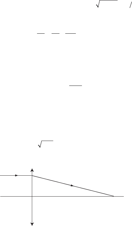
Figure 13–6. Geometry to calculate the radius of the nth half-period zone of
a zone plate illuminated by parallel light. The path from the nth zone to the
focus must be equal to f plus n/2 waves.

Chapter 13 Principles and Applications of Zone Plate X-Ray Microscopes 845
focus well for a range of wavelengths, although the focal length will
vary inversely with wavelength. Thus the chromatic aberration of a
zone-plate lens is much larger than that of a refractive lens and, to get
a good focus, the zone plate needs to be illuminated by monochromatic
light. The required degree of monochromaticity for achievement of the
diffraction-limited resolution is roughly given by ∆λ/λ ≤ 1/n (Thieme,
1988).
Some useful quantities follow from the fundamental zone-plate
Eq. (4). First we can take the difference between the nth and (n − 1)th
equations to get the outer zone width ∆r
n
∆r
f
r
n
n
=
λ
2
. (6)
This allows the conclusion that all of the zones have equal area and
also gives us the numerical aperture
NA
r
fr
n
n
≡=
λ
2∆
, (7)
and thence the Rayleigh resolution
δ
λ
Rayleigh n
NA
r==
061
122
.
. ∆
. (8)
Thus we see that a given zone plate can be specifi ed by its r
n
and ∆r
n
from which the resolution (which is independent of wavelength) and
the focal length and numerical aperture at any given wavelength,
follow from Eq. (8), Eq. (6) and Eq. (7) respectively. So far we have been
discussing the fi rst-order focus but in general, beams of all integral
orders may be produced. Thus there is a zero-order (unfocused) beam,
a series of positive-order converging beams with focal distance f/m and
a series of negative-order diverging beams with focal distance −f/m
(Figure 13–7). In mth order the numerical aperture is m times larger
ZP
(m=-5)
(m=-3)
(m =-1)
(m = -5)
(m=-3)
(m=-1)
OSA
λ
f
5
5nλ
2
+
f
3
3nλ
2
+
nλ
2
f +
f
f
5
f
3
Figure 13–7. Diagram showing orders number
−5, −3, −1, 1, 3, 5 of a zone plate. Compared to
the incident beam, the negative orders diverge,
the positive orders converge and the zero order
(omitted for clarity) has the same shape. For
plane-wave illumination, the virtual foci of the
negative order beams and the real foci of the
positive order beams are at distances | f |/m from
the zone plate where | f | is the focal distance of
fi rst order. (From Attwood, © 1999, with permis-
sion of Cambridge University Press.)

846 M. Howells et al.
and hence the resolution is m times smaller (better) than in fi rst order.
As we will see, if the open and opaque zones are of equal width, then
the even orders are missing.
2.2 Zone Plate Image Quality
2.2.1 Optical Path Function Analysis
We consider the imaging of a general point A (x,y,z) by a planar zone
plate lying in the y − z plane (Figure 13–8) (Kamiya, 1963). We will use
the method of the optical path function so we start by calculating the
optical path from A to a general point B (x′,y′,z′) that we will later iden-
tify as the Gaussian image point. Without loss of generality we set y =
y′ = 0. We calculate the path APB where P (0,w,l) is a general point in
the zone plate. The expression for the optical path will be a power
series in the aperture coordinates, w and l, and the fi eld angle, z/x and
each term in the series will represent a specifi c aberration. Evidently
AP =
++−xw zl
22 2
()
(9)
so expanding the square root and keeping terms up to fourth order,
we have
AP =+
+
+−− +
()
++
{
−
x
wl
x
z
x
zl
xx
wl z
zl z
1
1
2
1
2
2
2
1
8
1
44
22
2
2
22 4
22
2
4
22 3322222
222lwlz wlzl++
()
−+
()
]
+
}
(10)
There is an identical series for PB except that, for PB, the x, y and z are
replaced by x′, y′, and z′ (Figure 13–8). We are now in a position to write
down the optical path function, F. Before doing so we drop terms
z
x
x
y
l
w
x'
z'
z
P
A
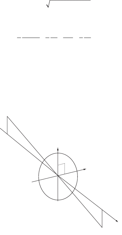
B
O
Figure 13–8. Notation for the optical-path-function analysis of a zone plate.

Chapter 13 Principles and Applications of Zone Plate X-Ray Microscopes 847
which do not depend on w, l or z/x because they do not represent aber-
rations, and introduce the term −nmλ/2 as we did to get Eq. (1). We
choose to analyze the case of a parabolic zone plate (that is one built
according to Eq. (4)) so initially, we use Eq. (4) to write the optical path
function up to third order only, as follows.
F
nm
wl
fm
wl
xx fm
l
z
x
z
=+− =+−
+
=
+
+
′
−
−+
′
AP PB AP PB
λ
22
1
2
11 1
22
22
′′
+
x
(11)
Specializing to the case when B is the Gaussian image point, the fi rst
(defocus) term vanishes and by considering the ray AOB we obtain
z/x = −z′/x′ (12)
so that the second term also vanishes. This leaves only the fi ve fourth-
order terms
F
wl
xx
lz
xfm
lz
x
z
x
wl
=−
+
()
+
′
(
)
−++
′
′
−
+
(
22
2
33
22
2
3
3
3
3
22
8
11
2
1
2
))
+
+
()
+
′
(
)
+
4
1
2
11
2
2
22
22
z
xfm
wl
lz
xx x
(13)
These are the fi ve Seidel aberrations; spherical aberration, astigmatism,
distortion, fi eld curvature and coma respectively. Because of Eq. (12),
the distortion term vanishes identically which is a useful property of
zone-plate lenses and we can therefore turn our attention to the remain-
ing aberrations.
2.2.2 Ray Aberrations
We need to know the ray pattern delivered by the zone plate for a given
point object. That is, we want the ray aberrations ∆y′ and ∆x′ relative
to the Gaussian image point (coordinates identifi ed by subscript zero).
For a normal-incidence optic these are given by the following expres-
sion (Born and Wolf, 1999)
∆∆
′
=
′′
=
′
yx
F
w
zx
F
l
00
∂
∂
∂
∂
,
(14)
We now apply this to the Seidel aberrations individually.
2.2.3 Spherical Aberration
We can rearrange the spherical aberration term using the magnifi cation
M = x′
0
/x
F
wl
f
M
M
sp ab
=−
+
()
+
+
22
2
3
3
3
8
1
1()
(15)
The last term of this expression, which we will denote by Θ, approaches
unity in the cases of interest to us namely M large (a microscope) or
M small (a microprobe). If we consider the case that Θ does approach
unity, then Eq. (15) reduces to the fourth-order term of Eq. (3). This is
expected because we are reverting to the conjugates used to derive (3)
