Hawkes P.W., Spence J.C.H. (Eds.) Science of Microscopy. V.1 and 2
Подождите немного. Документ загружается.


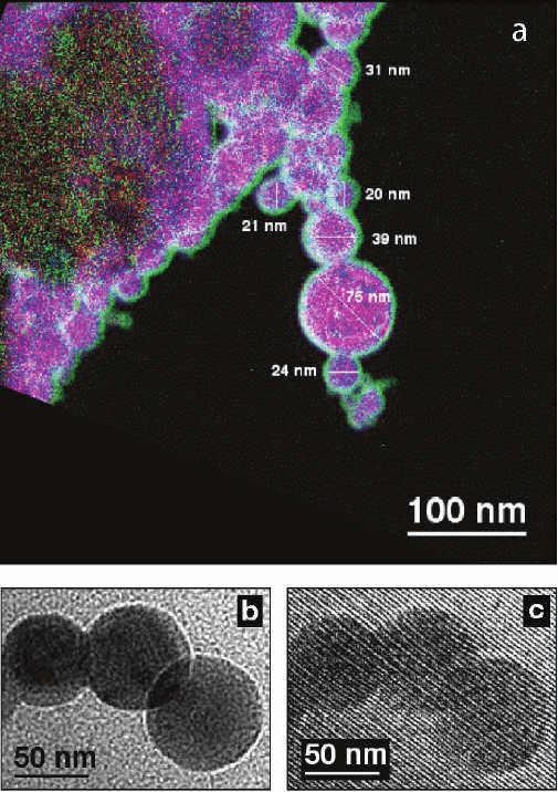
Figure 18–10. (a) Chemical map of Fe
0.56
Ni
0.44
nanoparticles, obtained using three-window back-
ground-subtracted elemental mapping with a Gatan imaging fi lter, showing Fe (red), Ni (blue), and
O (green). (b) Bright-fi eld image and (c) electron hologram of the end of a chain of Fe
0.56
Ni
0.44
particles.
The hologram was recorded using an interference fringe spacing of 2.6 nm. (Reprinted from Dunin-
Borkowski et al., 2004b.)

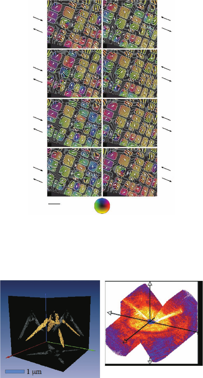
Figure 19–5. (A) Tomographic reconstruction from a soft X-ray diffraction pattern shown in (B). The
object consists of gold balls (50 nm diameter) lying along the edges of a pyramidal-shaped silicon
nitride structure. This is one image from a rotation series. From the complete series, three-dimensional
surfaces of constant density can be constructed. (B) The volume of soft X-ray diffraction data collected
to obtain the three-dimensional reconstruction in (A).
Figure 18–13. Magnetic phase contours from the region shown in Figure 8–12, measured using elec-
tron holography. Each image was acquired with the specimen in magnetic fi eld-free conditions. The
outlines of the magnetite-rich regions are marked in white, while the direction of the measured mag-
netic induction is indicated both using arrows and according to the color wheel shown at the bottom
of the fi gure (red = right, yellow = down, green = left, blue = up). Images (a), (c), (e), and (g) were
obtained after applying a large (>10,000 Oe) fi eld toward the top left, then the indicated fi eld toward
the bottom right, after which the external magnetic fi eld was removed for hologram acquisition.
Images (b), (d), (f), and (h) were obtained after applying identical fi elds in the opposite directions.
(Reprinted from Harrison et al., 2002.)
1340 Oe
628 Oe
225 Oe
0 Oe
0 Oe
225 Oe
628 Oe
1340 Oe
after
saturating
after
saturating
after
saturating
after
saturating
after
saturating
after
saturating
200 nm
ab
cd
ef
gh
after
saturating
after
saturating
A
B
