Hawkes P.W., Spence J.C.H. (Eds.) Science of Microscopy. V.1 and 2
Подождите немного. Документ загружается.

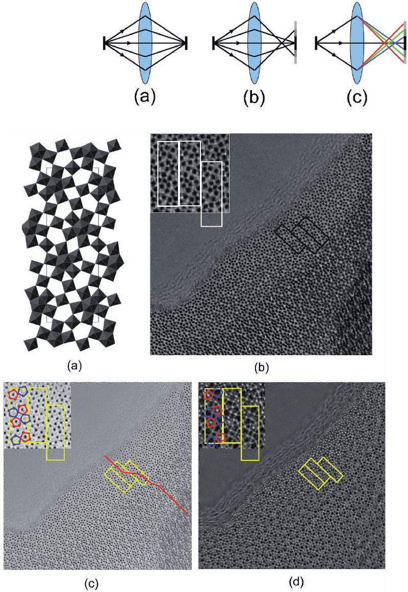
Figure 1–14. Illustration of
certain lens aberrations. (a)
A perfect lens focuses a
point source to a single
image point. (b) Spherical
aberration causes rays at
higher angles to be overfo-
cused. (c) Chromatic aber-
ration causes rays at
different energies (indi-
cated by color) to be focused
differently.
Figure 1–18. (a) Structural model of the complex oxide Nb
16
W
18
O
94
projected along [001]. (b) Conven-
tional axial HRTEM image recorded at the Scherzer defocus of a thin crystal. (c) Reconstructed
modulus of the exit-plane wavefunction of Nb
16
W
18
O
94
with the marked area enlarged (inset), which
directly shows the cation positions (black) with improved resolution compared to the axial image. The
line indicates a stacking fault with a shift of a third of a unit cell along [010]. (d) Reconstructed phase
of the exit-plane wavefunction with the marked area enlarged (inset). The cation sites in the phase are
recovered with positive (white) contrast and additional weak between the cation atomic columns
which indicate the positions of the oxygen anions are also resolved. The reconstructed phase and
modulus are shown at the same scale.
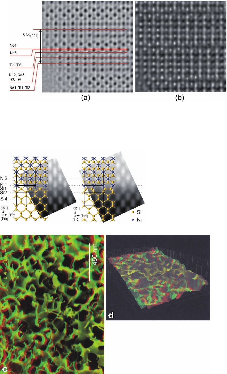
Figure 2–14. An ADF image of an
NiS
2
/Si(001) interface with the struc-
ture determined from the image over-
laid. [Reprinted with permission from
Falke et al. (2004). Copyright (2004)
by the American Physical Society.]
Figure 3–56. Stereo pair of SE micrographs (a and
b) of the hydrogel poly-(N-isopropylacrylamide)
(PNIPAAm) in the swollen state recorded at 2 keV
with the “in-lens” FESEM. The specimen was rapidly
frozen, freeze dried, and ultrathin rotary shadowed
with platinum/carbon (for details see Matzelle et al.,
2002). (c) Red–green stereo anaglyph prepared from
(a and b). The tilt axis has a vertical direction. (d)
Red–green stereo anaglyph in a “bird view.”
Figure 1–20. (a) Enlarged region taken from the reconstructed modulus calculated from a tilt azimuth
dataset of an Nd
4
SrTi
5
O
17
crystal edge in the [010] projection showing a small difference in positions
of the Nd(4) and Nd(5) cations at the interface between adjacent perovskite slabs. (b) Reconstructed
phase of the same region as (a) showing details of the oxygen anion sublattice between the Ti sites.
The experimental data were recorded using the tilt series geometry described in the text with a JEOL
JEM-3000F FEGTEM, 300 kV, C
3
= 0.57 mm with an injected tilt of 1.9 mrad.
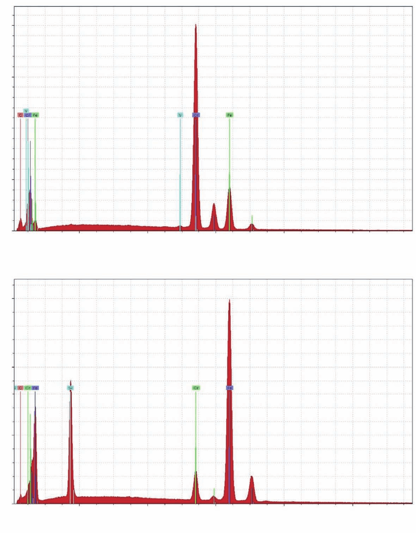
Figure 3–66. X-Ray microanalysis of a Cr–Fe-alloy with a Si phase. The EDX spectra (a and b) were
recorded with the Röntec XFlah3001 from locations “Punkt1” and “Punkt2” marked in the SE micro-
graph of the specimen (c). The positions of the characteristic X-ray energies for the various elements
emerging in the spectra are indicated by thin colored lines, which are labeled with the chemical
symbol of the corresponding chemical element. The elements iron, chromium, and vanadium occur
with one K
α
peak each in the energy range from 4.95 to 6.40 keV and with one less intense L
α
peak
each in the energy range from 0.51 to 0.71 keV. Elemental distribution maps of Fe (d), Cr (e), Si (f), and
Ti (g) were recorded using the K
α
lines. (h) Mixed micrograph obtained by superimposition of the SE
image and the maps of the distribution of Fe, Cr, Si, and Ti within the fi eld of view. Experimental
conditions: SEM, LEO 438VP. For recording spectra: E
0
= 20 keV; count rate, ≈ 3 × 10
3
cps; acquisition
time, 300 s. For recording maps: E
0
= 25 keV; count rate ≈ 1.5 × 10
5
cps; acquisition time 600 s. (EDX
spectra, SE micro graph, and elemental distribution maps were kindly provided by Röntec GmbH,
Berlin, Germany.)
x 1E3 Pulses/eV
4.0
3.0
2.0
1.0
0.0
2
a
b
46
- keV -
810
x 1E3 Pulses/eV
3.0
2.0
1.0
0.0
24 6
- keV -
810
(continued)
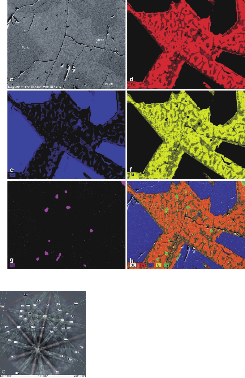
Figure 3–66. Continued
Figure 3–68. ESBD patterns from an as-cast niobium specimen.
(c) EBSD pattern from (a) with colored pairs of Kikuchi lines
generated by automatic indexing. [EBSD patterns were kindly
provided by Dr. S. Zaefferer, Max-Planck-Institut für Eisenforsc-
hung, Düsseldorf, Germany. (a and b) From Zaefferer, 2004; with
kind permission from JEOL (Germany) GmbH, München,
Germany.
5.00 µm = 100 steps
colour coding:
ND
hatched areas:
austenite
111
001
b) c)
grain boundary character
rotation angle fraction
41˚ 42˚ 0.051
42˚ 43˚ 0.112
43˚ 44˚ 0.140
44˚ 45˚ 0.132
45˚ 46˚ 0.097
46˚ 47˚ 0.018
deviation to
centre grain
orientation
orientation class
<001> ll ND (max. 20˚)
all other orientations
austenite
101
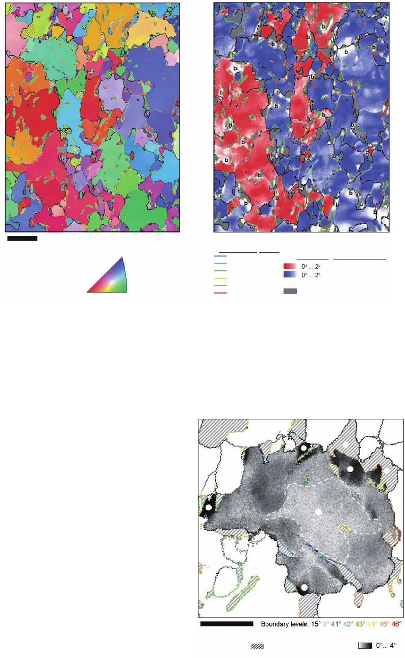
Figure 3–69. Secondary electron micrograph of austenite (b) Orientation map of (a) measured by
automated crystal orientation mapping and color coded for the crystal direction parallel to the normal
direction (ND) of the sheet. Hatched areas correspond to austenite grains. (c) The boundary character
between γ- and α-grains, different orientation components [(001)||ND, red; all others blue] and the
orientation variations inside of each grain (color shading; b, bainite). The micrograph and the maps
are recorded with thermal FESEM JSM-6500F. Note: the extension toward the top and bottom of the
measured are in (b) and (c) is larger than the area marked in (a). (Reprinted from Zaefferer et al., 2004;
copyright 2004, with permission from Elsevier.)
2.50 µm = 50 steps
austenite
b
b
b
a
a
b
f
Angular deviation
to orientation in
grain centre
Figure 3–70. Orientation map of one grain from
the microstructure in Figure 3–69a. Color code:
angular deviation of every mapping point to
one orientation in the center of the grain. Bainite
appears in conjunction with a steep orientation
gradient in ferrite. The white line marks the
maximum extension of austenite at austeniza-
tion temperature. f, ferrite; a, austenite (hatched);
b, bainite. (Reprinted from Zaefferer et al.,
2004; copyright, 2004, with permission from
Elsevier.)
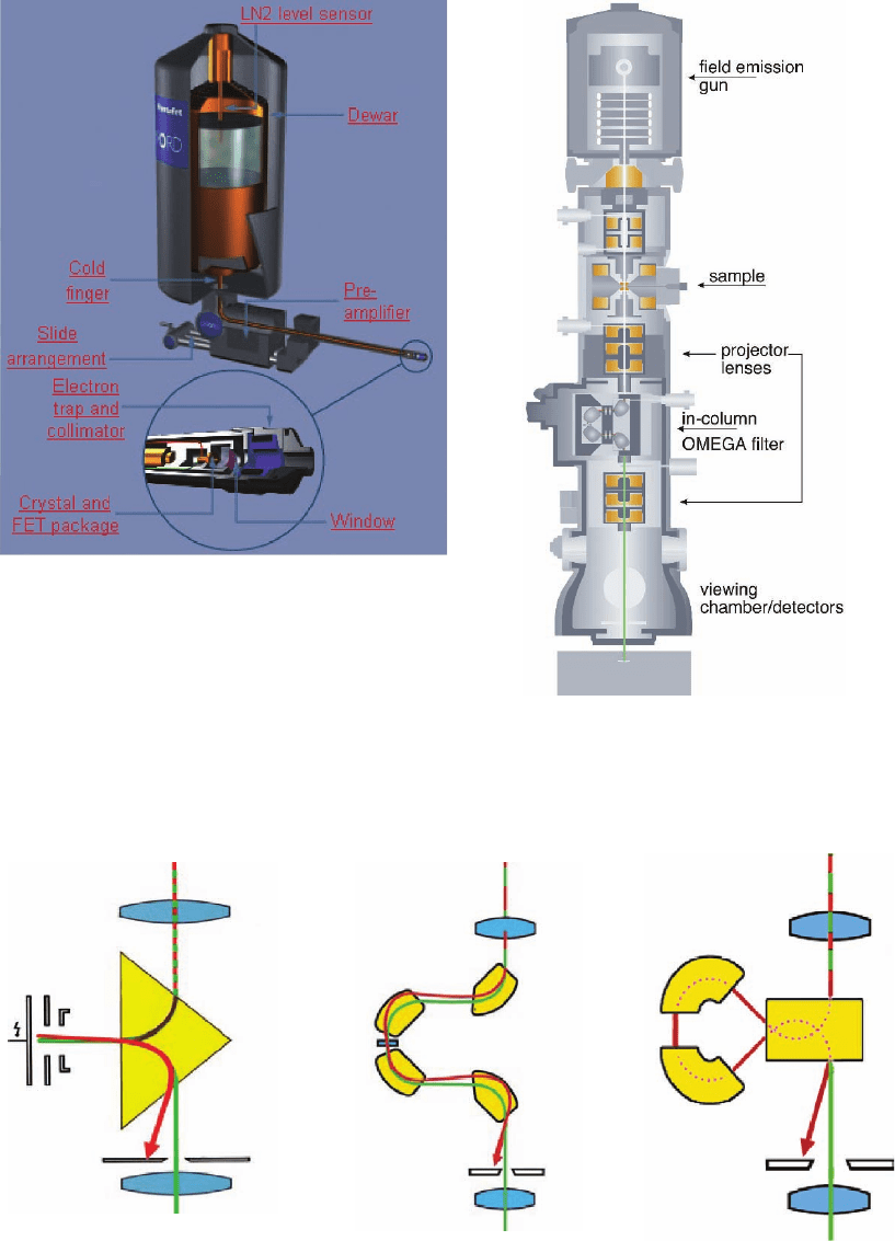
Figure 4–19. Schematics of a commercial
EDXS detector showing the detector front, the
Dewar system to cool the detector, and various com-
ponents that are interfaced in the electron micro-
scope. (Courtesy of N. Rowlands, Oxford
Instruments.)
abc
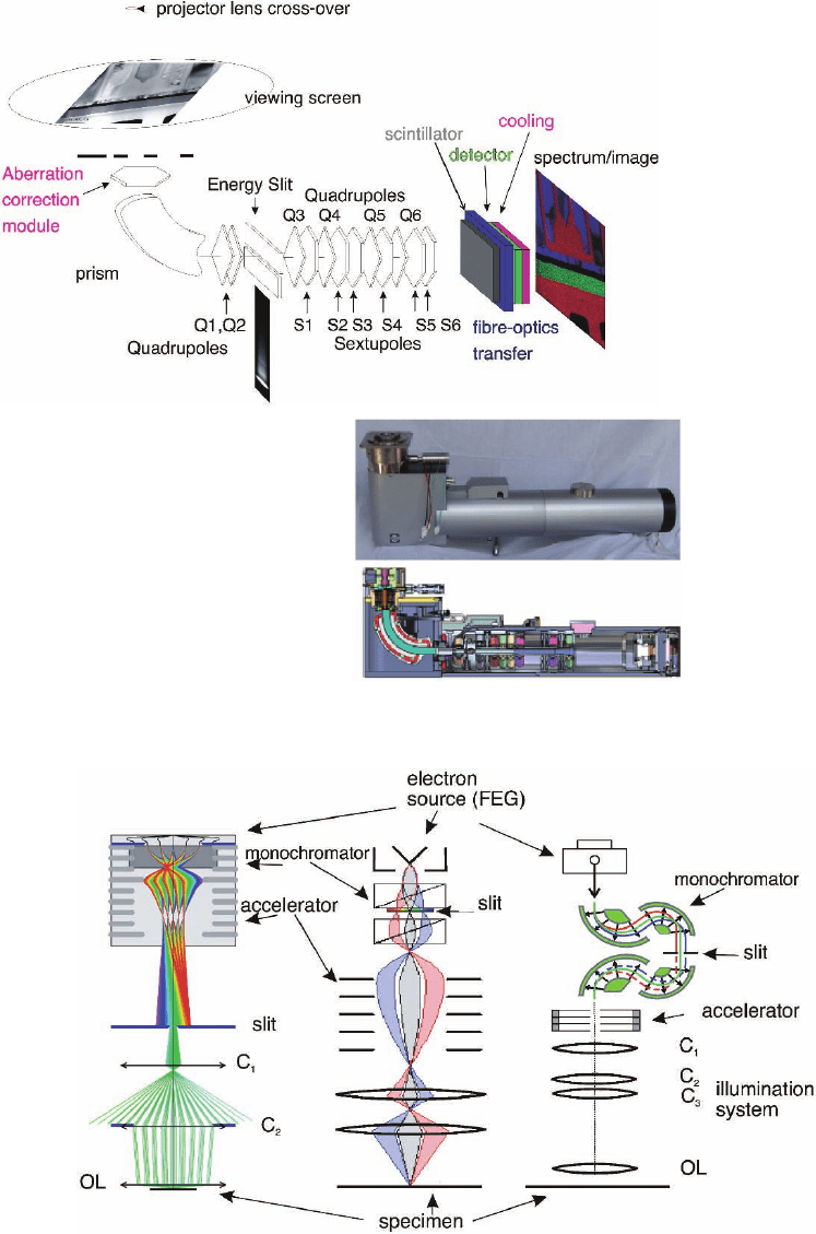
Figure 4–26. Various in-column spectrometer confi gurations. (a) Mirror-prism spectrometer,
(b) OMEGA fi lter, and (c) Mandolin fi lter. (Courtesy of P. Schlossmacher, Zeiss SMT.)
Figure 4–27. Schematic diagram of the in-
column energy-fi ltered microscope (Zeiss-Libra
200). (Courtesy of P. Schlossmacher, Zeiss SMT.)
a)
b)
Figure 4–29. (a) Postcolumn imaging
fi lter (Gatan Imaging Filter GIF);
schematic diagram of the electron
optics components and detection
system (top diagram). (Adapted from
Krivanek, Gubbens et al., 1991a.) (b)
Actual spectrometer [Gatan’s GIF
2000 series spectrometer (Tridiem
model)] and components (bottom
diagram). (Courtesy of M. Kundman,
Gatan.)
Figure 4–31. Various implementations of monochromators in commercially available instruments.
Left: The FEI monochromator, single Wien fi lter. (Courtesy of P. Tiemeijer, FEI Company.) Center: The
JEOL monochromator double-Wien fi lter. (Courtesy of JEOL Ltd.) Right: The lectrostatic omega fi lter
implemented in the Libra Zeiss microscope. (Courtesy of M. Haider, CEOS GmbH.)
0.7 Å probe ON column
16.3 Å
16.3 Å
max = 0.36 min = 0.00
300 Å
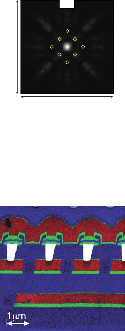
Figure 4–37. Real space intensity plots demonstrating the dispersion of the electron intensity when
the electron beam is located on top of the atomic column and when it is located between two atomic
columns. The bright empty circles indicate the position of the atoms in the cell closest to the point of
impact of the electron beam. Channeling is observed when the beam is positioned on the atomic
column (a) while much stronger dispersion is observed when the electron beam is not on the atomic
column (b). (Data courtesy of C. Dywer and J. Etheridge.)
Figure 4–74. Color-coded elemental map of a device showing the distribution of elements in the ras-
tered area: red, Al-rich area; blue, Si-rich area; green, Ti-rich area. White, W. Interdiffusion of Si into
the Al is noted through the bottom barrier layer containing Ti.
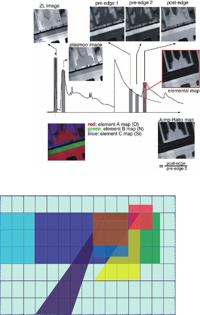
Figure 4–77. Various appr-
oaches to EFTEM imaging.
Zero-loss (ZL) fi ltered imaging
(selecting only electrons that
have lost no signifi cant
amounts of energy), plasmon
imaging (selecting only elec-
trons that have lost energy in
the 10–30 eV range), and core
loss imaging with the three-
windows technique (extrapo-
lation of the background
under the edge) and the jump-
ratio technique.
Figure 5–1. Phenomena classifi ed by spatial and temporal resolution. Spatial resolution, is defi ned as
follows: (1) if the technique is an imaging method, the low end of the bounding box would be defi ned
by the smallest resolvable feature and the high end by the typical fi eld of view or (2) for a nonimaging
technique, the bounding box would be defi ned by the range of probe or spot sizes. Time resolution is
defi ned as that for a single-shot investigation of irreversible processes. So time resolution is defi ned
as the single-shot exposure time to obtain data that demonstrate a particular spatial resolution.
10
10
3
10
10
4
10
10
5
10
10
6
10
10
7
10
10
8
10
10
9
10
10
10
10
10
10
0
10
10
2
10
10
4
10
10
6
10
10
8
10
10
10
10
10
10
12
12
10
10
14
14
Spatial Resolution (m
Spatial Resolution (m
-1
-1
)
Time Resolution (s
Time Resolution (s
-1
-1
)
Diffraction of
Phase
Transformations
structural
changes in
biology
nucleation
and
growth
of damage
Imaging of
Phase
Transformations
Making
and
Breaking
of Bonds
Dislocation
Dynamics at
Conventional
Strain Rates
magnetic
switching
melting
and
resolidification
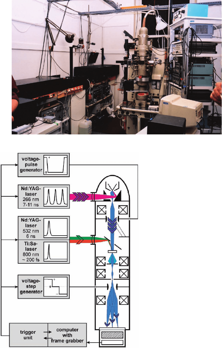
Figure 5–2. The DTEM at TU Berlin. Cathode and sample drive lasers are at left.
Figure 5–3. TU Berlin dynamic trans-
mission electron microscope.
