Hawkes P.W., Spence J.C.H. (Eds.) Science of Microscopy. V.1 and 2
Подождите немного. Документ загружается.

Chapter 3 Scanning Electron Microscopy 215
possibly, such as applications in which high emission currents (cf. Table
3–1) or high electron beam current stability are indispensable. Because
vacuum conditions in FESEMs are more strict concerning the pressure
in the specimen chamber (at least one order of magnitude less than in
CSEM) as well as the content of gaseous hydrocarbons and hydrocar-
bons at the specimen, some specimens may not meet the requirements
for cleanness and very low partial pressure. However, if high-
resolution FESEM is applied instead of CSEM, more information about
the specimen will be obtained due to the higher resolution as soon as
the magnifi cation used exceeds approximately 10,000× to 20,000×. That
means that lateral resolutions requiring magnifi cations clearly beyond
about 20,000× belong to the dedicated domain of high-resolution
SEM.
The following few applications selected from an almost unlimited
quantity should domonstrate the strength of HRSEM in several fi elds
of research. It is clearly beyond the scope of this section to discuss in
this context specifi c details about the specimens and imaging
techniques.
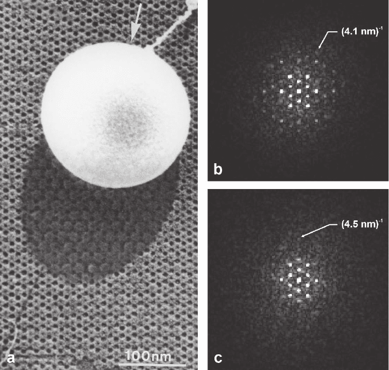
Figure 3–40a shows the secondary electron micrograph of a regular
protein surface layer of a bacterial cell envelope. The specimen was
unidirectional shadowed with an ultrathin tungsten layer leaving an
uncoated region behind the latex bead. Comparison of the regular
structure of the HPI layer in the coated and the uncoated region shows
that the contrast in the uncoated area is signifi cantly lower than in the
coated region, though the resolution of structural details is very similar
as verifi ed by the related power spectra. This example also demon-
strates that coating with the ultrathin very fi ne-grain metal fi lm does
not mask fi ne structural features. On principle, a similar resolution can
also be obtained with nonregular organic specimens, however, it
remains more diffi cult to quantify unambiguously the resolution
obtained.
An extremely important application of HRSEM, as yet unrivaled by
other surface imaging techniques, is the localization of molecules on
surfaces by immunolabeling techniques (for review see, e.g., Griffi th,
1993; Hayat, 1989/1991, 2002; Polak and Varndell, 1984; Verkleij and
started more than 20 years age (de Harven et al., 1984; Gamliel and
Polliack, 1983; Hicks and Molday, 1984; Molday and Maher, 1980;
Walther and Müller, 1985, 1986; Ushiki et al., 1986). Since then efforts
microscopy in terms of localization precision, contrast, and SNR (e.g.,
Albrecht et al., 1988; Hermann et al., 1991; Hirsch et al., 1993; Simmons
et al., 1990). While the colloidal gold can be localized directly in the
BSE image, the precision of the indirect localization of the antigen
depends on the type of labeling and the size of the colloidal gold and
ranges from less than 5 to about 30 nm (Baschong and Wrigley, 1990;
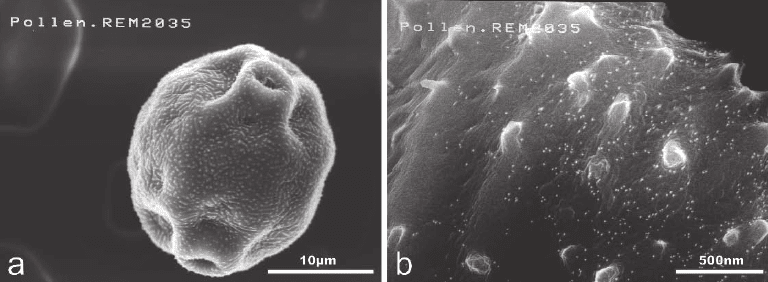
Müller and Hermann, 1990). Figure 3–41b demonstrates the unambig-
uous detection of immunogold-labeled calcium-binding birch pollen
allergen Bet v4 in birch pollen using 10 nm colloidal gold (Grote et al.,
1999b). The BSE micrograph shows the topographic features and the
were made to optimize the technique of immuno-scanning electron
Leunissen, 1989). The use of HRSEM for immunoelectron microscopy
216 R. Reichelt
small colloidal gold beads as sharp bright spots due to the material
microscopy has been established as a trusted technique and, with the
commercial availability of high-quality gold probes (available in sizes
ranging from 1 to 40 nm), is used in many electron microscopic labo-
ratories for various studies (e.g., Apkarian and Joy, 1988; Erlandsen
et al., 1995; Grote et al., 2000; Müller and Hermann, 1992; Ris and
Malecki, 1993; Yamaguchi et al., 1994).
The interior structure of biological specimens is accessible by HRSEM,
if samples are rapidly frozen and opened by cryofracturing or cryoul-
Figure 3–40. Secondary electron micrograph of a regular protein surface layer [hexagonally packed
intermediate (HPI) layer (Baumeister et al., 1982)] of Deinococcus radiodurans recorded with an “in-lens”
FESEM at 30 kV (a). The specimen was unidirectional shadowed (see arrow) at an elevation angle of 45°
with a 0.7-nm-thick tungsten layer leaving an uncoated region behind the latex bead. The power spectra
of a coated (b) and an uncoated (c) region of the HPI layer reveal the resolution obtained (outermost dif-
fraction spots are indicated and the corresponding reciprocal values of resolution are given). The con-
trast in the uncoated region is about 15–20% of that from the coated region. [Micrograph kindly provided
by Dr. R. Wepf; from Reichelt (1995); with kind permission of GIT Verlag, Darmstadt, Germany.]
contrast. However, for more than a decade immuno-scanning electron
Chapter 3 Scanning Electron Microscopy 217
tramicrotomy. After partial freeze-drying and double-layer coating of
the block face, the specimen can be directly analyzed in the cryo-SEM
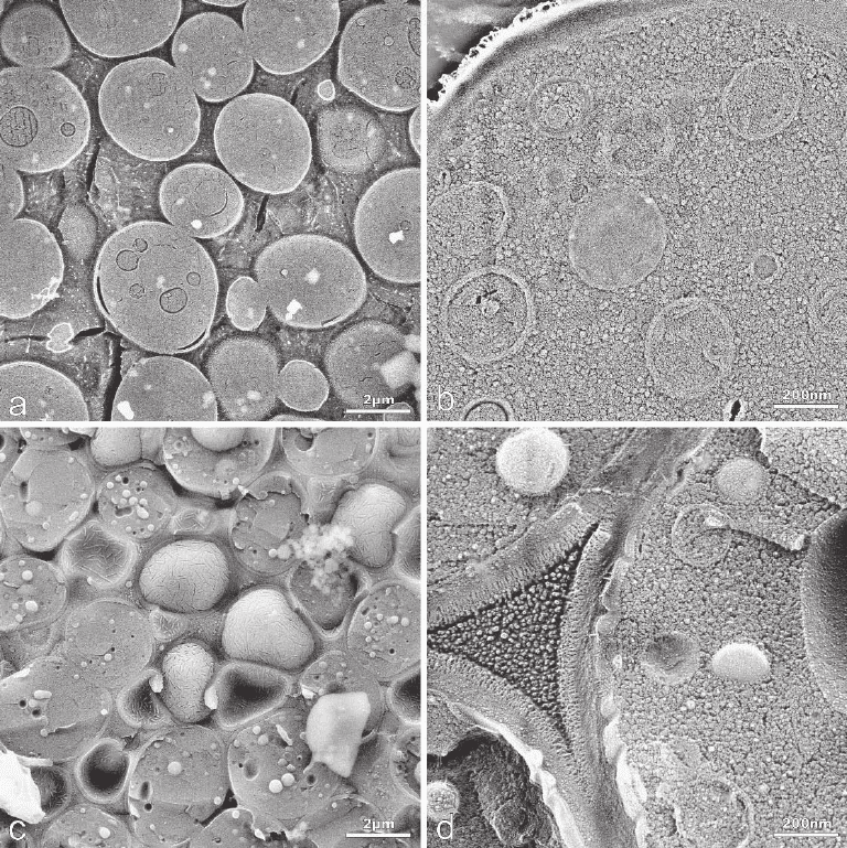
(Echlin, 1971; Hermann and Müller, 1993; Walther et al., 1995). Figure
3–42 shows for comparison the BSE micrograph of the cryosectioned
and the cryofractured block face of high-pressure frozen yeast cells
(Walther and Müller, 1999). At low magnifi cation the cryosectioned
block face appeared very fl at in the image (Figure 3–42a), whereas the
cryofractured face exhibits the typical rough fracture pattern (Figure
3–42c). At high magnifi cation (Figure 3–42b and d) the cytoplasm
appeared densely packed with different classes of particle. Particles as
small as 25 nm can be visualized clearly. It is important to mention that
the typical artifacts of cryosections such as compression and crevasses
are not visible on the block face.
Both strengths of the HRSEM, namely the high resolution and the
high depth of focus, are required to resolve surface structures at the
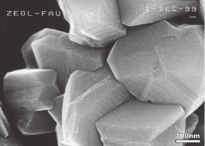
nanometer scale on tilted surfaces randomly oriented. One typical
example with submicrometer-sized crystalline zeolite particles is shown
in Figure 3–43. The HRSEM is the tool most suited to characterize the
habit of the individual particles as well as to visualize the fi ne surface
structure such as growth steps of terraces (see, e.g., González et al.,
2004). The growth step-edge height, usually in direct relation to the unit
cell dimension and important in understanding the crystal growth
mechanism of this novel microporous material, cannot be measured
precisely by HRSEM, but the atomic force microscope (AFM) enables
measurement of direct height with subnanometer resolution. Thus, the
combination of the two high-resolution surface-imaging methods
HRSEM and AFM is strongly advisable to obtain more complete
information as demonstrated by different applications (e.g., González
et al., 2004; Huang et al., 2004; Keller and Chih-Chung, 1992; Lian et al.,
2005; Stracke et al., 2003; Wang et al., 2004; Z. X. Zhao et al., 2005).
Figure 3–41. Secondary electron micrograph from a birch pollen at low magnifi cation (a) and the
backscattered electron micrograph at high magnifi cation recorded with an “in-lens” FESEM at 10 kV.
Immunolabeling of the calcium-binding birch pollen allergen Bet v4 in dry and rehydrated birch
pollen was performed using 10-nm colloidal gold (for more details see Grote et al., 1999a,b). The BSE
image shows superimposed topographic and material contrast. The colloidal gold beads are unam-
biguously detected in the BSE micrograph as tiny bright spots.
218 R. Reichelt
HRSEM is also a very valuable tool for the evaluation of mechanical
properties of structural materials. For example, most structural materi-
als are strengthened by fi ne particles of second phases usually having
diameters less than 500 nm. The strengthening effect is primarily gov-
erned by the mean size, the size distribution, and the volume fraction
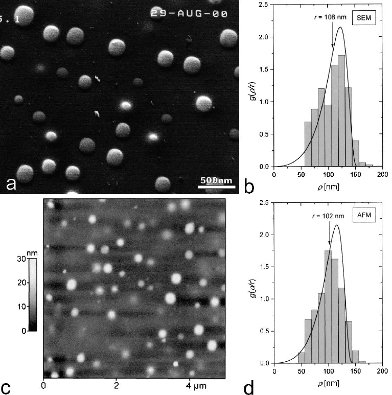
of the particles. Both HRSEM and AFM allow for the precise determi-
nation of the mean size, size distribution, and volume fraction of the
particles as demonstrated by Fruhstorfer et al. (2002). Figure 3–44
shows the SEM micrograph (Figure 3–44a) and the AFM topograph
(Figure 3–44c) of the electrolytically polished surface of the superalloy
NIMONIC PE16 with the protruding caps of the second phase parti-
Figure 3–42. BSE micrographs of high-pressure frozen yeast cells recorded in an “in-lens” cryo-
FESEM at 10 kV. (a and b) Block face after cryosectioning and (c and d) block face after freeze facturing.
The block faces were double-layer coated with 2.5 nm platinum/carbon and subsequently with 5–10 nm
carbon. The cytoplasm from both the sectioned (b) and the fractured (d) sample appears to consist of
particles with variable size. [From Walther and Müller (1999); with kind permission of Blackwell
Publishing Ltd., Oxford, U.K.]
Chapter 3 Scanning Electron Microscopy 219
cles. In contrast to AFM, where corrections were necessary to take into
account the exact tip radius, corrections for the very small electron
probe diameter are not urgently required in HRSEM. The size distribu-
tion function and mean radius of the second phase particles calculated
from HRSEM (Figure 3–44b) and AFM (Figure 3–44d) data are in excel-
lent agreement with those gained earlier by TEM (Nembach, 1996). The
distinct advantages of HRSEM in this application are that micrographs
are readily recorded and the data can be processed without additional
correction procedures.
The characterization of porous materials such as porous silicon or
porous aluminum oxide gains increasing attention because of impor-
tant potential applications (see, e.g., Anglin et al., 2004; Galca et al.,
2003; Pan et al., 2004; Yamazaki, 2004; Z. X. Zhao et al., 2005; Y. C. Zhao
et al., 2005). Among others, HRSEM is an indispensable tool for struc-
tural characterization of porous materials taking advantage of the large
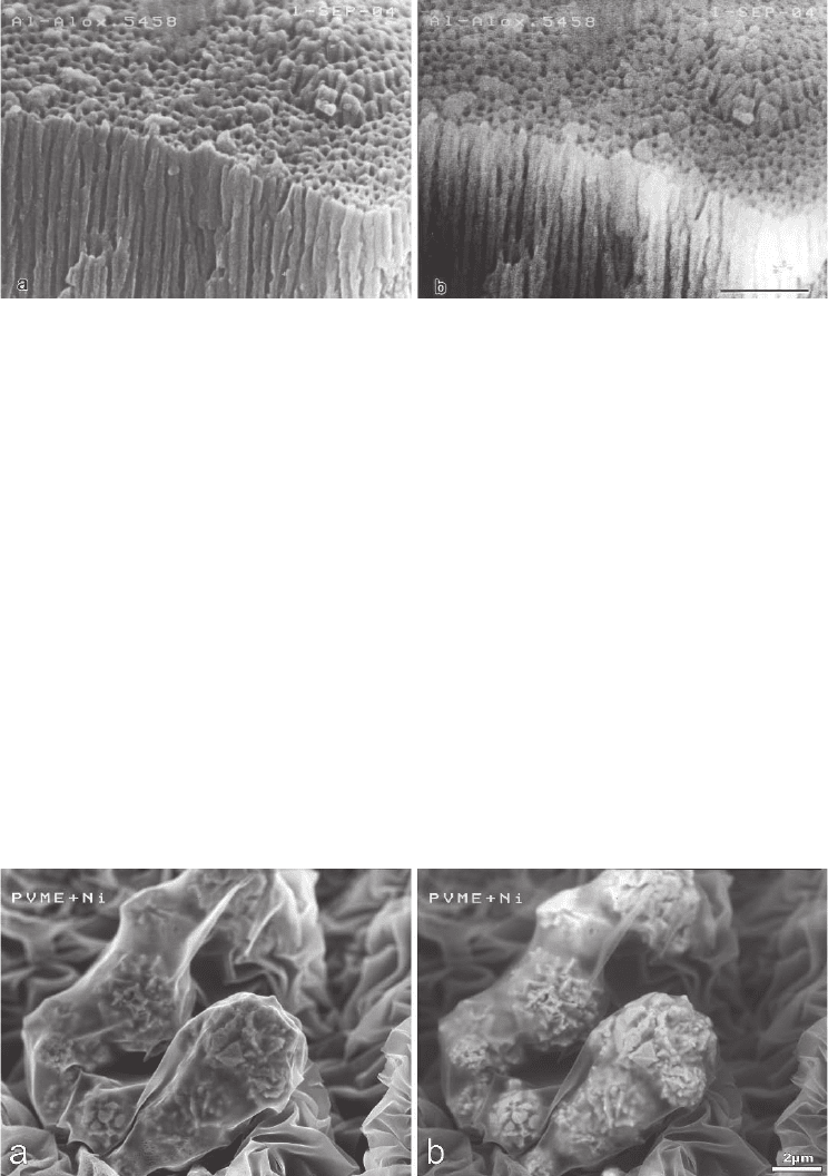
depth of focus and the high resolution obtainable. Figure 3–45 shows
high-resolution SE and BSE micrographs of the surface and cross
section of porous aluminum oxide, which exhibits a network with
randomly distributed, but almost perfectly aligned cylindrical pores
perpendicular to the substrate. The simultaneous imaging of the surface
and the cross section reveals information about the three-dimensional
specimen structure. Under the conditions given the SE mode yields
higher resolution than the BSE mode.
However, the BSE mode is of signifi cant importance if greater infor-
mation depth and material differentiation are required. Figure 3–46
shows SE and BSE micrographs of tem perature-sensitive hydrogels,
based on poly (vinylmethyl ether) (PVME), with ferromagnetic proper-
ties due to incorporated nickel particles used as ferromagnetic fi ller.
The contrast in the SE micrograph (Figure 3–46a) is mainly caused by
Figure 3–43. Secondary electron micrograph of zeolite FAU (faujasite) parti-
cles recorded with an “in-lens” fi eld emission SEM at 10 kV. The particles are
adsorbed to a thin hydrophilic amorphous carbon fi lm and rotary shadowed
with 1.5 nm platinum/carbon (Pt/C) at an elevation angle of 65° and, addition-
ally, unidirectional shadowed with 2 nm Pt/C at an elevation angle of 10°. The
habit, intergrowth of particles, and growth steps at the surface are clearly
visible. (Specimen kindly provided by Dr. G. Gonzaléz, Instituto Venezolano
de Investigaciones Científi cas, Caracas, Venezuela.)
220 R. Reichelt
Figure 3–44. Surface of electrolytically polised superalloy NIMONIC PE16. Secondary electron micro-
graph recorded with an “in-lens” FESEM at 10 kV (a) and AFM topograph (c). The related distribution
functions g of the true radii ρ are shown for the HRSEM in (b) and for the AFM in (c). [Adapted
from Fruhstorfer et al. (2002); with kind permission of Taylor & Francis Ltd., http://www.tandf.co.uk/
journals.
the very thin membrane-like PVME, which envelops the nickel parti-
cles, whereas the BSE image (Figure 3–46b) has a strong material con-
trast component due to the nickel particles underneath the PVME
membrane. This new class of hydrogels is of great interest for delivery
of materials at the micro- and nanometer scale.
As mentioned in Section 2.2.3, the high-resolution “in-lens” FESEM
equipped with an annular dark-fi eld detector is capable of mass mea-
surements on thin specimens (Engel, 1978; Wall, 1979) at a resolution
close to that of a dedicated STEM (Reichelt et al., 1988; Krzyzanek and
Reichelt, 2003; Krzyanek et al., 2004). Mass measurement of molecules
Chapter 3 Scanning Electron Microscopy 221
and molecular assemblies are of great importance in biophysics and
structural biology (for review see, e.g., Müller and Engel, 2001).
Finally, nanotechnology and “nanoelectromechanical systems”
(NEMS) are additional fi elds in which HRSEM is used as a tool for
monitoring processes, detecting defects, or measuring sizes and dis-
tances, e.g., in nanodevices, which will contain nanotubes, nanoparti-
cles, nanowires, and other particles (see, e.g., Aoyagi, 2002; Nagase and
Kurihara, 2000; Nagase and Namatsu, 2004).
3.2 Low- and Very-Low-Voltage Scanning Electron Microscopy
Scanning electron microscopy with electron energies below 5 keV is
usually designated as scanning low-energy electron microscopy
Figure 3–45. High-resolution micrographs of the surface (upper part of image) and the cryosectioned
cross section (lower part of image) of porous aluminum oxide recorded with an “in-lens” FESEM at
8 kV with SE (a) and BSE (b). The specimen is rotary shadowed with 1.5 nm platinum/carbon. The bar
corresponds to 200 nm. (Specimen kindly provided by Drs. C. Blank and R. Frenzel, Institut für Poly-
merforschung Dresden e.V., Dresden, Germany.)
Figure 3–46. High-resolution micrographs of poly(vinyl methyl ether) (PVME) hydrogel with ferro-
magnetic properties fi lled with submicrometer nickel particles in the swollen state. The hydrogel was
rapidly frozen, freeze dried, and rotary shadowed with an ultrathin layer of platinum/carbon. The SE
(a) and BSE (b) micrograph were recorded with an “in-lens” FESEM at 10 kV, (Specimen kindly pro-
vided by Dr. K.-F. Arndt, Institut für Physikalische Chemie und Elektrochemie, Technische Universität
Dresden, Dresden, Germany.)
222 R. Reichelt
(SLEEM) or, related to the acceleration voltage, LVSEM. The energy of
5 keV can be considered as some threshold energy because the monotonic
dependence of the BSE coeffi cient on the atomic number breaks below
this (cf. Section 2.2.2). A second prominent energy threshold is at about
50 eV, which corresponds to the electron energy with minimum inelastic
mean free path of electrons in matter (Seah and Dench, 1979). Therefore,
scanning microscopy with electron energies below 50 eV is designated as
scanning very low-energy electron microscopy (Müllerova and Frank,
2003) or, related to the acceleration voltage in the scanning mode, very
low-voltage scanning electron microscopy (VLVSEM).
What is the motivation for low electron energy operation in SEM?
What are the advantages expected at low energies and what are the
inherent disadvantages?
Clearly, almost all of the advantages for working at low energy
derive directly from the energy dependence of the electron–specimen
interaction (see Section 2.2). The advantages include the following:
1. The penetration depth of the impinging electrons decreases with
decreasing energy due to the reduced electron range R [Eq. (2.29)], i.e.,
the excitation volume in the specimen shrinks (cf. Figure 3–13) and the
volume emitting SE2 and BSE2 approaches the volume emitting SE1
and BSE1 (cf. Figure 3–14). As a result the edge effect, i.e., overbrighten-
ing of edges, is strongly reduced or even suppressed completely.
2. The SE yield δ increases because of the reduced electron range
and the SE are generated near the surface, where they can escape (cf.
Figure 3–15). As a result, the SNR of the SE signal increases with
decreasing energy as low as E
0,m
.
3. As the SE yield increases, the total amount of emitted electrons
approaches unity (cf. Figure 3–16). Because of the conservation of elec-
tric charge [Eq. (2.40)] the amount of incoming and emitted charges is
balanced and, consequently, the specimen current equals zero. That
means that at this particular electron energy E
2
no electric conductivity
of the specimen is required. Ideally, imaging of electric insulators
without conductive coating becomes possible. For normal incidence, E
2
is within the range 0.5–5 keV for most of the materials. E
2
increases
with the increasing angle of beam incidence θ according to
E
2
(θ) = E
2
(0)/cos
2
θ (3.2)
where E
2
(0) = E
2
(θ = 0) (Joy, 1989), i.e., increases as θ increases.
4. As mentioned above, the monotonic de-pendence of the BSE coef-
fi cient on the atomic number breaks below 5 keV (Reimer and Tollkamp,
1980; Schmid et al., 1983). This behavior enables the material contrast
in the BSE image to be fi netuned by choosing the most suitable electron
energy (Müllerova, 2001).
5. There is a reduced depth of specimen radiation damage (see
Section 2.5). At very low electron energies, say less than 30 eV, the
elastic scattering dominates and radiation damage becomes negligible
(Müllerova and Frank, 2003).
The problems and disadvantages inherent to microscopy at low elec-
tron energy concern both the instrumentation and the specimen include
the following:
Chapter 3 Scanning Electron Microscopy 223
1. Reduced resolution due to chromatic aberration and diffraction
[see Eqs. (2.8)–(2.10)].
2. Stronger sensitivity of the electron beam to electromagnetic stray
fi elds.
3. Special detector strategies required for SE and BSE.
4. Enhanced contamination rate, which can be counteracted by
ultrahigh vacuum.
5. Reduced topographic contrast in SE and BSE micrographs. For
electron energies below 5 keV, the increase of SE yield δ(θ) with
increasing θ [cf. Eq. (2.31)] drops way down to 0.5 keV as shown for
different metals experimentally and by Monte Carlo simulation
(Joy, 1987a; Böngeler et al., 1993). Similarly, the backscattering co-
effi cient η(θ) shows less increase with θ than given by Eq. (2.34),
which is more pronounced at low electron energies (Böngeler et al.,
1993).
6. Reduced material contrast, because the differences of the back-
scattering coeffi cient between low and high atomic number material
become smaller (Darlington and Cosslett, 1972; Lödding and Reimer,
1981; Reimer and Tollkamp, 1980).
3.2.1 Electron Lenses
Modern commercial fi eld emission scanning electron microscopes can
operate usually from 30 keV down to 1 keV or even 0.5 keV, i.e., that
energy range covers conventional electron energies and most of the
low-energy region. Improved computer-aided methods enable electron
optical systems to be designed that have high performance within
the whole energy range mentioned above. Compared with the old-
fashioned thermionic gun scanning electron microscopes the aberra-
tion coeffi cients of the objective lens were improved dramatically for
modern fi eld emission instruments commercially available: C
s
was
reduced by a factor of about 30 down to C
s
= 1.6 mm (Uno et al., 2004),
and C
c
was reduced by a factor of about 10. With the ultrahigh-
resolution objective lens, the CFEG, improved electrical and mechani-
cal stability, as well as strongly reduced specimen contamination rate,
the resolution obtained with test specimens amounts presently to
0.5 nm (at 30 keV) and 1.8 nm (at 1 keV) (Sato et al., 2000). Those values
are exemplary for the high performance of commercial FESEM over an
energy range from 1 to 30 keV, though obtained with special test speci-
mens. Very recently, a new commercial FESEM became available
equipped with a spherical and chromatic aberration corrector, which
consists of four sets of a 12-pole component that corrects the spherical
and chromatic aberration simultaneously (Kazumori et al., 2004). Using
the spherical and chromatic correction, the resolution obtained with
the test specimen amounts to 1.0 nm (at 1 keV) and 0.6 nm (at 5 keV)
(Kazumori et al., 2004).
Electrostatic as well as combined magnetic and electrostatic lenses
in LVSEM are a very interesting alternative to the magnetic lenses
mentioned above. Microscopes equipped with this type of objective
lens permit nonconstant beam energy along the column, i.e., the beam
electrons pass the column with high energy and are decelerated to low
energy in the immersion electrostatic lens. First, the magnitude of the
224 R. Reichelt
aberrations of immersion electrostatic lenses corresponds to the high
energy at the entrance side. A more detailed treatment of the estima-
tion of electrostatic lenses is beyond the scope of this section (see, e.g.,
Lencová and Lenc, 1994; Lencová, 1997). Second, the high electron
energy in the column is advantageous because the gun brightness
increases with electron energy [see Eqs. (2.4) and (3.1)] and electromag-
netic stray fi elds result in less deterioration of the electron beam at high
energy. The combined magnetic–electrostatic objective lens (Frosien
et al., 1989) has aberration coeffi cients as low as C
s
= 3.7 mm and C
c
=
1.8 mm. Martin et al. (1994) achieved with this lens a resolution of
2.5 nm at 5 keV, 4.0 nm at 1 keV, and 5.0 nm at 0.5 keV.
Very low landing energies of the electrons can be realized with a
retarding-fi eld SEM. There are several retarding-fi eld confi gurations
described in the literature but basically in all of them the specimen is
connected to the adjustable bias supply U
sp
(e.g., Zworykin et al., 1942;
Paden and Nixon, 1968; Zach and Rose, 1988a,b; Munro et al., 1988;
Müllerova and Lenc, 1992). The landing energy of the beam electrons
simply is given by the difference E
0
− eU
sp
. Using retarding-fi eld SEM,
landing energies of a few electronvolts are achievable and recently
micrographs with refl ected electrons even at 0.5 eV were obtained
(Müllerova et al., 2001).
With the availability of magnetic materials having high coercive
force permanent rare-earth-metal magnets attract attention as replace-
ments for magnetic lens coils (Adamec et al., 1995). Khursheed (1998)
proposed a portable SEM column design, which makes use of perma-
nent magnets. The column of this miniature SEM amounts to a height
of less than 12 cm and is designed to be modular, so that it can fi t onto
different specimen chamber types, and can also be readily replaced.
Focusing of the electron beam onto the specimen can be achieved by
varying the specimen height or by an outer magnetic slip ring on the
objective lens, which controls the strength of the magnetic fi eld on the
axis. Scanning of the beam is performed by defl ection coils, which are
located above and within the permanent magnet objective lens. A high-
resolution miniature SEM with a total height of less than 5.5 cm, pro-
posed by Khursheed (2000), uses a permanent magnet objective lens
that lies outside the vacuum with spherical and chromatic aberration
coeffi cients (parameters: E
0
= 1 keV, WD = 7. 5 m m ) of 0.36 and 0.6 mm,
respectively. These aberration coeffi cients are about an order of mag-
nitude smaller than those for conventional SEMs with comparable
working distance conditions.
Miniaturization of the SEM column has advantages such as micro-
lenses with small aberration coeffi cients, reducing the infl uence of
electromagnetic stray fi elds and of the electron–electron interaction,
improving the mechanical stability, and reducing the demands on
space for the microscope. Chang et al. (1990) proposed a miniaturized
electron optical system consisting of a fi eld emission microsource and
an electrostatic microlens for probe forming with performance, exceed-
ing that of a conventional system over a wide range of potentials (0.1–
10 kV) and working distances (up to 10 mm). Liu et al. (1996) proposed
another design that has a column length of only 3.5 mm and can be
