Hawkes P.W., Spence J.C.H. (Eds.) Science of Microscopy. V.1 and 2
Подождите немного. Документ загружается.


Chapter 3 Scanning Electron Microscopy 195
specimen is tilted by approximately 40–60°, however, the maximum
contrast also depends on the takeoff angle of the BSE detector (Wells,
1978; Yamamoto et al., 1976). The BSE signal modulation due to mag-
netic fi elds inside the specimen is typically less than 1% of the collected
current and unwanted topographic contrasts can be reduced in com-
parison with this magnetic contrast by a lock-in technique (Wells,
1979).
2.4 Specimen Preparation
The specimen preparation procedures required for optimum results of
scanning electron microscopic investigations are of crucial importance.
The dedicated preparation of the specimen under study is an essential
prerequisite for the reliability of the experimental data obtained and
has a signifi cance comparable to the performance of the SEM used for
the investigation. Unfortunately, the importance of specimen prepara-
tion is often underestimated. In principle, the preparation required
depends signifi cantly on the properties of the specimen to be investi-
gated as well as on the type of SEM study, i.e., whether imaging of the
surface or of cross sections of the sample (cf. Sections 2.2, 2.3, and 3–6),
crystallographic characterization by electron diffraction techniques (cf.
Section 7), or X-ray microanalytical investigations (cf. Section 6) are
considered. Bearing in mind the variety of specimens having unknown
properties on the one hand and the multitude of possible investigation
techniques on the other hand, it is obvious that the choice of the most
promising preparation procedures can be a rather complex matter.
Although the preparation techniques are described in a variety of
books (e.g., Reimer, 1967; Hayat, 1974–1976, 1978; Revel et al., 1983; Polak
ETD
external
magnetic field
Specimen
F
m
F
m
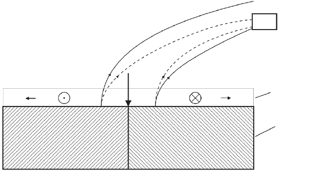
Figure 3–28. Scheme of type-1 magnetic contrast formation between two domains having oppositely
directed external magnetic fi elds. The dashed lines indicate the SE trajectories for the most probable
SE energy to the positively biased Everhart–Thornley detector (ETD) without magnetic fi eld and the
solid lines the trajectories with magnetic fi elds. The effect of the magnetic force F
m
on the SE tilts the
trajectories by a small angle toward or away from the ETD, respectively.

196 R. Reichelt
and Varndell, 1984; Müller, 1985; Steinbrecht and Zierold, 1987; Albrecht
and Ornberg, 1988; Edelmann and Roomans, 1990; Grasenick et al.,
1991; Echlin, 1992; Malecki and Romans, 1996), collections of methods
(e.g., Schimmel and Vogell, 1970; Robards and Wilson, 1993) with
updates, and publications, the successful preparation still also depends
in many cases on experience and skillful hands.
It is beyond the scope of this chapter to discuss the wide fi eld of
preparation techniques. Therefore, a brief rather general outline of
specimen preparation with reference to specifi c literature will be given.
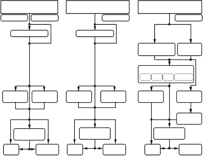
Figure 3–29 schematically outlines some important preparation proce-
dures used for inorganic and organic materials. As a general rule, a
successful investigation by SEM requires specimens, which have clean
surfaces, suffi cient electrical conductivity, are not wet or oily, and
possess a certain radiation stability, to resist electron irradiation during
imaging. An exception of this rule is allowed only for SEMs working
at ambient pressure (say at low vacuum; see Section 4), which permits
direct imaging of dirty, wet, or oily samples, although radiation damage
occurs with radiation-sensitive specimens. The goal of an ideal prepa-
Inorganic Material
Organic Material
(without water)
Organic Material
(with water)
Nonconduct.
Water withdrawal / substitut.
Ultra-
microtomy
Ultra-
microtomy
Ultra-
microtomy
FIB /
IBSC
Cryo ultra-
microtomy
Cryo ultra-
microtomy
Freeze
etching
Evaporation /
Sputtering
SEM /
Cryo-SEM
SEM
SEM /
Cryo-SEM
SEM
SEM /
Cryo-SEM
SEM
Evaporation /
Sputtering
Evaporation /
Sputtering
AD CPD
FD Embedd.
Rapid
freezing
Chemical Fixation
Nonconduct.
Nonconduct.
Conductive
Surface treatment
Surface treatment
Figure 3–29. Schematic drawing of important pre paration procedures for SEM used for inorganic and
organic materials with and without water. AD, air drying; CPD, critical point drying; FD, freeze
drying; FIB, focused ion beam; IBSC, ion beam slope cutting.
Chapter 3 Scanning Electron Microscopy 197
ration consists in making specimens accessible for high vacuum SEM
studies without changing the relevant properties under investigation.
Many inorganic samples with suffi cient electrical conductivity, such
as metals, alloys, or semiconductors, can be imaged directly with little
or no specimen preparation (Figure 3–29). This is one very useful
feature of scanning electron microscopy. In some cases a surface treat-
ment may be required, e.g., to clean the specimen surface with an
appropriate solvent, possibly in an ultrasonic cleaner, and with low-
energy reactive gas plasma for the removal of hydrocarbon contamina-
tion (Isabell et al., 1999). The cleanings are suitable to prepare electrically
conductive specimens for surface imaging in SEM. In case of noncon-
ductive samples, such as ceramics, minerals, or glass, a conductive
coating (e.g., Willison and Rowe, 1980) with a thin metal fi lm (e.g., gold,
platinum, tungsten, chromium) or a mixed conductive fi lm (e.g., gold/
palladium, platinum/carbon, platinum/iridium/carbon) is required
for good-quality imaging. For X-ray microanalysis carbon coating is
preferred because of its minimum effect on the X-ray spectrum. The
coating can be performed by evaporation (e.g., Reimer, 1967; Shibata et
al., 1984; Hermann et al., 1988; Robards and Wilson, 1993), by diode
sputtering (Apkarian and Curtis, 1986), or by planar magnetron sput-
tering (Nagatani and Saito, 1989; Müller et al., 1990). High-quality con-
ductive thin-fi lm coating for high-resolution SEM (see Section 3) can be
performed in an oil-free high vacuum by both evaporation, using e.g.,
tungsten, tantalum/tungsten, platinum/carbon, or platinum/iridium/
carbon, and rotary shadowing methods (Gross et al., 1985; Hermann et
al., 1988; Wepf and Gross, 1990; Wepf et al., 1991) as well as by ion beam
and by penning sputtering with, e.g., chromium, tantalum, and niobium
(Peters, 1980).
For the study of microstructural features (see Section 7) and for
microanalytical investigations (see Section 6) a fl at surface is required,
therefore rough specimen surfaces have to be fl attened by careful
grinding and subsequent polishing according to standard metallo-
graphic methods (Glauert, 1973). To remove mechanical deformations
caused by grinding and mechanical polishing, a fi nal treatment with
electrochemical polishing or ion beam polishing may be necessary. In
case of polycrystalline and heterogeneous material, selective etching
by ion bombardment may be used, which generates a surface profi le
caused by locally different sputtering yields, thus giving rise to topo-
graphic contrast of grains and the individual materials (Hauffe, 1971,
1995).
Often, specimens need to be characterized and analyzed both above
and below the surface, e.g., if the subsurface composition of the
material, process diagnosis, failure analysis, in situ testing, or three-
dimensional reconstruction of the spatial microstructure is required.
Flat cross sections through the specimen can be obtained by ultrami-
crotomy (Reid and Beesley, 1991; Sitte, 1984, 1996; the block face can be
used for SEM imaging), ion beam slope cutting (Hauffe, 1990; Hauffe
et al., 2002), an FIB technique (Kirk et al., 1988; Madl et al., 1988; Ishitani
and Yaguchi, 1996; Shibata, 2004; Giannuzi and Stevie, 2005), or by a
combination of an FIB system with a fi eld emission SEM (SEM/FIB),

198 R. Reichelt
which allows the precise positioning of the cross section and, most
importantly, realtime high-resolution SEM imaging of the cutting
process, which enables, among other things, the examination of the
spatial structure (Sudraud et al., 1987; Gnauck et al., 2003a, b; McGuin-
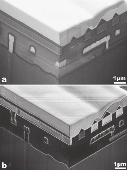
ness, 2003; Holzer et al., 2004; Sennhauser et al., 2004). An example for
two perpendicular vertical cross sections into an integrated circuit is
shown in Figure 3–30. The combined SEM/FIB additionally can be
equipped with analytical techniques such as energy-dispersive X-ray
spectroscopy, wavelength-dispersive X-ray spectroscopy, Auger elec-
tron spectroscopy, and secondary ion mass spectrometry allowing for
three-dimensional elemental analysis of the interior of the specimen.
The other class of samples consists of organic material, which usually
has an insuffi cient electrical conductivity for scanning electron micros-
copy. Although biological specimens contain water—the water content
ranges in human tissues from approximately 4 to 99% (Flindt, 2000)—
many other organic materials do not, e.g., numerous polymers. The
preparation strategies to be applied to specimens with and without
water differ (cf. Figure 3–29), although there are also some similarities
between them.
Figure 3–30. Integrated circuit with two perpendi cular vertical cross sections
into the interior. FIB sectioning was performed with the CrossBeam® tool
from Carl Zeiss NTS. The secondary electron images using the EDT were
obtained by the fi eld emission SEM (a) at 3 kV and by the FIB system (b) at
5 kV. Both micrographs reveal the site-specifi c internal structure of the inte-
grated circuit, although some features occur with different contrast caused by
different mechanisms of SE generation by electrons (a) and ions (b) (Courtesy
of Carl Zeiss NTS, Oberkochen, Germany.)
Chapter 3 Scanning Electron Microscopy 199
The surface treatment of organic specimens without water, such as
cleaning, grinding, polishing, and etching by dissolution, chemical
attack, or ion bombardment, has many similarities to the surface treat-
ment of inorganic materials. A detailed discussion of and the recipes
for specifi c preparation procedures for polymers are given in the
chapter “Specimen preparation methods” in the book by Sawyer and
Grubb (1996). Analogous to nonconductive inorganic materials, con-
ductive coating with a thin metal fi lm (e.g., gold, platinum, tungsten,
chromium) or a mixed conductive fi lm (e.g., gold/palladium, plati-
num/carbon, platinum/iridium/carbon) is required for good-quality
imaging. If the subsurface structure of the material has to be studied,
fl at cross sections usually are prepared by ultramicrotomy or cryoul-
tramicrotomy, depending on the cutting behavior of the specimen
under study. In principle, cutting with ions and imaging and analysis
with electrons by using a combined FIB/SEM tool seem possible also
with polymers. It was recently shown that ion milling is possible, e.g.,
with rubber (Milani et al., 2004; cf. also Figure 3–31) and with plastic
material (cf. Figure 3–32). As yet, the application of FIB for cutting and
milling of organic specimens is rare.
Most of the organic specimens that contain water are biological
samples. A small fraction of water-containing specimens is nonbiologi-
cal, e.g., hydrogels. Caused by the high vacuum in the SEM the water-
containing specimens cannot be investigated in the wet state. In
principle, three different preparation strategies exist to make wet speci-
mens accessible to SEM investigations:
1. Withdrawal of the water;
2. replacement of the water by some vacuum-resistant material such
as resins or freeze substitution (Feder and Sidman, 1958; Hess, 2003)
of the ice of the rapidly frozen specimen by some organic solvent;
and
3. rapid freezing of the water.
Irrespective of the preparation strategy used the native spatial struc-
ture of the specimen should be maintained. Air drying, which is the
most simple method of drying, is not suitable for drying soft specimens
because the surface tension induces remarkable forces during the
process of air drying, deforming the specimen irreversibly (Kellen-
berger and Kistler, 1979; Kellenberger et al., 1982). Figure 3–29 shows
different paths, which can be used, even though the degree of struc-
tural preservation depends on the preparation procedures applied.
The different preparation procedures have been described in detail
(Kellenberger and Kistler, 1979; Robards and Sleytr, 1985; Steinbrecht
and Zierold, 1987; Dykstra, 1992; Echlin, 1992; Kellenberger et al., 1992;
Robards and Wilson, 1993). Among the different preparation methods
rapid freezing is the method of choice for preparing biological speci-
mens in a defi ned physiological state (Echlin, 1992). In case of chemical
fi xation, which may create artifacts (Kellenberger et al., 1992), the water
of the sample has to be withdrawn or replaced afterward. If the surface
structure of the specimen has to be studied, then the specimen surface
has to be coated with a thin conductive fi lm prior to SEM investigation.
If the interior of the specimen has to be studied, the sample has to be

200 R. Reichelt
opened by sectioning with the ultramicrotome or possibly FIB and
subsequently coated. In case of physical fi xation, i.e., rapid freezing, the
specimen has to be opened by freeze fracturing (for review see Severs
and Shotton, 1995; Walther, 2003), cryosectioning, or now possibly by
ion milling the frozen-hydrated sample [ion milling in ice is possible
(McGuinness, 2003)]. After short partial freeze drying (also called
freeze etching), the fracture face or block face have to be properly
coated by a conductive fi lm and then can be directly analyzed in the
cryo-SEM (Echlin, 1971; Hermann and Müller, 1993; Walther and
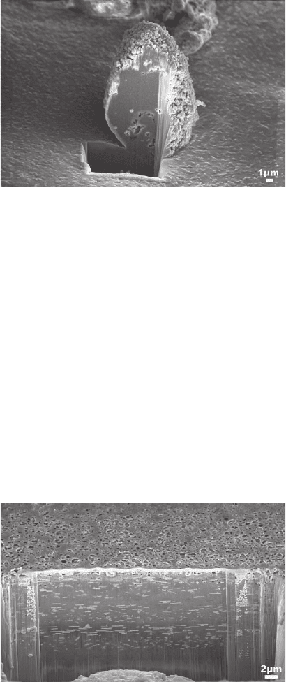
Figure 3–32. Secondary electron micrograph of an FIB cross-sectioned color
fi lm. The SE image is composed of the signals of two SE detectors, where the
“through-the-lens” detection contributes a fraction of 60% and the positively
biased ETD a fraction of 40%. The FIB sectioning and imaging at 5 kV were
performed with the CrossBeam® tool combining a focused ion beam system
with a fi eld emission SEM (cf. Gnauck et al., 2003a). The cross section in the
lower half of the micrograph reveals the interior of the fi lm (three color layers
corresponding to red, green, and blue and the submicrometer features as well),
whereas the upper half of the micrograph shows the porous outer surface of
the fi lm. (Courtesy of Carl Zeiss NTS, Oberkochen, Germany.)
Figure 3–31. Secondary electron micrograph (“though-the-lens” detection) of
a site-specifi c FIB cross-sectioned abrasive wear particle of a tyre supported
onto carbon. The FIB sectioning and imaging at 5 kV was performed with the
CrossBeam® tool combining a focused ion beam system with a fi eld emission
SEM (cf. Gnauck et al., 2003a). The cross section reveals the interior features
of the rubber particle. (Courtesy of Carl Zeiss NTS, Oberkochen, Germany.)

Chapter 3 Scanning Electron Microscopy 201
Müller, 1999; Walther, 2003). Another possible path is complete freeze-
drying and subsequent conductive coating of the sample, which then
can be analyzed at room temperature in the SEM.
As yet, FIB sample preparation and subsequent FIB or SEM imaging
are in early stages of application in the life sciences. Very recently, in
situ FIB sectioning was successfully performed with critical-point-
dried hepatopan creatic cells (Drobne et al., 2004) and some epithelium
cells (Drobne et al., 2005).
2.5 Radiation Damage and Contamination
The inelastic electron–specimen interaction inevitably damages the
irradiated specimen and can induce contamination at the specimen
surface. Once radiation damage, in particular of organic specimens,
has been extensively investigated for thin fi lms in transmission elec-
tron microscopy, comparatively little is systematically studied for
irradiation-sensitive samples in SEM. This may be due to the fact that
the interpretation of radiation damage in TEM is easier because of the
uniform ionization density through thin specimens. In bulk specimens,
however, the ionization density is a function of the depth (for a detailed
treatment of the depth dose function see, e.g., Shea, 1984) and a layer
below the surface at the maximum ionization density will be damaged
faster than others within the electron range R (cf. Figures 3–13 and 3–
14). According to the Bethe stopping power [see Eq. (2.26)], the damage
will be proportional to 1/E ln(1.166 E/J). Table 3–5 gives values of the
stopping power for carbon and protein for electron energies from 0.1 to
30 keV, which show the increase of the stopping power with decreasing
electron energy. It is commonly assumed that the shape of the depth–
dose curve is not a function of either the primary electron energy or
the material when normalized to the electron range (Shea, 1984). That
means that the layer with the maximum ionization density approaches
the surface as the electron energy decreases.
In organics, the radiation breaks due to the transfer of typically tens
of electron volts to an electron at the site of the interaction of many
intra- and intermolecular bonds, which generates free radicals (e.g.,
Bolt and Carroll, 1963; Dole, 1973; Baumeister et al., 1976). Many excited
Table 3 –5. Mean ionization potential J [Eqs. (2.27) and (2.28),
respectively] and the Bethe stopping power dE/ds [Eq. (2.26)] for
carbon and protein at different electron energies.
a
E (keV)
Sample Parameter 0.1 1.0 5.0 10 30
Carbon J (eV) 56.5 92.8 98.5 99.2 100.4
dE/ds (eV/cm) -56.4 -19.7 -6.4 -3.7 -1.5
Protein J (eV) 50.6 78.0 82.0 83.0 83.0
dE/ds (eV/cm) -43.8 -14.2 -4.5 -2.6 -1.1
a
The values listed for dE/ds have to be multiplied by 10
7
. The following values were
used for the calculation (Reichelt and Engel, 1984): carbon: Z = 6; A = 12; ρ = 2 g/cm
3
;
protein: mean atomic number <Z> = 3.836; A = 7.7; ρ = 1.35 g/cm
3
.
202 R. Reichelt
species will very rapidly recombine in 10
−9
to 10
−8
s and will reform the
original chemical structure dissipating the absorbed energy as heat.
Some recombinations will form new structures, breaking chemical
bonds and forming others. If the material was initially crystalline,
defects will form and gradually it will become amorphous. In addition
to these structural changes the generated free radicals will rapidly
diffuse to and across the surface or can evaporate, i.e., loss of mass and
composition change will occur (e.g., Egerton, 1989, 1999; Egerton et al.
1987; Engel, 1983; Isaacson, 1977, 1979a; Reimer, 1984b; Reichelt et al.,
1985). Bubbles may form at high dose rates when volatile products are
trapped. Not only the beam electrons damage the organic sample but
also fast secondary electrons (E
SE
> 50 eV) can produce damages outside
the directly irradiated specimen area (Siangchaew and Libera, 2000).
Furthermore, beam-induced electrostatic charging and heating can
also damage organic samples. Conductive coating of the organic speci-
men, as suggested for inorganic materials by Strane et al. (1988), can
keep trapped free radicals as well as reduce beam-induced tempera-
ture rise or electrostatic charging (Salih and Cosslett, 1977). Lowering
of the temperature of the specimen is a further measure to reduce the
sensitivity of an organic specimen to structural damage and mass loss.
However, the reduction factor depends considerably on the material of
the specimen.
The radiation damage mechanisms in semiconductors are different
from those described above. As mentioned in Sections 2.1.3.1 and 2.3.3.2,
the incident electrons generate electron hole pa irs, wh ich will be trapped
in the Si
2
O layer due to their decreased mobility. This can generate
space charges, which in turn can affect the electronic properties of the
semiconductor.
Beam-induced contamination is mass gain, which occurs when
hydrocarbon molecules on the specimen surface are polymerized by
the beam electrons. The polymerized molecules have a low surface
mobility, i.e., the amount of polymerized molecules increases in the
surface region where polymerization takes place. There are two main
sources for hydrocarbon contamination: (1) gaseous hydrocarbons
arising from oil pumps, vacuum grease, and possibly O-rings, and (2)
residual hydrocarbons on the specimen. Several countermeasures exist
to reduce the contamination to a tolerable level (see, e.g., Fourie, 1979;
Wall, 1980; Postek, 1996). The amount of gaseous hydrocarbons is sub-
stantially reduced when the SEM is operated with an oil-free pumping
system and a so-called cold fi nger located above the specimen. Further,
the contamination rate falls more rapidly as the specimen temperature
is lowered, and below −20°C contamination is diffi cult to measure
(Wall, 1980). This is caused by the reduced diffusion of hydrocarbons
on the specimen. In some cases, preirradiation of a large sur-
face area with the electron beam is helpful, which immobilizes (polym-
erizes) hydrocarbons around the fi eld of view to be imaged. Finally,
specimens are mostly exposed to the atmosphere before transfer into
the specimen chamber. Weakly bound molecules (e.g., hydrocarbons)
can be completely eliminated by gently heating the sample in the speci-
men exchange chamber (low vacuum) to 40–50°C for several minutes
by a spot lamp (Isaacson et al., 1979b). A detailed topical review on the

Chapter 3 Scanning Electron Microscopy 203
radiation damage and contamination in electron microscopy is given
by Egerton et al. (2004).
2.6 Applications
Scanning electron microscopy is an indispensable tool for investiga-
tions of a tremendous variety of specimens from very different fi elds
such as materials science, mineralogy, geology, semiconductor research,
microelectronics, in-dustry, polymer research, ecology, archeology, art,
and life sciences. Although the investigations are not restricted just to
imaging of surface structures, the majority of SEM studies apply the
imaging modes. As mentioned previously, considerable additional
information about the local elemental composition, electronic and
magnetic properties, crystal structure, etc. can be acquired when the
SEM is combined with supplementary equipment such as electron and
X-ray spectrometers to take advantage of the energy spectra of the
emitted electrons and X-rays. Table 3–6 surveys the information, which
can be obtained from inorganic and organic specimens not containing
Table 3 – 6. SEM applications on specimens from materials science, mine ralogy, geology,
polymer science, semiconductors, and microelectronics.
a
Specimen Information
Metals, alloys, and At the specimen surface:
intermetallics Topography (three-dimensional); microroughness; cracks;
fi ssures; fractures; grain size and shape; texture; phase
identifi cation; localization of magnetic domains; size and shape
of small particles; elemental composition; elemental map
Inside the specimen:
Grain and phase structures; three-dimensional microstructure;
cracks; fi ssures; material inclusions; elemental composition
Ceramics, minerals, At the specimen surface:
glasses Topography (three-dimensional); microroughness; cracks;
fi ssures; fractures; grain size and shape; pores; phase
identifi cation; size and shape of small particles; elemental
composition
Inside the specimen:
Grain and phase structures; three-dimensional microstructure;
cracks; fi ssures; material inclusions; pores; elemental
composition
Polymers, wood At the specimen surface:
Morphology; topography (three-dimensional); microroughness;
cracks; fi ssures; fractures; pores; size and shape of small fi bers
and particles; fi ber assemblage in woven fabrics; elemental
composition
Inside the specimen:
Cracks; fi ssures; fractures; pores; composite structure; elemental
composition
Semiconductors, integrated Dislocation studies (with CL); metallization and passivation
circuits, microelectronic integrity; quality of wire bonds; electrical performance; design
devices validation; fault diagnosis; testing
a
State-of-the-art preparation and image analysis techniques are required to take full advantage of the capabilities
of SEM.

204 R. Reichelt
water. Further, in situ scanning electron microscopy allows for differ-
ent specifi c specimen treatments in the specimen chamber (see, e.g.,
Wetzig and Schulze, 1995), which serves as a microlaboratory, and the
simultaneous observation of the specimen response (cf. Table 3–7).
The advancement of nanoscale science and technology demands the
manipulation of nanoobjects at the molecular level and ultimately the
manufacture of things via a bottom-up approach. Very recently, a four
nanoprobe system has been installed inside a fi eld emission SEM, which
may be used for gripping, moving, and manipulating nanoobjects, e.g.,
carbon nanotubes, setting up electric contacts for electronic measure-
ments, tailoring the structure of the nanoobject by cutting, etc. and for
making nanostructures (Peng et al., 2004). The SEM in this setup allows
for visualization of the four nanoprobes operating inside the specimen
chamber as well as the process of formation of microstructures.
Less spectacular, but nevertheless important, are applications of
scanning electron microscopy to image macroscopic samples in the
millimeter range at very low magnifi cation (about 10× to 100×), which
cannot be seen clearly by the eye or by the light microscope for some
reasons. Two examples from very different fi elds are shown in Figures
3–33 and 3–34 taking advantage of the large depth of focus as well as
distinct topographic and material contrast.
Working in the low magnifi cation range, the depth of focus limit in
the SEM (see Section 2.1.5.2) can be overcome by recording stacks of
through-focus images (as in conventional and confocal optical micros-
copy), which are digitally postprocessed to generate an all-in-focus
image (Boyde, 2004). The application of the technique is advantageous
when BSE imaging of spongy specimens is required, as demonstrated
with examples from the study of human osteoporotic bone (Boyde,
2004).
Table 3–7. In situ treatments in SEM and available information about specimens from
materials science.
Treatment Information
Static and dynamic deformation, Kinematic processes during deformation; submicrometer
e.g.,by tension, compression, cracks visible only under load; localized deformation
bending, machining centers, e.g., slip bands, crack nucleation; deformation-
induced acoustic emission
Laser irradiation, e.g., in pulse Phase transformations; structural modifi cations; crack
mode, Q-switch mode formation due to thermal shock; diffusion processes; laser-
induced surface melting and evaporation processes; vapor
deposition on substrates; cumulative effects of multiple
laser pulses
Ion beam irradiation Depth profi le/cross section; grain boundaries; spatial
microstructure; internal grain and phase structures
Electrical and magnetic effects Reversible and irreversible breakdown of voltage barriers;
size distribution of magnetic and ferroelectric domains;
orientation distribution of magnetic and ferroelectric
domains; effects accessible by EBIC, EBIV, and CL
