Hawkes P.W., Spence J.C.H. (Eds.) Science of Microscopy. V.1 and 2
Подождите немного. Документ загружается.


Chapter 3 Scanning Electron Microscopy 185
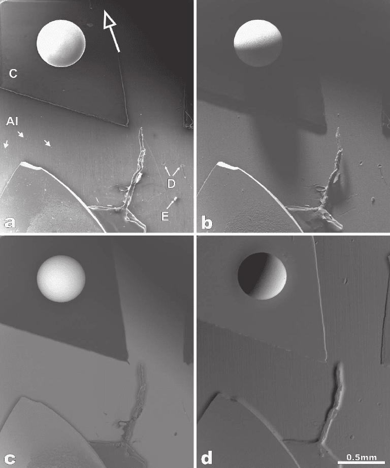
Figure 3–23. Secondary (a) and backscattered electron micro graphs (b–d) of a steel ball recorded at
30 kV and normal beam incidence. The arrow in (a) indicates the direction of the laterally located ET
detector. The BSE micrographs shown in (c and d) were acquired using a four-quadrant semiconductor
detector mounted below the objective pole piece, which records BSE over a large solid angle. The steel
ball is mounted on carbon (marked by C), which is supported by aluminum (marked by Al). The small
arrowheads in (a) indicate small particles with enhanced SE emission (bright blobs in the SE image).
Elevations (E) and depressions (D) are also marked by small arrowheads.

186 R. Reichelt
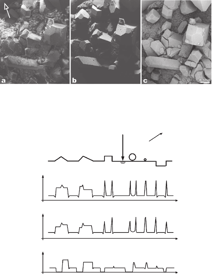
Figure 3–24. Secondary (a) and backscattered electron micrographs (b and c) of 10-nm gold-coated
crystal-like tartar (tartar contains mostly potassium–hydrogen–tartrate and calcium–tartrate) recorded
at 30 kV and normal beam incidence. The arrow in (a) indicates the direction of the laterally located ET
detector. The BSE micrograph shown in (c) was acquired using a four-quadrant semiconductor detector
mounted below the objective pole piece, which records BSE over a large solid angle. The small arrowheads
in (a) indicate small particles with enhanced SE emission (bright blobs in the SE image).
(a)
e
-
ETD
(b)
(c)
(d)
0
S
SE
0
S
BSE
0
n
SE
Figure 3–25. Schematic specimen surface profi le of an assumed topography having elementally
shaped elevations and a depression (a). Those elemental shapes are present in the samples shown in
Figures 3–23 and 3–24. The size of the excitation volume of the electron beam is drawn in relation to
the local topographic structures. The amount n
SE
of locally emitted SE is shown qualitatively in (b)
and the corresponding SE signal S
SE
in (c). The BSE signal S
BSE
collected by the negatively biased ET
detector is schematically presented by the graph in (d). ETD, Everhart–Thornley detector.
Chapter 3 Scanning Electron Microscopy 187
lines of magnetic fl ux until they reach the collecting fi eld of the ETD
(Lukianov et al., 1972).
It should be mentioned that the laterally located ETD also register
those BSE, which are within the small solid angle of collection defi ned
by the scintillator area and the specimen–scintillator distance. The BSE
contribute in the order of 10–20% to the SE signal (Reimer, 1985) and
are the same as those collected by the negatively biased ET detector.
Furthermore, BSE that are not intercepted by the detector strike the
pole piece of the objective lens and the walls of the specimen chamber.
These stray BSE generate so-called SE3 emitted from the interior sur-
faces of the specimen chamber. The SE3 carry BSE information and
form a signifi cant fraction of the SE signal for specimens with an inter-
mediate and high atomic number (Peters, 1984).
The contrast in BSE images is formed by the following
mechanisms:
1. Dependence of the BSE coeffi cient η on the angle of incidence θ
of the electron beam at the local surface element [cf. Eq. (2.34)].
2. Dependence of the detected signal on the angular orientation of
the local surface element related to the BSE detector (see Section 2.1.3).
BSE emitted “behind” local elevations, in holes, or in cavities, which do
not reach the BSE detector on nearly straight trajectories, are not
acquired. This causes a pronounced shadow contrast (cf. Figure 3–7b).
3. Increase of the BSE signal when diffusely scattered electrons pass
through an increased surface area. This is the case at edges or at pro-
truding surface features, which are smaller than the excitation
volume.
The BSE leave the specimen on almost straight trajectories and only
those within the solid angle of collection of the BSE detector can be
recorded. Thus, dedicated BSE detectors have a large solid angle of
collection to record a signifi cant fraction of the BSE and to generate a
signal with a suffi cient SNR. The larger the solid angle of collection the
less pronounced the shadow effects.
Contributions (1) to (3) mentioned above are illustrated by BSE
micrographs from two specimens used for SE imaging and are shown
in Figures 3–23b and c and 3–24b and c as well as schematically by
profi les of the topography and the related BSE signals in Figure 3–25.
Two different types of BSE images are shown: the highly directional
image recorded with the negatively biased ETD (Figures 3–23b and 3–
24b) and the “top-view” image recorded with the four-quadrant semi-
conductor detector mounted below the objective pole piece (Figures
3–23c and 3–24c). The ball in Figure 3–23b shows a contrast, which is
mainly due to the varying angle θ of beam incidence across the ball
[cf. (1) above] and the angular position of the local surface elements
related to the negatively biased ETD [cf. (2) above]. A pronounced
sharp shadow occurs at the back of the ball and behind the ball (shad-
owed oblong area of the support). Whereas the intensity of the BSE of
the ball reveals radial symmetry, the effect of detection geometry
causes the nonradial symmetric image intensity distribution of the ball
(cf. Figure 3–25a and d). The fade contour of the ball at its back is due
188 R. Reichelt
to BSE redirected toward the negatively biased ETD by scattering on
some interior surfaces of the specimen chamber. The pronounced
directional shadow contrast in the image allows for unambiguous
identifi cation of elevations and depressions (cf. Figure 3–23b). More-
over, if the detection geometry of the BSE is known, the length of the
shadow can be used in some cases to obtain a rough estimate of the
height of elevations or depth of depressions.
The BSE micrograph of the ball recorded with the four-quadrant semi-
conductor detector (Figure 3–23c) shows an almost radial symmetric
image intensity distribution. It is obvious that the increase of the BSE
coeffi cient η with the increasing angle of incidence θ (see Figure 3–18)
toward the rim of the ball is superimposed by the stronger counteracting
effect of the directed asymmetric distribution, for a large θ refl ection-
like angular distribution of BSE for nonnormal beam incidence (cf.
Section 2.2.2). The shadow-like hem along the contour of the elevations
refl ects the fact that BSE emitted from the lower surrounding areas
toward elevations can be absorbed or redirected; thus those BSE do not
reach the BSE detector. In case of depressions there is also a shadow-like
hem but it is located inside the contour of the depression. A comparison
of the different types of BSE images in Figure 3–23b and c clearly shows
that the topography of the sample is pr on ou n ce d i n Fi g u re 3 –2 3b wh i l e —
as we shall see later—the atomic number contrast is pronounced in
Figure 3–23c.
The BSE micrographs of large crystal-like particles (Figure 3–24b and
c) basically show the same contrast mechanisms as discussed previ-
ously [no orientation anisotropy of the electron backscattering and SE
emission (Reimer et al., 1971; Seiler and Kuhnle, 1970) is involved].
Figure 3–24b recorded with the negatively biased ETD shows large
shadowed regions (containing almost no information) and some high-
lighted individual fl at surface planes of the crystal-like particles that
occur with almost constant brightness because of the constant angle of
beam incidence of the constant detection geometry. Figure 3–24b dem-
onstrates that the detection geometry used for recording was not
optimum. The micrograph obtained with the four-quadrant semicon-
ductor detector is shown in Figure 3–24c, which depicts exactly the
same area as Figure 3–24b. Due to the large solid angle of collection of
this BSE detector the effects mentioned above in (1) and (2) do generate
just small differences in the image intensity of differently oriented
surface planes of the crystal-like particles. The effect of shadowing is
not substantial in that micrograph. The increase of the BSE signal at
edges, at surface steps, and small protruding particles [cf. (3) above]
located on the fl at surface planes due to enhanced BSE emission is
signifi cant. The fi ssures on some surface planes of the crystal-like par-
ticles (cf. Figure 3–24a) occur in the BSE micrograph also as rather dark
features because just a minor fraction of the BSE can escape from inside
the fi ssures.
The SEM micrographs are closely analogous to viewing a macro-
scopic specimen by eye. In the light optical analogy the specimen is
illuminated with light from the side of the detector and viewed from
the position of the electron beam (see, e.g., Reimer et al., 1984). When
Chapter 3 Scanning Electron Microscopy 189
a rather diffuse illumination is used then all surface elements are illu-
minated but those directed to the light source are highlighted. This
corresponds to the situation for the positively biased ETD. The light
optical analogy shows a pronounced shadow contrast if a directional
light source illuminates the specimen surface from a suitable direction.
This situation closely resembles BSE images recorded with a positively
biased ETD. The strong light optical analogy very likely explains the
fact that SEM micrographs of objects with a distinct topography can
be readily interpreted even without extensive knowledge of the physics
“behind” the imaging process.
As briefl y mentioned in Section 2.1.3.2, the topographic and the
material contrast can be pronounced or suppressed, respectively, by a
combination of the signals of two oppositely placed detectors, A and
B. Two BSE semiconductor detectors were fi rst used by Kimoto et al.
(1966) and they showed that the sum A + B results in material and
the difference A − B in topographic contrast. Volbert and Reimer (1980)
used a BSE/SE converter system and two opposite ET detectors for
that kind of contrast separation in the SEM. The four-quadrant semicon-
ductor detector used for recording Figures 3–23c and 3–24c allows for
signal mixing of the four signals acquired simultaneously. Figures 3–23c
and 3–24c represent the sum of the four signals (S
Q1
, . . . , S
Q4
), thus
both micrographs show a pronounced material contrast. By addition of
the signals of two adjacent quadrants at a time (i.e., S
Q1
+ S
Q2
= A; S
Q3
+
S
Q4
= B) the effect of two semiannular detectors A and B is obtained.
The difference image A − B shows a pronounced topographic contrast
(Figure 3–23d). The directionality in Figure 3–23d can be varied readily
by using a different combination of the individual signals of the
quadrants, e.g., A = S
Q2
+ S
Q3
and B = S
Q1
+ S
Q4
. Difference SE and BSE
images recorded at exactly defi ned detection geometry allow for the
reconstruction of the surface topography (Lebiedzik, 1979; Reimer,
1984a; Kaczmarek, 1997; Kaczmarek and Domaradzki, 2002; see also
Sections 2.1.3.2 and 2.1.5.2). However, special care is required for the
reconstruction of the surface topography using BSE images because
of artifacts in the reconstructed image (Reimer, 1984a). To demonstrate
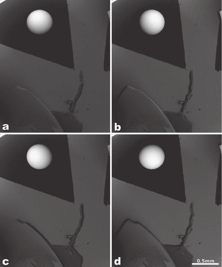
the effect of directionality for four different detection Figure 3–26
shows four individual BSE micrographs each recorded with another
quadrant of the semiconductor detector. The individual BSE images
contain superimposed topographic and compositional contrast
components.
2.3.2 Material Contrast
The material or compositional contrast arises from local differences in
chemical composition of the object investigated. As shown in Figure
3–17, the SE yield δ increases weakly with increasing atomic number
but the increase is signifi cantly less than that of the BSE coeffi cient η.
Experimental values of δ (see, e.g., the data collection by Joy 2001)
scatter strongly around a mean curve. The increase of δ(Z) with Z
is mainly due to SE [cf. Eq. (2.30)] generated by emitted BSE near
the specimen surface (SE2). At electron energies larger than 5 keV the
SE images usually show the same compositional contrast as the

190 R. Reichelt
Figure 3–26. BSE micrographs of a steel ball on carbon (cf. Figure 3–23) each recorded with another
individual quadrant of the four-quadrant semiconductor detector. (a) (−X)-quadrant, (b) (+Y)-quad-
rant, (c) (−Y)-quadrant, (d) (+X)-quadrant. The micrographs were recorded at 30 kV and normal beam
incidence. The angular position of the four-quadrant semiconductor detector is rotated clockwise
against the x–y coordinates of the images by 34°. Shadows of the surface step of the feature at the
bottom left of each image help to identify the position of the active quadrant visually.

Chapter 3 Scanning Electron Microscopy 191
corresponding BSE image. This situation is illustrated in Figure 3–23a–
c where at normal beam incidence carbon (Z = 6) is darker than alu-
minum (Z = 13) in both the SE and BSE image. Table 3–4 gives some
numerical values for the compositional contrast for carbon, aluminum,
and iron calculated with the related BSE coeffi cients for normal beam
incidence, which qualitatively agrees with the contrasts obtained in
Figure 3–23c.
2.3.3 Other Contrasts
2.3.3.1 Voltage Contrast
The secondary electron image intensity varies if the potential of the
specimen is positively or negatively biased with respect to the ground.
In principle, a positively biased surface area shows decreased image
intensity because low-energy SE are attracted back to the specimen by
the electric fi eld. Conversely, a negatively biased surface area shows
enhanced image intensity because all SE are repulsed from the speci-
men. This voltage-dependent variation in contrast is designated as
voltage contrast and dates back to the late 1950s (Rappaport, 1954;
Oatley and Everhart, 1957; Everhart et al., 1959). Strictly speaking all
emitted electrons are infl uenced to some extent by the potential of the
sample, but only the SE and, in principle, the Auger electrons can be
used for voltage contrast studies (Werner et al., 1998). The use of Auger
electrons is more diffi cult than that of SE because of the very low yield
of AE and the ultrahigh vacuum requirements of AE analysis.
The voltage contrast depends on the energy of the beam electrons
and on the properties of the specimen, being most pronounced in the
low electron energy region where the SE yield is highest (cf. Figure 3–
12). Biasing the specimen positively or negatively by a few volts not
only affects the amount of emitted SE but also their trajectories. This
is caused by the fact that the majority of the SE have energies of a few
electron volts in contrast to BSE and Auger electrons. The effect of
specimen voltage on the SE trajectories is rather complex because it
depends on the SE detection geometry, the sample position in the
specimen chamber, the properties of the sample, and the operation
conditions of the SEM. However, voltage contrast is a valuable tool for
the investigation of a wide range of simple faults in microelectronic
devices or studies of the potential distribution at grain boundaries
obtained on the cross section of varistors at applied low DV voltage
and their breakdown behavior at elevated voltage (Edelmann and
Wetzig, 1995). The voltage contrast is also used to characterize the
surface charge distribution of ferroelectrics (Uchikawa and Ikeda, 1981;
Table 3 –4. Compositional contrast calculated according to Eq. (2.33)
for normal beam incidence q = 0 for the elements C, A1, and Fe.
a
Element 1 (Z) Element 2 (Z) h
1
h
2
C = (h
1
- h
2
)/h
2
Aluminum (13) Carbon (6) 0.1530 0.0641 0.581
Iron (26) Carbon (6) 0.2794 0.0641 0.771
Iron (26) Aluminum (13) 0.2794 0.1530 0.452
a
Compare Figures 3–23b and c and 3–26. E
0
= 30 keV.
192 R. Reichelt
Hesse and Meyer, 1982; Roshchupkin and Brunel, 1993) and piezoelec-
trics (Bahadur and Parshad, 1980).
Voltage contrast measurements can also be performed in a dynamic
mode on semiconductor devices by pulsing the electron beam [called
electron stroboscopy (Spivak, 1966)] synchronously with the device
signal as shown by Plows and Nixon (1968). This dynamic mode allows
for quantitative voltage contrast measurements on semiconductor
devices at high-frequency operation conditions known as electron
beam testing widely used by the electronics industry for the develop-
ment, fault diagnosis, and debugging of innovative integrated circuits.
High-frequency electron stroboscopy requires high-speed electrostatic
beam blanking systems with subnanosecond time resolution. For very
high-frequency electron stroboscopy in the gigahertz range a special
transverse-longitudinal combination gate system (Hosokawa et al.,
1978) or microwave structure-based beam blanking techniques have
been employed (Fujioka and Ura, 1981). A comprehensive treatment of
the fundamentals of voltage contrast and stroboscopy has been pub-
lished by Davidson (1989) and the state of the art of voltage contrast
has been review by Girard (1991). Furthermore, improvements of
voltage contrast detectors as well as of detection strategies are dis-
cussed in detail by Dubbeldam (1991). Voltage contrast is now of a
mature age, but the extension to future microelectronics also presup-
poses an extension in the domain of in situ testing methods and
techniques.
2.3.3.2 Electron Beam-Induced Current
The electron beam generates a variety of signals emitted from the
specimen as shown in Figures 3–2 and 3–14. In semiconductors the
primary electrons generate electron hole pairs or minority carriers
within the excitation volume. The mean number of electron hole pairs
is given by E
0
/E
exm
[cf. Eq. (2.14)], where E
exm
is the mean energy per
electron hole pair forming event. For example, E
exm
amounts to 3.6 eV
for Si and 2.84 eV for Ge, i.e., one 10-keV electron generates on average
approximately 2.7 × 10
3
electron hole pairs in Si and 3.5 × 10
3
in Ge
(McKenzie and Bromely, 1959). The charge collection (CC) signal is
detected between two electric contacts; one of these contacts collects
the electrons and the other one collects the holes. If electromotive
forces caused by electron voltaic effects are generated by the beam
electrons in the specimen then a charge collection current I
CC
desig-
nated as an electron beam-induced current (EBIC) fl ows through the
ohmic contacts. If no electron voltaic effects occur, the beam electrons
cause local β-conductivity, where the separation of charge carriers
results in an electron beam-induced voltage (EBIV). The most impor-
tant type of signal of the two charge-collecting modes is EBIC. A
detailed treatment of the basic physical mechanisms and applications
of the charge collection mode is given by Holt (1974, 1989), Deamy
(1982), Reimer (1985), Shea et al. (1978), Alexander (1994), and Yakimov
(2002).
EBIC can be observed in SEM simply by connecting a high-gain large
bandwidth amplifi er across the specimen using the amplifi ed EBIC
signal as a video signal. The input impedance of the amplifi er must be
Chapter 3 Scanning Electron Microscopy 193
very low relative to that of the specimen to measure the true EBIC. For
usual electron probe currents of some nanoamperes the charge collec-
tion currents are in the order of microamperes since for many materials
the mean energy per electron hole pair is between approximately 1 and
13 eV (Holt, 1989). In contrast to EBIC, for the measurement of the true
EBIV an amplifi er with a very high input resistance is necessary.
The resolution obtained in the charge-collecting modes depends on
the size of the excitation volume within the specimen, which readily
can be extracted from Monte Carlo simulation data (see Section 2.2).
For the CC mode, a depth and a lateral resolution have to be defi ned.
The depth-dose function, which represents the energy loss per unit
depth in the electron beam direction, determines the depth resolution.
The lateral-dose function, which represents the energy loss per unit
distance perpendicular to the electron beam direction, determines the
lateral resolution. There are also empirical (Grün, 1957) and semiem-
pirical expressions (Everhart and Hoff, 1971) as well as several analyti-
cal models (Bishop, 1974; Leamy, 1982) for the depth-dose and for the
lateral-dose function as well (Bishop, 1974; Leamy, 1982).
Electron beam chopping and time-resolved EBIC can enhance the
accuracy of measurements in several cases, e.g., for the estimation of
the depth of p–n junction parallel to the surface (Georges et al., 1982)
or allows for quantitative analysis of electrical properties of defects in
semiconductors (Sekiguchi and Sumino, 1995) and interesting applica-
tions for the failure analysis of VLSI circuits (Chan et al., 2000).
2.3.3.3 Crystal Orientation Contrast
As previously mentioned, the backscattering coeffi cient η of a single
crystal varies with the direction of the incident beam electrons related
to the crystallographic orientation (cf. Section 2.2). This effect is caused
by the variation of the atomic density, which the incident electrons
encounter when penetrating into the crystal. In certain crystallographic
directions the beam electrons penetrate more deeply. Those directions
represent “channels” for the incident electrons. Changing the direction
of the incident electrons relative to the crystallographic orientation
causes the so-called crystal orientation or channeling contrast of the
BSE image, which amounts to a maximum of approximately 5%. Crystal
orientation contrast arises if a large single crystal is imaged at very low
magnifi cation using a small electron probe aperture of about 1 mrad.
Scanning at low magnifi cation both moves the electron probe and
changes the angle of incidence across the fi eld, thereby generating an
electron channeling pattern (ECP). At higher magnifi cation the angle
of beam incidence varies just insignifi cantly across the small scanned
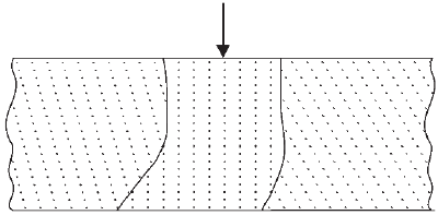
fi eld and channeling contrast is obtained in polycrystalline samples
from small grains with different crystal orientations (Figure 3–27). The
information depth of the crystal orientation contrast is in the order of
a few nanometers only (Reimer, 1985) and therefore the contrast is very
sensitive to distortions of the crystal at the surface. The channeling
contrast reaches the maximum at energies between 10 and 20 keV
(Reimer et al., 1971; Drescher et al., 1974).
An orientation anisotropy also occurs for the secondary yield (Reimer
et al., 1971), which gives rise to an SE orientation contrast.

194 R. Reichelt
2.3.3.4 Magnetic Contrasts
Basically, two different types of magnetic contrast can arise from the
interaction of the emitted electrons with the magnetic fi eld of small
domains of the specimen.
Type-1 Magnetic Contrast. Secondary electrons are defl ected after emis-
sion by an external magnetic fi eld, thus generating a magnetic contrast
(Dorsey, 1969). External magnetic fi elds can exist in natural or synthetic
engineered ferromagnetic materials such as magnetic tape, magnetic
cards, and computer disks. The fringe fi elds near the surface are highly
inhomogeneous and the SE trajectories are affected by the Lorentz
force, which is proportional to v × B where v is the velocity vector of
the SE and B the magnetic fi eld. The most probable velocity corre-
sponds to the electron energy of a few electronvolts (see Section 2.2.1).
The acting Lorentz force defl ects the trajectories of the SE and the
resulting effect can be approximated to a tilt of Lambert’s angular SE
emission characteristics of the SE (see Section 2.2.1; Reimer, 1985).
Figure 3–28 illustrates this effect for two domains in the specimen
having oppositely directed external magnetic fi elds. To observe type-1
magnetic contrast in case of weak magnetic fi elds an ETD with a high
angular sensitivity, a two-detector system (Dorsey, 1969; Wardly, 1971)
or digital image processing (Szmaja, 2000, 2002) has been employed.
For the type-1 magnetic contrast low beam electron energies are favor-
able because of the enhanced SE yield and therefore an increased
signal-to-noise ratio (SNR). The actual problems related to the compli-
cated mechanism of type-I magnetic contrast and its relatively low
resolution were discussed by Szmaja (2002).
Type-2 Magnetic Contrast. This type of contrast arises from the defl ec-
tion of backscattered electrons by the internal magnetic fi eld within the
specimen (Philibert and Tixier, 1969; Fathers et al., 1973). Depending
on the direction of the magnetic fi eld inside the sample, the BSE are
bent toward or away from the surface between consecutive scattering
events, i.e., the BSE coeffi cient is increased in domains where trajecto-
ries are bent toward the surface and decrease when bending the BSE
trajectories in an opposite direction. To observe a suffi cient type-2 mag-
netic contrast the beam electrons need an energy of at least 30 keV and
a relatively high beam current. The type-2 contrast is maximized if the
Figure 3–27. Cross section of a polycrystalline sample having grains with
different crystal lattice orientation relative to the electron beam.
