Hawkes P.W., Spence J.C.H. (Eds.) Science of Microscopy. V.1 and 2
Подождите немного. Документ загружается.

Chapter 3 Scanning Electron Microscopy 165
inner shells, respectively. After an inelastic scattering event the electron
trajectory has a slightly different direction than before scattering (typi-
cally the inelastic scattering angles are of the order of a few milliradians
only) and less kinetic energy. If the lost energy was transferred to elec-
trons in the valence or conduction band then the excitation of plasmons
(a plasmon is a longitudinal charge-density wave of the valence or con-
duction electrons) or inter- and intraband transitions may occur. Both
the energy of plasmons and the energy differences of inter- and intra-
band transitions are in the order of about 5–50 eV. The physics of the
latter processes is reviewed by Raether (1980). If the lost energy was
transferred to atomic electrons of inner shells then, for example, K-, L-,
or M-shell ionization may occur. In this case the energy loss typically is
higher than 50 eV. The differential inelastic electron scattering cross
section with a free electron (which is an approximation for an electron
in the valence or conduction band) is given (Reimer, 1985) as
dσ
in
/dW = πe
4
/[(4πε
0
)
2
EW
2
] (2.22)
where W is the energy loss. The equation shows that the differential
inelastic scattering cross section is inversely proportional to electron
energy E and to W
2
and that small energy losses occur with a larger
probability. In a more complex approach for the differential inelastic
scattering cross section the energy loss function Im(−1/ε) is used taking
the dielectric properties of the material into account (e.g., Powell, 1984).
An impinging electron can be inelastically scattered passing the atom
even in a distance of a few nanometers, thus the inelastic scattering is
delocalized to a certain extent (Isaacson and Langmore, 1974; Zeitler,
1978; Reichelt and Engel, 1986; Müller and Silcox, 1994, 1995).
In the case of inner shell excitation the electron interaction is local-
ized to an electron shell. The corresponding inelastic scattering, also
called ionization cross section, is the probability of bringing a scatter-
ing atom to a given excited state through an inelastic process. The
related cross sections are typically at least two orders of magnitude
smaller than those for the electrons in the valence or conduction band.
Calculations of the ionization cross sections of the K-, L-, and M-shell
have been published (see, e.g., Leapman et al., 1980; Inokuti and
Manson, 1984; Egerton, 1986).
The total inelastic scattering cross section σ
in
can be obtained by
integration
σπσ ϕϕ
π
in
dd d=
∫
2
0
(/)sin
in
Ω
(2.23)
The mean free path for inelastic scattering Λ
in
is given analogous to Eq.
(2.20) as
Λ
in
= 1/Nσ
in
(2.24)
(for detailed data and calculation of the electron inelastic mean free
path see Powell and Jablonski, 2000).
The total scattering cross section then is given as
σ = σ
el
+ σ
in
(2.25)
166 R. Reichelt
In bulk specimens multiple scattering of the impinging electrons
takes place. Mainly the multiple elastic scattering causes a successive
broadening of their angular distribution and can, after numerous scat-
tering events, result in beam electrons leaving the specimen. The beam
electrons, which leave the specimen, are designated as backscattered
electrons and carry an important class of information about the local
specimen volume through which they have been passing. Multiple
inelastic scattering along the electron trajectories results in a slowing
down and the beam electron can come to a standstill if it cannot leave
the specimen as BSE. The majority of beam electrons are scattered both
elastically as well as inelastically. Therefore, the majority of BSE have
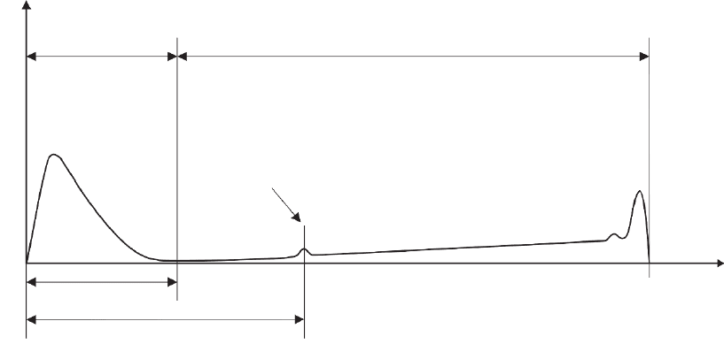
energies smaller than E
0
(cf. Figure 3–12). The broadening of the angular
distribution can be calculated analytically using the autoconvolution
of the single scattering distribution expanded in terms of Legendre
polynomials (Goudsmit and Saunderson, 1940).
Another method to treat multiple scattering is the simulation of the
successive scattering events by Monte Carlo calculations for about
10
3
–10
5
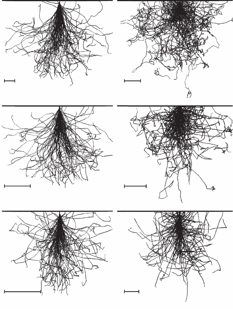
electron trajectories (cf. Figure 3–13; for Monte Carlo simula-
tions of electron scattering see, e.g., Reimer and Krefting, 1976; Kyser,
1984; Reimer and Stelter, 1986; Joy, 1987b; Reimer, 1968, 1996; Drouin
et al., 1997; Hovington et al., 1997a,b).
In this method, the most important scattering parameters, such as
scattering angle, mean free electron path, and energy loss, are simu-
lated for each individual scattering event along the trajectory by a
computer using random numbers and probability functions of the scat-
tering parameters. The energy loss along the trajectory can be described
by the Bethe continuous-slowing-down approximation (Bethe, 1930)
dE/ds[eV/cm] = −7. 8 × 10
10
(Zρ/AE)ln(1.166 E/J) (2.26)
n
el
SE
BSE
AE
50 eV
E
AE
E
0
E
Figure 3–12. Schematic energy distribution of electrons emitted from a surface as a result of its bom-
bardment with fast electrons with energy E
0
. AE, Auger electrons; BSE, backscattered electrons; SE,
secondary electrons; E
AE
, energy of AE; n
e1
, energy-dependent number of emitted electrons.
Chapter 3 Scanning Electron Microscopy 167
C
C
C
2 µm
200 nm
20 nm
2nm
200 nm
20 nm
Figure 3–13. Monte Carlo simulation of the trajectories of 100 electrons for carbon (atomic number Z = 6)
and gold (Z = 79) for electron energies E
0
= 30, 5, and 1 keV. For simulation of the electron trajectories the
Monte Carlo program MOCASIM (Reimer, 1996) was used. Note the different scales across three orders
of magnitude indicated by bars and the variation of the shape of the local volume where electron scatter-
ing takes place. That local volume is usually denominated as the excitation volume.
Au
Au
Au
E
0
=30keV E
0
=30keV
E
0
=5keV
E
0
=5keV
E
0
=1keV E
0
=1keV
168 R. Reichelt
where ρ is the density (g/cm
3
), E the electron energy (eV), and J the
mean ionization potential (Berger and Seltzer, 1964) given by
J[eV] = 9.76Z + 58.8Z
−0.19
(2.27)
The limitations of the Bethe expression at low electron energy can be
overcome by using an energy-dependent value J* for the mean ioniza-
tion potential (Joy and Luo, 1989)
J* = J/(1 + kJ/E) (2.28)
where k varies between 0.77 (carbon) and 0.85 (gold). The total travel-
ing distance of a beam electron in the specimen—the Bethe range
R
B
—can be obtained by integration over the energy range from E
0
to
a small threshold energy and extrapolation to E = 0.
The practical electron range R (cf. Figure 3–14) obtained by fi tting
experimental data of specimens with different Z over a wide energy
range is given by the power law
R = aE
0
n
(2.29)
where n is in the range of about 1.3–1.7 and the parameter a depends
on the material (Reimer, 1985). Characteristic values for R, σ
el
, σ
in
, Λ
el
,
CL
X-ray
SE1
R
Specimen thickness
BSE1
t
SE
t
BSE
BSE2
SE2
AE
e
-
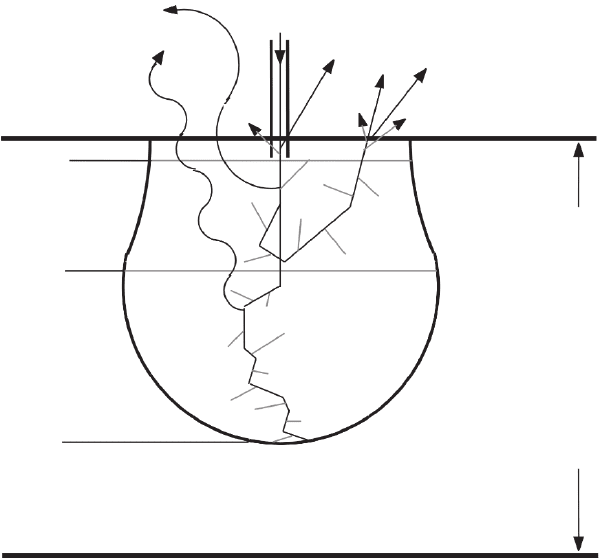
Figure 3–14. Schematic illustration of the generation of secondary electrons
SE1 and SE2, backscattered electrons BSE1 and BSE2, Auger electrons AE,
cathodoluminescence CL, and X-rays in a bulky specimen. t
SE
and t
BSE
indicate
the escape depth for SE and BSE, respectively. R is the electron range.

Chapter 3 Scanning Electron Microscopy 169
Table 3 –3. Characteristic values for R, s
el
, s
in
, L
el
, and L
in
.
a
Element Parameter E
0
= 1 keV E
0
= 5 keV E
0
= 10 keV E
0
= 30 keV
Carbon s
el
(nm
2
) ¥ 10
2
0.65 0.11 0.055 0.018
Z = 6 s
in
(nm
2
) ¥ 10
2
1.95 0.33 0.165 0.054
L
el
(nm) 1.5 9.0 18 55
L
in
(nm) 0.5 3.0 6 18
R (mm) 0.033 0.49 1.55 9.7
Copper s
el
(nm
2
) ¥ 10 1.84 0.64 0.37 0.15
Z = 29 s
in
(nm
2
) ¥ 10
2
1.10 0.38 0.22 0.09
L
el
(nm) 0.64 1.8 3.2 7.8
L
in
(nm) 1.07 3.0 5.3 13
R (mm) 0.007 0.11 0.35 2.26
Gold s
el
(nm
2
) ¥ 10
2
3.93 1.6 1.05 0.52
Z = 79 s
in
(nm
2
) ¥ 10
2
0.79 0.32 0.21 0.10
L
el
(nm) 0.43 1.0 1.6 3.3
L
in
(nm) 2.15 5.0 8.0 16.5
R (mm) 0.003 0.05 0.17 1.0
a
Values are listed for four different electron energies between 1 and 30 keV and three elements having a low (C),
medium (Cu), and high atomic number (Au), respectively. For calculation, the following densities were used: C,
ρ = 2 g cm
−3
; Cu, ρ = 8.9 g cm
−3
; Au, ρ = 19.3 g cm
−3
.
and Ι
in
are shown in Table 3–3. It shows that independent on the elec-
tron energy the electron range for carbon is about one order of magni-
tude larger than for gold. The decrease of the electron energy from 30
to 1 keV, i.e., a factor of 30, reduces the electron range by a signifi cantly
higher factor of roughly 300.
The mean free path lengths indicate after which traveling distance
on average elastic and inelastic collisions will occur. For example, in a
thin organic specimen having a thickness of 50–100 nm only a few col-
lisions on average will take place with 30-keV electrons but about seven
times more with 5-keV electrons. Specimens, which have thicknesses
of about t ≤ 10[Λ
el
Λ
in
/(Λ
el
+ Λ
in
)] can also be imaged in the transmission
mode (cf. Figures 3–1 and 3–2) using unscattered, elastically or inelasti-
cally scattered electrons, respectively. The angular and energy distri-
bution of the scattered electrons can be calculated by Monte Carlo
simulations if the elemental composition and the density of the speci-
men are known (Reichelt and Engel, 1984; Krzyzanek et al., 2003).
The inelastic electron scattering events in the specimen cause second-
ary electrons, Auger electrons, cathodoluminescence, and X-rays, which
carry a wealth of local information about the topography, the electronic
structure, and the composition of the specimen. The signals, resulting
from inelastic electron scattering, can also be calculated by Monte Carlo
simulations.
2.2.1 Secondary Electrons
The energy spectrum of the electrons emitted from a specimen irradi-
ated with fast electrons consists of secondary electrons, backscattered
2
170 R. Reichelt
electrons, and Auger electrons (cf. Figure 3–12). The SE show a peak at
low energies with a most probable energy of 2–5 eV. By defi nition the
maximum energy of SE amounts 50 eV. Secondary electrons are gener-
ated by inelastic scattering of the beam electrons along their trajectories
within the specimen (Figure 3–14). The physics of secondary electron
emission is reviewed by Kollath (1956) and Dekker (1958) but is beyond
the scope of this chapter. Due to the low energy of the SE only those SE
are observable that are generated within the escape depth from the
surface. The actual escape depth of SE for pure elements varies with
their atomic number (Kanaya and Ono, 1984). A general rule for their
escape depth is t
SE
= 5 Λ
SE
(Seiler, 1967), where Λ
SE
is the mean free path
of the SE. t
SE
amounts to about 5 nm for metals and up to about 75 nm for
insulators (Seiler, 1984). The angular distribution of SE follows Lam-
bert’s Law, i.e., is a cos ζ distribution, where ζ represents the SE emission
angle relative to the surface normal (Jonker, 1957; Oppel and Jahrreiss,
1972). The angular distribution of the SE is not important for the image
contrast in SEM because the extraction fi eld of the ET detector normally
collects the emitted SE. The situation, however, is different in case of
magnetic “through-the-lens” detection where no electric extraction fi eld
is applied (cf. Figure 3–6).
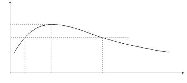
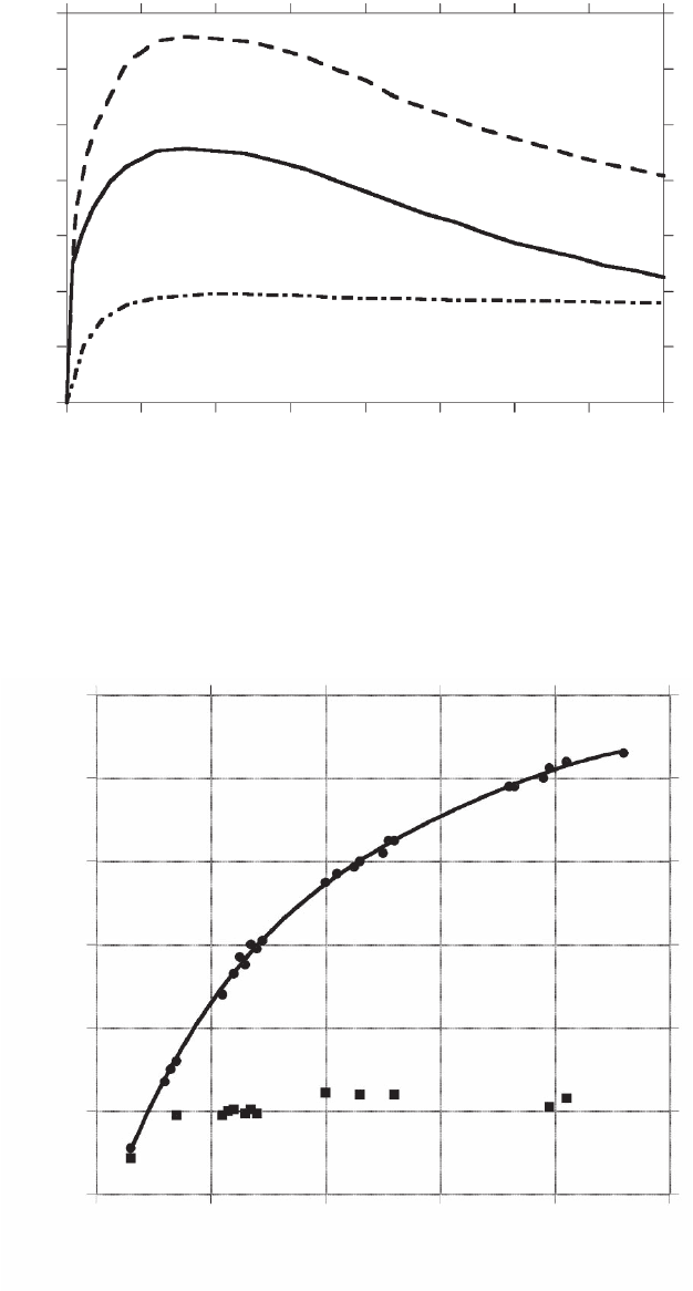
Figure 3–15 shows schematically the SE yield δ versus the energy of
the beam electrons, which is the number of SE produced by one beam
electron. δ increases with E
0
, reaches its maximum δ
m
at E
0,m
, and then
decreases with further increasing E
0
. Typical values for metals are
0.35 ≤ δ
m
≤ 1.6 and 100 eV ≤ E
0,m
≤ 800 eV and for insulators 1.0 ≤ δ
m
≤
10 and 300 eV ≤ E
0,m
≤ 2000 eV (Seiler, 1984). For E
0
>> E
0,m
δ is propor-
tional to E
0
−0.8
(Drescher et al., 1970), which indicates that δ is signifi -
cantly smaller at 30 than at 5 keV. Both parameters, δ
m
at E
0,m
depend
on the ionization energy of the surface atoms (Ono and Kanaya, 1979).
Figure 3–16 shows the SE yield δ versus the energy E
0
for the element
copper.
There is no monotonic relation between δ and the atomic number as
shown in Figure 3–17. However, published data of δ scatter which
1
0
0
E
0,1
E
0,m
E
0,2
E
0
δ
m
δ
Figure 3–15. Schematic representation of the SE yield δ vs. the energy E
0
of
beam electrons.
Chapter 3 Scanning Electron Microscopy 171
0
0
1
0.2
0.4
0.6
0.8
1.2
1.4
η + δ
η
δ
0.5 1.5
E
0
(keV)
1
2.5
2
3.5
3
4
Figure 3–16. SE yield δ, BSE yield η, and δ + η versus the energy E
0
for poly-
crystalline copper at θ = 0°. (Data from Bauer and Seiler, 1984.)
δ, η
δ
η
0.6
Eo = 30 keV
0.5
0.4
0.3
0.2
0.1
0
0
20 40
Atomic number Z
60 80 100
Figure 3–17. SE yield δ and BSE yield η versus atomic number Z at E
0
= 30 keV
and θ = 0°. (Data from Heinrich, 1966; Wittry, 1966.)
172 R. Reichelt
indicates that the specimen surface conditions and the quality of the
vacuum can signifi cantly affect the secondary yield (cf. “Data Base on
Elector-Solid Interactions” by Joy, 2001).
Secondary electrons generated by the incident beam electrons are
designated SE1 (Drescher et al., 1970). The SE1 carry local information
about the small cylindrical volume that is given approximately by the
cross section of the beam (π/4)d
pe
2
and the escape depth t
SE
. For a beam
diameter about ≤1 nm the SE1 deliver high-resolution information.
Those beam electrons, which are multiply scattered and emerge from
the specimen as BSE, also generate secondary electrons within the
escape depth. These secondary electrons are designated SE2 (Drescher
et al., 1970). Their origin is far from the point of incidence of the beam
caused by the spatial distribution of BSE. Changes of the amount of
SE2 correlate with corresponding changes of BSE, thus SE2 carry infor-
mation about the volume from which the BSE originate. The size of the
volume depends on the electron range R and is much larger than the
excitation volume of the SE1 for electron energies E
0
> 1 keV (cf. Figure
3–14 and Table 3–3); thus SE2 deliver low-resolution information. The
SE yield δ consists of the contributions of SE1 and SE2 given as
δ = δ
SE1
+ ηδ
SE2
(2.30)
where η is the BSE coeffi cient and δ
SE2
the SE2 yield, i.e., the number
of SE2 generated per BSE. For E
0,m
< E
0
< 5 kV the ratio δ
SE2
/δ
SE1
amounts
to about 4 and for E
0
≥ 10 kV about 2 (Seiler, 1967). For an increasing
angle of incidence θ, this ratio decreases (Seiler, 1968).
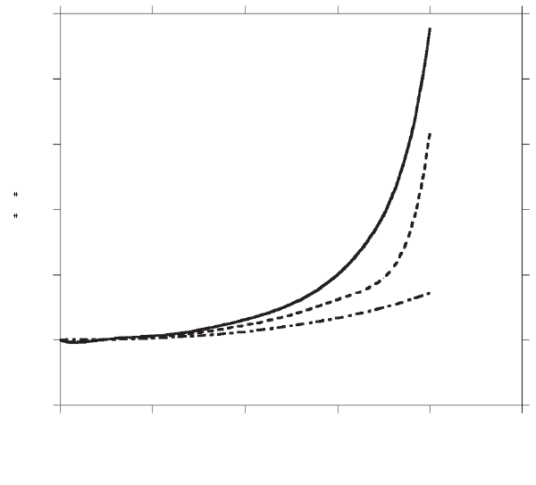
The SE yield increases with increasing angle of incidence θ according
to
δ(θ) = δ
0
/cos θ; δ
0
= δ(θ = 0) (2.31)
(Figure 3–18). This relation is valid for a specimen with a mean atomic
number, for E
0
≥ 5 keV, and θ up to a few degrees below 90°. The
increase of δ with θ is greater for specimens with a low atomic number
For crystalline objects, the increase of δ with θ is superimposed by
electron channeling and crystalline orientation contrast (see Section
2.3). The distinct dependence of the SE yield on θ provides the basis
for the topographic contrast in secondary electron micrographs.
2.2.2 Backscattered Electrons
The majority of BSE is due to multiple scattering of the beam electrons
within the specimen (Figure 3–14). The energy spectrum of the back-
scattered electrons is shown schematically in Figure 3–12. By defi nition
the energy of BSE is in the range 50 eV < E
BSE
≤ E
0
. The BSE spectrum
has a small peak consisting of elastically scattered electrons at E
0
(this
peak is not visible in Figure 3–12). Toward energies lower than E
0
there
is a broad peak, which covers the range down to about 0.7E
0
for high
atomic numbers and further down to about 0.4E
0
for low atomic
numbers. The majority of BSE are within this broad peak. For high
atomic number elements such as gold, the maximum of the distinct
peak is at about 0.9E
0
, whereas for low atomic numbers, e.g., carbon,
the maximum of the less distinct peak is located at about (0.5–0.6)E
0
.
and smaller for samples with high Z (Reimer and Pfefferkorn, 1977).
Chapter 3 Scanning Electron Microscopy 173
The cumulative fraction of 50% of BSE is reached for carbon at E
BSE
/E
0
= 0.55 and for gold at 0.84, respectively (Goldstein et al., 2003). It seems
worth mentioning that the energy distribution of BSE is shifted toward
higher energy if the angle of incident electrons is larger than 70° (Wells,
1974).
As shown in Figure 3–14, the BSE can originate either from the small
area directly irradiated by the electron beam—they are denoted as
BSE1—or after multiple elastic and inelastic scattering events from a
signifi cantly larger circular area around the beam impact point, which
are designated BSE2. The lateral distribution of BSE2 has been calcu-
lated by Monte Carlo simulation for different materials (see, e.g.,
Murata, 1974, 1984). It shows that the BSE-emitting surface area
increases with electron energy E
0
. For a given energy E
0
the size of the
BSE-emitting area increases with descending atomic number. As with
SE1, the BSE1 carry local information about the small volume and
deliver high-resolution information for a beam diameter of about
≤1 nm. As a consequence of lateral spreading the BSE2 carry informa-
tion about a much larger region, thus fi ne structural details on the scale
of the beam diameter cannot be resolved.
Figure 3–14 also shows that the beam electrons travel in a small
subsurface volume before they return to the surface to escape as BSE2.
The escape depth of BSE is much larger than t
SE
and depends on—in
0
0
1
2
3
4
5
Cu
Au
6
20
40
Θ (1°)
60 80 100
δ , η
δ
∗
η
∗
η
∗
Figure 3–18. Normalized SE yield δ* and BSE yield η* versus the angle of
incidence θ of the electron beam. δ* = δ(θ)/δ
0
, η* = η(θ)/η
0
. η was calculated
for gold and copper according to Eq. (2.34).

174 R. Reichelt
contrast to t
SE
—the electron energy E
0
. Experimental data for different
materials show that t
BSE
amounts to about half of the electron range R
(Drescher et al., 1970; Seiler, 1976).
Knowledge of the angular distribution of BSE is of great importance
for understanding and optimization of BSE detection geometry. For
normal beam incidence the angular distribution can be approximated
by a cos ζ distribution (Drescher et al., 1970), where ζ represents the
BSE emission angle relative to the surface normal. Due to the fact that
the emitted BSE move on nearly straight trajectories, the angular detec-
tor position has a strong infl uence on the collection effi ciency of the
detector. For nonnormal beam incidence the distribution is asymmetric
and a refl ection-like emission maximum is observed. The angular dis-
tribution consists for large angles of incidence θ of cosine distribution
approximately directed to −θ and a superimposed fraction at smaller
emission angles (Drescher et al., 1970).
The BSE coeffi cient η is defi ned by
η = n
BSE
/n
b
(2.32)
where n
BSE
is the number of BSE and n
b
is the number of incident elec-
trons. η is approximately independent of the electron energy E
0
in the
range of about 10–30 keV. For low atomic numbers and beam energies
below 5 keV η increases as E
0
decreases, whereas for medium and high
atomic numbers η decreases with E
0
(cf. Figure 3–16) (Reimer and
Tollkamp, 1980). However, at low energies η depends in a complex
manner on the atomic number (Heinrich, 1966; cf. “Data Base on
Electron-Solid Interactions” by Joy, 2001).
The BSE coeffi cient monotonically increases with the atomic number
as shown for 30 keV in Figure 3–17. Because of the approximate inde-
pendence of the electron energy E
0
, the graph of the BSE coeffi cient is
valid for beam energy ranging from 30 down to about 5 keV. The graph
of η versus Z can be approximated by a polynomial (Reuter, 1972)
η(Z) = 0.0254 + 0.016 Z − 1.86 × 10
−4
Z
2
+ 8.31 × 10
−7
Z
3
(2.33)
For energies below 5 keV the dependence of η on Z is more complicated
(for details see Hunger and Kuchler, 1979; Joy, 1991; Zadrazil et al.,
1997). The distinct dependence of the BSE coeffi cient on the atomic
number Z provides the basis for the atomic number contrast (see
Section 2.3).
Like the SE yield, the backscattering coeffi cient also increases
monotonically with increasing angle of incidence θ according to (Arnal
et al., 1969)
ηθ θ() ( cos)
/
=+
−
1
9 Z
(2.34)
Figure 3–18 shows the graphs η(θ) versus θ for Cu (Z = 29) and Au
(Z = 79). The graphs indicate the strong infl uence of the atomic number,
in particular for θ > 50°. The monotonic increase of η with θ provides
the basis for the topographic contrast in BSE micrographs. For the sake
of completeness it should be mentioned that Drescher et al. (1970)
derived from experimental data at 25 keV an analytical expression for
η(θ, Z) other than the one given by Eq. (2.34).
