Hawkes P.W., Spence J.C.H. (Eds.) Science of Microscopy. V.1 and 2
Подождите немного. Документ загружается.


Chapter 3 Scanning Electron Microscopy 145
LaB
6
requires a vacuum better than 10
−4
Pa to avoid cathode contamina-
tion (tungsten cathode: about 10
−3
Pa) and (2) its alignment is critical.
Characteristic values of the triode gun with thermionic tungsten and
the LaB
6
cathode are summarized in Table 3–1.
2.1.2 Electron Lenses
As discussed in Section 2.1.1 the electrons emerge from the electron
gun as a divergent beam. Two or three electromagnetic lenses and
apertures in the microscope column (cf. Figure 3–1) reconverge and
focus the beam into a demagnifi ed image of the fi rst crossover gener-
ated by the gun. The fi nal lens—the objective lens—focuses the beam
into the smallest possible spot of 4–10 nm on the sample surface, i.e.,
the total demagnifi cation is about 5000×.
Rotationally symmetric electromagnetic lenses consist of a coil with
N ⋅ I ampere windings inside an iron pole piece. Typically, N ⋅ I is in the
order of 10
3
A for the condenser and objective lenses. The iron pole piece
has a small gap in its axial bore. The current in the coil generates a
magnetic fi eld carried by the iron, which also appears at the gap forming
a bell-shaped stray fi eld distribution on the optical axis with a radial
and axial fi eld component. Off-axis electrons move due to the Lorentz
force along screw trajectories because the radial component of the fi eld
results in a rotation around the optical axis. Electrons emerging diver-
gently from a point in front of the lens are focused in an image point
behind the lens. The lenses of SEMs can usually be considered weak
lenses (because the pole piece is not saturated). In this case, the princi-
pal planes of the lens coincide with its optical center and the formulas
for thin light optical lenses can be used. In close analogy to light optics
the strength of an electromagnetic lens can be characterized by its focal
length f. Using the thin lens formulas we can write
Table 3 –1. Characteristic parameters of different electron guns.
Source
Schottky
Thermionic LaB
6
- emission
Parameters W-cathode
a
cathode
a,b
Cold FEG
c
Hot FEG
c
cathode
c
Brightness 10
5
10
6
10
7
–10
8
10
7
–10
8
10
7
–10
8
(A cm
-2
sr
-1
Energy 1–3 0.5–2 0.2–0.4 0.5–0.7 0.8
spread (eV)
Vacuum (Pa) ~10
-3
~10
-4
10
-8
–10
-9
10
-8
–10
-9
10
-8
Emission ~100 1–>50 ~10 ~10 30–70
current (mA)
Life time ~40 h ~200 h >1 year >1 year >1 year
~150 h
d
~8 years
d
a
Reimer (1985).
b
DeVore and Berger (1996).
c
Reimer (1993).
d
Reichelt (unpublished).
)
146 R. Reichelt
1/f = 1/p + 1/q (2.5)
where p is the distance from the object (= crossover) and q is the dis-
tance to the image. Both, p and q are related to the center of the lens.
The magnifi cation M is given simply by
M = q/p (2.6)
where M < 1 for p > 2f, i.e., a demagnifi ed image of an object is obtained
at these imaging conditions. Therefore, a strong demagnifi cation of
about 5000× of the fi rst crossover can be obtained for p >>
2f for each
of the two or three lenses by successive demagnifi cation of each inter-
mediate crossover. In case of two condenser lenses they usually are
combined and adjusted by one control only.
The pole pieces of condenser lenses are symmetrical, i.e., the diam-
eters of the axial bores in the upper and lower half of the pole piece
are identical. In contrast to that the pole piece of the objective lens is
very asymmetric (1) to limit the magnetic fi eld at the specimen level
and (2) to house the beam defl ections coils, the adjustable objective
aperture, and the stigmator (not shown in Figure 3–1). The asymmetric
objective lens (called the pinhole or conical lens) adapts for the wide
range of the WD of about 5–30 mm by an adjustable focal length.
However, working at a large WD inevitably degrades the electron
optical properties of the objective lens and enlarges the fi nal spot size
d
p
. For a detailed description of the electron optical properties of elec-
tromagnetic lenses and defl ection coils the reader is referred to books
about electron optics (e.g., Glaser, 1952; Grivet, 1972; Klemperer, 1971;
Reimer, 1998).
All electromagnetic lenses involved in successive demagnifi cation
suffer from an imperfect rotational symmetry and aberrations, which
degrade their electron optical performance. The effects of lens aberra-
tions cannot be compensated, however, they can be minimized, which
is most effective for the fi nal—the objective—lens. Let us consider
briefl y the three signifi cant effects.
1. Spherical Aberration. The spherical aberra tion constant C
s
causes an
error disc of the diameter (Cosslett, 1972)
d
s
= 1/2 C
s
α
p
3
(2.7)
2. Chromatic Aberration. The chromatic aberration caused mainly by
the energy spread of the electrons from the gun is characterized by
the constant C
c
causes an error disc of the diameter
d
c
= C
c
⋅ ∆E/E
o
⋅ α
p
(2.8)
where ∆E/E
o
represents the relative energy spread of the beam
electrons.
3. Diffraction. The diffraction of electrons on the objective aperture
results in a further error disc—the Airy disc—of diameter
d
f
= 0.6λ/α
p
(2.9)
where λ is the wavelength of the electrons.
Chapter 3 Scanning Electron Microscopy 147
In a fi rst approximation it is possible to superpose the squared diam-
eters of the individual discs to estimate the effective electron probe
diameter
d
pe
2
= d
p
2
+ d
s
2
+ d
c
2
+ d
f
2
(2.10)
d
p
2
is given by Eq. (2.3) as d
p
2
= (4I
p
/π
2
β)α
p
−2
. More precise, but at the
same time more complicated relations for the effective probe diameter
were derived by Barth and Kruit (1996) and Kolarik and Lenc (1997).
Under the conditions normally used in conventional SEM (i.e., E
o
=
10–30 keV) the chromatic aberration as well as the effect of the diffrac-
tion are relatively small compared to the remaining contributions and
can be neglected (Reimer, 1985). The optimum aperture α
opt
, which
allows the smallest effective electron probe diameter d
min
, can be
obtained by the fi rst derivative ∂d
pe
/∂α
p
= 0 and is given as
α
opt
= (4/3)
1/8
[(4I
p
/π
2
β)
1/2
/C
s
]
1/4
(2.11)
By using the approach mentioned above, i.e., d
pe
2
= d
p
2
+ d
s
2
, and Eqs.
(2.3), (2.7), and (2.11), the minimum effective electron probe diameter
is
d
p,min
= (4/3)
3/8
[(4I
p
/π
2
β)
3/2
C
s
]
1/4
(2.12)
It is obvious that d
p,min
increases as I
p
increases or β decreases. Both, I
p
and β are parameters depending on the performance of the electron
gun [cf. Eq. (2.3)]. C
s
is a parameter characterizing the performance of
the objective lens and should be as small as possible. As previously
mentioned, the operation of the SEM at a large WD inevitably degrades
the electron optical properties of the objective lens, i.e., C
s
increases as
the WD increases. Just to provide a rough idea about values for d
p,min
and α
opt
at usual electron energies (10–30 keV), a moderate WD and a
probe current I
p
of about 10
−11
A, which gives a suffi cient S/N ratio, d
p,min
typically amounts to approximately 10 nm and α
opt
to 5–10 mrad.
It is also of interest to know the maximum probe current I
p,max
under
these conditions. Using the Eqs. (2.12) and (2.3) one obtains
I
p,max
= 3π
2
/16 ⋅ β C
s
−2/3
d
p,min
8/3
(2.13)
Interestingly, it becomes obvious from Eq. (2.13) that including the
effect of the spherical aberration, I
p
is now proportional to d
p
8/3
instead
of d
p
2
as before [cf. Eq. (2.3)].
The electron probe current in a SEM equipped with a thermionic
gun can be increased several orders of magnitude above 10
−11
A as
required, e.g., for microanalytical studies (cf. Section 6). It is clear from
the considerations above that an increase in the probe current inevita-
bly increases the probe size. A rough estimate for 30 keV electrons
shows that an increase of I
p
to 10
−9
A requires a probe size of about
60 nm. However, due to the electron–specimen interaction the lateral
resolution of X-ray microanalysis is limited to about 1 µm for thick
samples. Therefore, a probe diameter of 100 nm or even several hundred
nanometers can be tolerated without disadvantage for X-ray micro-
analysis in this case.
148 R. Reichelt
When considering the effective electron probe diameter the chro-
matic aberration of the objective lens could be neglected for energies
>10 keV. Because d
c
is inversely proportional to E
o
[cf. Eq. (2.8)] there is
a signifi cant increase for energies below 10 keV, in particular for the
low-voltage range below 5 keV. For example, for 1 keV electrons the
diameter of the chromatic error disc increases by a factor of 30 com-
pared to 30 keV! When using a thermionic cathode with a tungsten fi la-
ment and a probe current of about 10
−11
A the energy spread is about
2 eV (cf. Table 3–1) and d
c
contributes dominantly to the enlargement
of the probe diameter [cf. Eq. (2.10)]. Therefore, the ther mionic source
is inappropriate for imaging in the low-voltage range. As we shall see
in Section 3, fi eld emission guns with a one order of magnitude smaller
energy spread and about fi ve orders of magnitude larger brightness
are very well suited for low-voltage SEM (LVSEM).
In the context of the objective lens the existence of a stigmator was
mentioned, which usually is located near the pole-piece gap. Due to
imperfect rotational symmetry of the pole-piece bores, magnetic inho-
mogeneities of the pole piece, or some charging effects in the bore or
at the objective aperture, the magnetic fi eld in the objective lens
becomes asymmetric. This causes different focal lengths in the sagittal
and meridional planes, which leads to low image quality degraded by
astigmatism. The astigmatism can be compensated for by adding a
cylinder lens adjustable in its strength and azimuth. The effect of a
cylinder lens is realized by the stigmator consisting of a pair of quad-
rupole lenses.
2.1.3 Detectors and Detection Strategies
Electron detectors specifi cally collect the signals emerging from the
specimen as a result of electron–specimen interaction. The effi ciency
of the signal collection depends on the type of the detector, its perfor-
mance, and its detection geometry, i.e., its position related to location
of the signal emitting area. For an understanding of the recorded
signals, knowledge of the infl uence of these parameters is critical.
2.1.3.1 Detectors
To detect electrons in SEM three different principles are commonly
used. One principle is based on the conversion of signal electrons to
photons by a scintillation material. Then, the photons are converted
into an electric signal by a photomultiplier, which is proportional to
the number of electrons impinging on the scintillator. The second
principle is based on the conversion of electrons to electron hole pairs
by a semiconductor, which can be separated before recombination
causing an external charge collection current. This current is propor-
tional to the number of electrons impinging on the semiconductor.
While the principle of scintillation detection is used for secondary,
backscattered, and transmitted electrons (in case of thin specimens),
the semiconductor detector is mostly used for backscattered electrons
only. Finally, the third principle is based on the electron channel mul-
tiplier tube, which converts the signal electrons by direct impact at its
Chapter 3 Scanning Electron Microscopy 149
input to secondary electrons and multiplies them inside the tube. The
output signal is proportional to the number of impinging signal
electrons.
As we shall see later, Auger electrons (AE) have a characteristic
energy, which is related to the atomic number of the element involved
in their generation. For recording of AE in the energy range from 50 eV
to several keV, spectrometers with a high energy resolution and high
angular collection effi ciency are needed in combination with the scin-
tillation detection used, e.g., for secondary and backscattered electrons.
The most widely used spectrometer for AE spectroscopy is the cylin-
drical mirror analyzer. Only those electrons, which pass the energy-
selecting diaphragm of the spectrometer, can impinge on the scintillator,
thus contributing to the signal.
Besides of electrons the electron–specimen interaction can also
produce electromagnetic radiation, namely cathodoluminescence (CL)
and X-rays (cf. Figure 3–2). Cathodoluminescence shows a close analogy
to optical fl uorescence light microscopy (FLM) where light emission is
stimulated by irradiation with ultraviolet light (photoluminescence). In
principle, for the detection of emitted light, which has a wavelength in
the range of about 0.3–1.2 µm, a photomultiplier is very well suited (see
above) and therefore most often used. However, the commonly low
intensity of the CL signal requires, for a suffi cient S/N ratio, a high
collection effi ciency of the emitted light. Table 3–2 presents the most
common detector types for SE, BSE, and CL. The detectors for X-rays
will be described in Section 6 of this chapter.
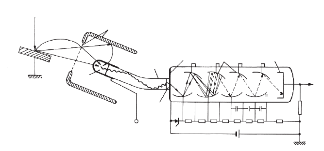
Scintillation Detector. The scintillation detector for SE—the Everhart–
Thornley (ET) detector (Everhart and Thornley, 1960)—is shown sche-
matically in Figure 3–5. The generated SE are collected by a positively
biased collector grid, then they pass the grid and are accelerated by
about 10 kV to the conductive coated scintillator. The scintillation mate-
rial converts electrons to photons, which are guided by a metal-coated
quartz glass to the photocathode of a photomultiplier where photoelec-
trons are generated and amplifi ed by a factor of about 10
6
. Usually the
electronic signal at the output of the photomultiplier is further ampli-
fi ed. Several scintillator materials, such as plastic scintillators, lithium-
activated glass, P-47 powder, or YAG and YAP single crystals, are in
use, which differ in their performance (for details see, e.g., Reimer,
1985; Autrata, 1990; Autrata and Hejna, 1991; Autrata et al., 1992a,b;
Schauer and Autrata, 2004).
When the collector grid of the ET detector is negatively biased
by <−50 V SE are not collected. In this case only the BSE can reach
the scintillator on almost straight trajectories because of their higher
energies. The detected fraction of BSE is very low because of the
small solid angle of collection, i.e., small angular collection effi ciency
(CE). However, for an effi cient detection of BSE the solid angle of
collection of BSE detectors is signifi cantly larger by using a larger
scintillator and at the same time a shorter distance to the specimen.
The BSE detector does not require the collector grid used for the SE
(cf. Figure 3–5).

150 R. Reichelt
Table 3 –2. Most common electron detectors for SEM.
a
Signal Type of detector Principles Specifi cations References
SE Everhart–Thornley Scintillator–LP–PM High CE; positively biased Everhart and Thornley (1960);
collector grid Reimer (1985)
Solid state Electron hole pair generation SE are accelerated to>10 keV before Crewe (1970);
detection Reimer (1985)
MCP Electron multiplier tube Positively biased from plate Postek and Keery (1990);
Reimer (1985)
BSE Everhart–Thornley Scintillator–LP–PM Very low CE; negatively biased Everhart and Thornley (1960);
collector grid Reimer (1985)
Autrata Scintillator–LP–PM High CE; E
BSE
≥0.8 keV Autrata et al. (1992)
Robinson Scintillator–LP–PM High CE; E
BSE
≥0.9 keV Robinson (1990)
Solid state Electron hole pair generation High CE; E
BSE
≥1.5 keV Stephen et al. (1975)
bandwidth about ≤2 MHz
MCP Electron multiplier tube High CE; E
BSE
≥1 keV; negatively Postek and Keery (1990)
biased front plate
CL Ellipsoidal or parabolic Mirror–PM; mirror– High CE; normally simultaneous Autrata et al. (1992);
mirror with parallel or spectrometer–PM; BSE detection is not possible Bond et al. (1974);
focused light output or mirror–LP–PM (for exceptionsee Autrata et al., Rasul and Davidson (1977);
coupled to an LP 1992) Reimer (1985)
a
MCP, Microchannel plate; LP, light pipe; PM, photomultiplier; CE, collection effi ciency; E
BSE
, energy of backscattered electrons.

Chapter 3 Scanning Electron Microscopy 151
Semiconductor Detector. The semiconductor detector—often denoted as
a solid state detector—generates from an impinging electron with the
energy E a mean number of electron hole pairs given by
n
m
= E/E
exm
(2.14)
where E
exm
= 3.6 eV is the mean energy per excitation in silicon (Wu and
Wittry, 1974). The electron hole pairs can be separated before recombina-
tion, in this way generating an external charge collection current, which
is proportional to the number of impinging electrons. Because of the
energy dependence of n
m
the BSE with higher energy contribute with a
larger weight to the signal than the BSE having low energies. The semi-
conductor detector can be used only for the direct detection of BSE
because impinging SE are absorbed in its thin electrical conductive layer.
However, a special detector design for accelerating the SE to energies
above 10 keV also allows for detection of SE (Crewe et al., 1970).
Microchannel Plate Detector. A microchannel plate (MCP) consists of a
large number of parallel very small electron multiplier tubes (diameter
about 10–20 µm, length of a few millimeters) covering an area of about
25 mm in diameter (e.g., Postek and Keery, 1990). Thus this detector is
thin and, when placed between objective pole piece and specimen,
enlarges the work distance by only about 3.5 mm. The MCP detector
system is effi cient at both high and low accelerating voltages, and is
capable of both secondary electron and backscattered electron detec-
tion. The MCP becomes of increasing interest for studies with low
currents and in low-voltage scanning electron microscopy (Russel and
Manusco, 1985). However, as yet the MCP detector is not as common
as the other detector types described above.
Cathodoluminescence Detectors. In the few cases of strongly luminescent
specimens a lens or a concave mirror is suffi cient for light collection
(Judge et al., 1974). As mentioned above, mostly the intensity of the CL
signal is low, thus a high collection effi ciency of the emitted light is
PE
SE
BSE
BSE
Collector
Grid and screen
1e
-
± 200 V
Specimen
Scintillator
hv
SE
Light pipe
Photomultiplier
Photocathode
Dynodes
Anode
Signal
R
1 MΩ
U
PM
= 500 – 1000 V
10 nF
10
6
e
100 kΩ
Optical
contact
+10 kV
Figure 3–5. Schematic drawing of Everhart–Thornley detector (scintillator–photomultiplier combina-
tion) for recording secondary electrons (SE). BSE, backscattered electrons; PE, primary electrons; PM,
photomultiplier; hν, energy of photons. [From Reimer (1985); with kind permission of Springer-Verlag
GmbH, Heidelberg, Germany.]
152 R. Reichelt
indispensable. This requires a solid angle of collection as large as pos-
sible, an optimum transfer of the collected light to a monochromator
or directly to the photomultiplier, and a photomultiplier with a high
quantum effi ciency in the spectral range of the CL (Boyde and Reid,
1983). Commercial CL collector and imaging systems allow for inves-
tigations with a wavelength from less than 200 nm to about 1800 nm in
the imaging and spectroscopy mode. The following are the most com-
monly used collection systems.
1. Parabolic or elliptic mirrors. The light-emitting area of the speci-
men is located at the focus of the mirror and is formed into a parallel
beam for a parabolic mirror (Bond et al., 1974) or focused to a slit of a
spectrometer for an elliptic mirror (e.g., McKinney and Hough, 1977).
The solid angle of collection is in the order of π sr but SE detection with
an ET detector is still feasible.
2. Rotational ellipsoidal mirror. The light-emitting area of the speci-
men is located at one focus of the half of the ellipsoid of rotation (Hörl,
1972). The emitted light is focused to a light pipe or to the focal point
of an optical microscope objective at the second focal point of the ellip-
soid. Although the ellipsoidal mirror has the largest collection angle,
the effective collection angle is limited by the acceptance angle of the
light pipe or the optical microscope objective, respectively, to about
0.75 π sr. The limitation by the acceptance angle can be avoided by
placing a parabolic mirror below the second focal point of the ellipsoid
(Hörl, 1975). Very recently Rau et al. (2004) proposed an ellipsoidal
confocal system collecting the emitted light, which enables CL microto-
mography in SEM. In principle, the proposed system allows for CL
studies at high resolution, which is well below the size of the light-
emitting volume.
3. Optical microscope objective. The CL of an optically transparent
specimen can be studied by an optical microscope objective positioned
below the specimen. The collection angle of this setup amounts to
about 1.4 π sr (Ishikawa et al., 1973).
2.1.3.2 Detection Strategies
Generally, the detectors for the various signals can be combined and
each of them should have an optimum position to make the best use
of the electron–specimen interaction. The use of electron spectrometers
for AE, BSE, and SE can provide supplementary information about the
specimen surface but additional space is needed for a spectrometer. As
a matter of fact, the space for detectors is limited in particular with a
short WD or with an in-lens position of the specimen for higher resolu-
tion. Very recently a proposal was made to improve this situation in
scanning electron microscopes with a new design (Khursheed and
Osterberg, 2004). The suggested arrangement allows for the effi cient
collection, detection, and spectral analysis of the scattered electrons on
a hemispherical surface that is located well away from the rest of the
SEM column.
A conventional SEM commonly is equipped with an ET detector
located laterally above the specimen and a BSE detector (for different
types see Table 3–2) located centrally above the specimen (top posi-
tion). Additional ports at the specimen chamber of the SEM enable
Chapter 3 Scanning Electron Microscopy 153
additional detectors to be installed. Because of limited space not all of
the installed detectors may be used simultaneously, however, there are
retractable detectors (e.g., BSE detectors) available, which can be kept
in the retracted position when not needed (providing space for another
detector or allowing for a shorter WD) and can readily be moved into
working position if required for signal recording. Numerous multide-
tector systems have been proposed for BSE and SE (for review see
Reimer, 1984a, 1985). In the top position, e.g., two semiannular semi-
conductor detectors (Kimoto et al., 1966; Hejna and Reimer, 1987) allow
for separation of topographic and material contrast; with a four-
quadrant semiconductor detector (Lebiedzik, 1979; Kaczmarek, 1997;
Kaczmarek and Domaradzki, 2002) the surface profi le can be recon-
structed and the distinction between elements with different atomic
numbers is improved. Even a six-segment semiconductor detector is of
interest (Müllerová et al., 1989).
A combination of two opposite ET detectors, A and B, allows two SE
signals, S
A
and S
B
, to be recorded simultaneously. The difference signal
S
A
− S
B
illustrates the topographic contrast whereas the sum S
A
+ S
B
signal illustrates the material contrast (Volbert and Reimer, 1980;
Volbert, 1982). The mixing of the analog electronic signals at that time
was performed by electronic circuitry. After analog signal mixing the
two original signals were lost. Today, modern SEMs usually record
digital images, which are stored in a PC. Thus the mixing of images
(their raw data are stored in a memory) can be performed readily after
image recording by means of image processing software available from
numerous software companies.
For high-resolution and LVSEM the work distance should be as short
as possible (say below 5 mm) because both the focal length and the
aberrations of the objective lens increase with the WD (see also Sec-
tions 2.1.2 and 3). In contrast to the asymmetric objective lens (large
focal length) where the region above the specimen is a magnetic fi eld
free space, the specimen is immersed in the fi eld of the objective lens
with a short focal length. In this case the specimen is very close to the
lower objective pole piece or is placed directly inside the pole-piece gap
[as in a transmission electron microscope (TEM); see Chapters 1, 2, 6,
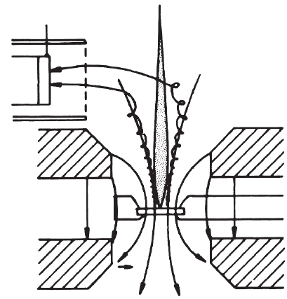
and 7]. For the latter lens type—the specimen has an “in-lens” position
and is limited in size to a few millimeter only—the collection of SE
takes advantage of the fact that they can spiral upward in the magnetic
fi eld of the objective lens due to their axial velocity component. The SE
have to be defl ected off the axis to be recorded by an ET detector
located laterally above the lens (cf. Figure 3–6).
The separation of the downward moving beam electrons and the
upward moving secondary electrons can be done most effi ciently by
an E × B system, which employs crossed electric and magnetic fi elds.
The forces of these fi elds compensate each other for the beam electrons,
but add for the opposite moving secondary electrons. This magnetic
“through-the-lens” detection (for review see Kruit, 1991) of SE has
several advantages: (1) SE are separated from BSE, which do not reach
the detector because their higher kinetic energy causes different tra-
jectories; (2) very high collection effi ciency for real SE emerging from
the specimen and a suppression of SE created on the walls of the

154 R. Reichelt
+10 kV
+200 V
ETD
SE
B
Figure 3–6. Schematic drawing of the magnetic “through-the-lens” detection
of secondary electrons (SE) for the “in-lens” position of the specimen. B, mag-
netic fi eld lines, ETD, Everhart–Thornley detector. [From Reimer (1993); with
kind permission of the International Society of Optical Engineering (SPIE),
Bellingham, WA.]
system by BSE; (3) improved collection effi ciency from inside a porous
specimen (in particular cavities or holes facing the electron beam)
(Lukianov et al., 1972); and (4) loss of directionality in the image
because the SE are detected irrespective of the direction of emission
(in contrast to the lateral position of the ET detector; cf. Figure 3–7a).
However, a combination of two opposite ET detectors to illustrate the
topographic or material contrast has not been tried as yet.
It seems worth mentioning that a real “through-the-lens” detection
system was incorporated in one of the early SEMs (Zworykin et al.,
1942). The magnetic “through-the-lens” detection of SE was established
by Koike et al. (1970) using a TEM with scanning attachment. Finally,
magnetic “through-the-lens” detection can be combined with any elec-
tron spectrometer as done in SE and Auger spectroscopy (for review
see Kruit, 1991).
Another type of “in-lens” detection of SE and BSE is used in the
electrostatic detector objective lens (Zach and Rose, 1988a,b; Zach,
1989). The detector is of the annular type and possesses a high collec-
tion effi ciency of SE of about 75%. Replacing the annular detector by a
combination of two semiannular detectors A and B (Figure 3–8) could
be used to illustrate the topographic or material contrast, respectively
(Reimer, 1993). Similarly, “in-lens” annular type detection of SE and BSE
is also used in combined magnetic-electrostatic objective lenses (Frosien
et al., 1989), known under the trade name of “Gemini lens.” Both types
of lens are advantageous for low-voltage SEM (see Section 3.2) because
they provide excellent image resolution at low electron energies.
