Hawkes P.W., Spence J.C.H. (Eds.) Science of Microscopy. V.1 and 2
Подождите немного. Документ загружается.

Chapter 3 Scanning Electron Microscopy 175
The backscattering coeffi cient of a single crystal depends sensitively
on the direction of the incident electrons related to the crystal lattice
(Reimer et al., 1971; Seiler, 1976). This dependence is caused by the
regular three-dimensional arrangement of the atoms in the lattice,
whose atomic density depends on the direction. The backscattering
coeffi cient is lower along directions of low atomic density, which
permits a fraction of the incident electrons to penetrate deeper than in
amorphous material before being scattered. Those electrons have a
reduced probability of returning to the specimen surface and leaving
the sample as BSE. The maximum relative variation of the backscatter-
ing coeffi cient is in the order of 5%.
2.2.3 Transmitted Electrons
When the thickness of a specimen approaches the electron range R or
becomes even smaller than R, an increasing fraction of beam electrons
is transmitted. Those transmitted electrons interact with the support,
thus generating non-specimen-specifi c signals, which superimpose the
specimen-specifi c signals. The spurious contribution of the support to
the signal, originating from the specimen, can be reduced signifi cantly
by replacing the solid support by a very thin (about 5–15 nm thick)
amorphous carbon fi lm. Such thin carbon fi lms supported by a
metallic mesh grid are commonly used in TEM and STEM as electron-
transparent support for thin specimens. To improve the stability of the
5-nm-thick carbon fi lm, the fi lm is placed onto a holey thick carbon
fi lm supported by a mesh grid. In contrast to a solid support, a 5-nm-
thick carbon fi lm contributes only insignifi cantly to the SE and BSE
signal (cf. Figure 3–19), thus particles deposited onto a thin support
fi lm can be imaged in the normal manner using SE and BSE,
respectively.
In addition to the reduction of spurious signal, the transparent
support fi lm enables use to be made of the transmitted electrons, which
carry information about the interior of the specimen (in some SEM the
specimen stage must be altered to make the transmitted electrons
accessible). As a result of electron–specimen interaction the transmit-
ted electrons can be unscattered or elastically or inelastically scattered
(cf. Figure 3–2). Due to their characteristic angular and energy distri-
bution, the transmitted electrons can be separated by placing suitable
detectors (preferentially combined with an electron spectrometer)
below the specimen. Frequently, a rather simple and inexpensive device
for observing an STEM image (Oho et al., 1986)—sometimes called
“poor man’s STEM in SEM detector”—is used. The transmitted elec-
trons are passing through an angle-limiting aperture, strike a tilted
gold-coated surface, and thus create a high SE and BSE signal, which
can then be collected by a conventional ET detector. The angle-limiting
aperture cuts off the transmitted, scattered electrons. In this case the
“poor man’s STEM in SEM detector” acquires those electrons, which
represent the bright-fi eld signal. The “poor man’s STEM in SEM detec-
tor” just cuts the transmitted scattered electrons without making use
of their inherent information. Both the elastically and the inelastically
scattered electrons are signals, which very sensitively depend on the

176 R. Reichelt
mass thickness ρt if the specimen thickness t ≤ [Λ
el
Λ
in
/(Λ
el
+ Λ
in
)]
(Reichelt and Engel, 1984, 1985) (Figure 3–20) can be used, e.g., for mass
determination of biomolecules and assemblies thereof (Zeitler and
Bahr, 1962; Lamvik, 1977; Engel, 1978; Wall, 1979; Feja et al., 1997).
Although most of the mass determination studies were made with
dedicated STEM, high-resolution FESEM equipped with an effi cient
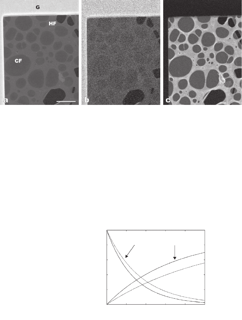
Figure 3–19. SE and annular dark-fi eld micrographs showing a particle on an ∼4-nm-thick amorphous
carbon fi lm (CF) attached to a holey carbon fi lm of ∼20 nm in thickness (HF), which is supported by
a Cu mesh grid (G). (a) SE micrograph recorded with a probe current of about 30 pA at 30 kV. A very
low probe current of ∼3 pA (pixeltime: 23 µs. i.e., ∼430 incident electrons/pixel) was used for simultane-
ously recording the SE micrograph (b) and the dark-fi eld micrograph with scattered transmitted elec-
trons (annular dark-fi eld mode) (c). The image intensity and the signal-to-noise ratio in (c) is much
higher than the intensity in (b) because the number of SE (almost only SE1 are generated) is signifi -
cantly smaller than the number of the collected transmitted scattered electrons. No usable BSE signal
can be detected from the thick and thin carbon fi lms. The scale bar corresponds to 20 µm.
1
0
020406080100
0.8
unscattered
scattered
t [nm]
0.6
Fraction of electrons
0.4
0.2
Figure 3–20. Fraction of transmitted electrons scattered into an angular range
of 25–300 mrad for carbon (ρ = 2 g cm
−3
; solid line) and protein (ρ = 1.35 g cm
−3
;
dashed line). Parameters: E
0
= 30 keV, α
p
= 10 mrad. The graphs show an
increasing fraction of scattered and a decreasing fraction of unscattered elec-
trons with increasing thickness. (Calculation according to Krzyzanek and
Reichelt, 2003.)
Chapter 3 Scanning Electron Microscopy 177
annular dark-fi eld detector capable of single-electron counting
and MHz counting rates would allow for such quantitative studies
(Reichelt et al., 1988; Krzyzanek and Reichelt, 2003; Krzyzanek et al.,
2004).
Another important application of STEM imaging is the measure-
ment of the physical probe size of the SEM using a thin carbon fi lm
(thickness below 10 nm, preferably containing nanoparticles of gold
or gold-palladium for better contrast). In this case the broadening
of the electron beam in the fi lm is negligible and so the resolution
of the STEM image is equal to the probe diameter. The image resolu-
tion can be determined either by analysis of the diffractogram
(power spectrum) of the STEM micrograph (Frank et al., 1970;
Reimer, 1985; Joy, 2002) or by cross-correlation function analysis
(Frank, 1980) of the phase noise in the bright-fi eld STEM image of
the carbon fi lm. The latter directly yields the probe diameter of the
SEM (Joy, 2002).
Combining the SE and BSE detectors above as well as the bright-fi eld
and dark-fi eld detectors below the specimen, its surface as well as its
internal structure can be observed simultaneously in the SEM.
2.2.4 Cathodoluminescence
Cathodoluminescence (CL) is the emission of light generated by the
electron bombardment of semiconductors and insulators (Muir and
Grant, 1974; cf. Figure 3–14). Those materials have an electronic band
structure characterized by a fi lled valence band and an empty conduc-
tion band separated by an energy gap ∆E
CV
= E
C
− E
V
. Electrons from the
valence band can interact inelastically with a beam electron and can be
excited to an unoccupied state in the conduction band. The excess
energy of the excited electron will be lost by a cascade of nonradiative
phonon and electron excitations. Most of the recombination processes of
excited electrons with holes in the valence band are nonradiative pro-
cesses, which elevate the sample temperature. There are different radia-
tive processes, which take place in inorganic materials, semiconductors,
and organic molecules.
In inorganic materials intrinsic and extrinsic transitions can take
place. The intrinsic emission is due to direct recombination of electron
hole pairs. Extrinsic emission is caused by the recombination of trapped
electrons and holes at the donor and acceptor level, respectively. The
trapping increases the probability of recombination. The extrinsically
emitted photons have a lower energy than intrinsically emitted
photons.
In semiconductors the radiative recombination can be due to the
direct collision of an electron with a hole with the emission of a phonon.
Depending on the nature of the band structure of the material, the
recombination can be either direct or indirect. In the latter case the
recombination must occur by simultaneous emission of a photon.
Indirect recombination is less likely than direct recombination. If
the material contains impurities, the process of recombination via
impurity level becomes important. The modifi cation of CL effi ciency
as a function of the purity and the perfection of the material is the
most important aspect of the use of this method in scanning electron
178 R. Reichelt
microscopy. It is because of such modifi cations that a contrast is
generated (for details see, e.g., Holt and Yacobi, 1989; Yacobi, 1990).
It was shown in some cases that the sensitivity of CL analyses can be
at least 10
4
times higher than that obtainable by X-ray microanalysis,
i.e., an impurity concentration as low as 10
14
cm
−3
(Holt and Saba,
1985).
In organic materials the excitation is inside an individual molecule.
Electrons go from a ground state to a singlet state at least two states
above. Then the deexcitation to the ground state is radiationless up to
the singlet state directly above the ground level and from this state the
deexcitation can be either radiationless or radiative with decay times
larger than 10
−7
s (fl uorescence). The CL spectra depend on the chemical
structure of the molecule (DeMets et al., 1974; DeMets, 1975). Cathodo-
luminescence of organic matter also can be caused by selective staining
with luminescent molecules (fl uorochromes). Typical fl uorochromes
are, e.g., fl uoresceine, fl uoresceine isothiocyanate (FITC), and acridine
orange.
Independent on the material the light generated by CL inside the
specimen has to pass the surface according to the Snell law (Bröcker
et al., 1977). The critical angle θ
t
of total internal refl ection is given as
sin θ
t
= n
1
/n (2.35)
where n
1
= 1 (vacuum) and n is the refractive index of the specimen
(1 < n < 5). As shown by Eq. (2.35) the fraction of emitted light is signifi -
cantly reduced by total internal refl ection for n > 2 (semiconductors).
2.2.5 X-Rays
The X-ray spectrum is considered to be that part of the electromagnetic
spectrum that covers the wavelengths λ
X
from approximately 0.01 to
10 nm. The energy of X-rays is given as
E
X
= hν = hc/λ
X
(2.36)
where h = 6.6256 × 10
−34
Js is Planck’s constant, c = 2.99793 × 10
8
m/s is
the speed of light, and ν is the frequency of X-rays. The X-rays are
generated by deceleration of electrons (X-ray continuum or brems-
strahlung) or by electron transition from a fi lled higher state to a
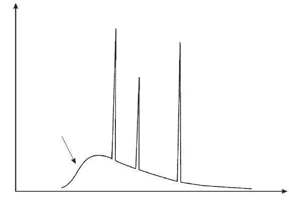
vacancy in a lower electron shell (characteristic X-ray lines) (Figure
3–21).
The X-ray continuum is made up of a continuous distribution of
intensity as a function of energy whereas the characteristic spectrum
represents a series of peaks of variable intensity at discrete element-
specifi c energies. As the electron energy increases the intensity of the
continuous spectrum also increases and the maximum of the distribu-
tion is shifted toward higher energies. The general appearance of the
continuous spectrum is independent of the atomic number of the speci-
men, however, the absolute intensity values are dependent on the
atomic number. The maximum possible energy E
X
is given by the elec-
tron energy E
0
, which corresponds to instantaneous stopping of an
electron at a single collision (Duane-Hunt limit). According to Kramers

Chapter 3 Scanning Electron Microscopy 179
(1923) the intensity of the continuous spectrum I
c
emitted in an energy
interval with the width dE
X
is given as
I
C
(E
X
)dE
X
= kZ(E
0
− E
X
)/E
X
⋅ dE
X
(2.37)
k represents the Kramers constant, which varies slightly with the
atomic number (Reimer, 1985). A detailed treatment of the continuous
X-ray emission is given by Stephenson (1957).
The characteristic X-ray spectrum consisting of peaks at discrete
energies is superimposed on the continuous X-ray spectrum (cf. Figure
3–21). Their positions are independent of the energy of the incident
electrons. The peaks occur only if the corresponding atomic energy
level is excited. The generation of characteristic X-rays consists of three
different steps. First, a beam electron interacts with an inner shell
electron of an atom and ejects this inner shell electron leaving that
atom in an excited state, i.e., with a vacancy on the electron shell.
Second, subsequently the excited atom relaxes to the ground state by
transition of an electron from an outer to an inner shell vacancy. The
energy difference ∆E
ch
between the involved shells is characteristic for
the atomic number. Third, this element-specifi c energy difference is
expressed either by the emission of an electron of an outer shell with
a characteristic energy (Auger electron) or by the emission of a char-
acteristic X-ray with energy E
X
= ∆E
ch
. The fraction of characteristic X-
rays emitted when an electron transition occurs is given by the
fl uorescence yield ω. This quantity increases with the atomic number
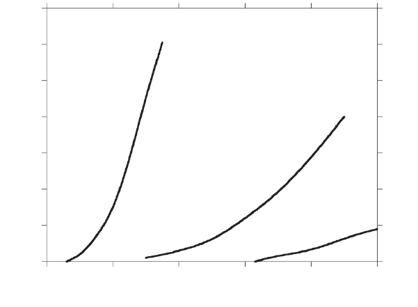
and depends on the inner electron shell involved (Figure 3–22). The
complement, 1 − ω represents the Auger electron yield, which gives the
corresponding fraction of Auger electrons produced. The fl uorescence
yield for the different shells and subshells can be calculated (for details
see Bambynek et al., 1972).
Characteristic
X-ray spectrum
X-ray
continuum
Intensity
E
x
Figure 3–21. Schematic representation of the X-ray spectrum emitted from a
specimen bombarded with fast electrons.

180 R. Reichelt
Moseley studied the line spectra in detail and found that the general
appearance of the X-ray spectrum is the same for all elements. The
energy of a characteristic X-ray line depends on the atomic shells
involved in the transition resulting in the emission of this line. The X-
ray lines can be classifi ed in series according to the shell where the
ionization took place, e.g., K-, L-, M-shell, etc. The quantum energies of
a series are given by Moseley’s law
E
X
= A(Z − B)
2
(2.38)
where A and B are parameters that depend on the series to which the
line belongs. The characteristic X-ray energy E
x
is denoted by symbols
that identify the transition that produced it. The fi rst letter, e.g., K, L,
identifi es the original excited level, whereas the second letter, e.g., α,
β, designates the type of transition occurring. For example, K
α
denotes
the excitation energy between the K- and L-shell, whereas K
β
denotes
the excitation energy between the K- and M-shell. Transitions between
subshells are designated by a number, e.g., a transition from the sub-
shell L
III
to K is denoted as K
α1
and from the subshell L
II
to K is denoted
as K
α2
, respectively. The transition from the subshell L
I
to K is forbid-
den. The characteristic X-ray energies and X-ray atomic energy levels
for the K-, L-, and M-shells are listed in tables (Bearden, 1967a,b). For-
tunately, the atomic energy levels are not strongly infl uenced by the
type and strength of the chemical bonds. However, chemical effects on
X-ray emission are observed for transitions from the valence electron
states, which are involved in chemical bonds. In such cases the narrow
lines show changes of their shape and their position (energy shift
<1 eV) as well (Baun, 1969).
0.7
K-shell
Fluorescence yield ω
1 - ω
L-shell
M-shell
0.6
0.5
0.4
0.3
0.2
0.1
0.4
0.3
0.5
0.6
0.7
0.8
0.9
1.0
0
020
Atomic number
40 60 80 100
Figure 3–22. Dependence of the X-ray fl uorescence yield ω and its comple-
ment (1 − ω) of the K-, L-, and M-shell from the atomic number. The comple-
ment (1 − ω) corresponds to the Auger electron yield.
Chapter 3 Scanning Electron Microscopy 181
2.2.6 Auger Electrons
As mentioned in Section 2.2.5, when an excited atom relaxes to the
ground state by transition of an electron from an outer to an inner shell
the energy difference ∆E
ch
between the involved shells can be expressed
by the emission of an electron of an outer shell with a characteristic
energy E
AE
. The emission is due to the Auger effect (Burhop, 1952;
Åberg and Howart, 1982) and the emitted electron is designated as an
Auger electron (AE). Its energy E
AE
is given by
E
AE
= ∆E
ch
− E
ionr
(2.39)
where the term E
ionr
contains the ionization energy of the AE emitting
outer shell and also considers relaxation effects. The shape and the
position of the AE peaks are infl uenced by the type and strength of
the chemical bonds (Madden, 1981). The AE peaks have an energy
width of a few electronvolts and are superimposed on the low-energy
range of the BSE spectrum up to energies of about 2.5 keV (Figure 3–12).
The identifi cation of the AE peaks on the BSE background (Bishop,
1984) can be improved by differentiation of the electron energy
spectrum.
The Auger electrons are generated within the excitation volume
(Figure 3–14). Due to their low energies only AE with a short pathway
to the specimen surface can escape. However, energy losses caused by
inelastic scattering on the pathway to the surface remove AE from the
AE peaks. The decrease of the AE peak is proportional to exp(−x/Λ
AE
),
where x denotes the length of the path inside the specimen and Λ
AE
the mean free path of the AE. Depending on their energy and the
atomic number of the specimen the mean free path Λ
AE
can have values
in the range of about 0.4 nm to a few nanometers (Palmberg, 1973; Seah
and Dench, 1979). If AEs are inelastically scattered on their path to the
surface, then they cannot be identifi ed as an AE in the BSE background.
Therefore, only atoms within a depth of about Λ
AE
can contribute to
the AE peaks. Since Auger electrons yield information on element
concentrations very near the surface, the specimen must be in an ultra-
high vacuum environment and special sample preparations are
required (e.g., ion sputtering in situ, cleavage in situ) to obtain clean
surfaces.
Because the AE yield of the K-shell 1 − ω
K
is much larger than ω
K
for
light elements (cf. Figure 3–22) AE spectroscopy is advantageous for
elements with atomic number Z = 4(ω
K
= 4.5 × 10
−4
) up to Z ∼ 30(ω
K
=
4.8 × 10
−1
) (Bauer and Telieps, 1988). Similar to SE, which are also
emitted only from a very thin surface layer, the AE can be generated
directly by the beam electrons and by BSE within a larger circular
area around the beam impact point (cf. Figure 3–14). Like the SE
yield, which increases with increasing angle of incidence θ according
to δ(θ) = δ
0
/cos θ [cf. Eq. (2.31)], the integral AE peak intensity is
proportional to 1/cos θ (Bishop and Riviere, 1969; Kirschner, 1976;
Reimer, 1985).
Recent developments in scanning Auger microscopy and AE spec-
troscopy are described by Jacka (2001).
182 R. Reichelt
2.2.7 Others
The incident electron beam bombards the specimen with electrons
thereby introducing a negative electric charge. A certain amount of
negative electric charge is leaving the specimen as secondary (I
SE
),
backscattered (I
BSE
) and Auger electrons (I
AE
). To avoid an accumulation
of charges a specimen current I
sp
must fl ow from the specimen to the
ground. The conservation equation for the electric charge is
I
p
= I
SE
+ I
BSE
+ I
AE
+ I
sp
(2.40)
where I
p
is the probe current. The specimen current changes the sign
when I
SE
+ I
BSE
+ I
AE
> I
p
. Because I
SE
+ I
BSE
>> I
AE
this means basically
that δ + η > 1 (cf. Figure 3–16). I
sp
depends on the angle of beam inci-
dence θ and the electron energy E
0
as expected from δ(θ, E
0
) and η(θ,
E
0
) (Reimer, 1985). The resolution of specimen current images is com-
parable to that of BSE images. One advantage of the specimen current
mode is that the contrast is independent on the detector position. A
critical review of this mode was published by Newbury (1976).
The electron–specimen interaction also generates acoustic waves. The
frequencies of these waves depend on the imaging conditions of the
SEM and the specimen studied. They cover a range from low sound up
to very high ultrasound frequencies. The electron acoustic mode, usually
denoted as scanning electron acoustic micro scopy (SEAM), was intro-
duced by Brandis and Rosencwaig (1980) and Cargill (1980). SEAM uses
a periodic beam modulation or short electron beam pulses that allow for
analysis of the SEAM frequency response (Balk and Kultscher, 1984;
Balk, 1986; Kultscher and Balk, 1986). The SEAM was reviewed by Balk
(1989) and applications of this method in semiconductor research
(Balk, 1989) and for the investigation of magnetic structures were
shown (Balk et al., 1984).
2.3 Contrast Formation and Resolution
Since the image formation is due to the image signal fl uctuation ∆S
from one point to another point, the contrast C is designated as in
television to be
C = (S − S
av
)/S = ∆S/S (2.41)
S
av
is the average value of the signal and S represents the signal of the
considered point (S > S
av
, i.e., C is always positive). The signal fl uctua-
tion may be caused by local differences in the specimen topography,
composition, lattice orientation, surface potential, magnetic or electric
domains, and electrical conductivity. The minimum contrast is obtained
if S = S
av
, whereas the maximum contrast is obtained for S
sv
= 0. This
is the case, e.g., when the signal S from a feature is surrounded by a
background with S
av
= 0. The contrast will be visible if C exceeds the
threshold value of about 5 × 10
−2
.
According to the point-resolution criterion two image points sepa-
rated by some horizontal distance (i.e., within the x–y plane perpen-
dicular to the optical axis) are resolved when the minimum intensity
Chapter 3 Scanning Electron Microscopy 183
between them is 75% or less of the maximum intensity. The point reso-
lution therefore corresponds to the minimum distance of two object
points, those superimposed image intensity distributions drop to 75%
of their maximum intensity between them. Due to the inherent noise
of each signal of the SEM characterized by its signal-to-noise ratio
(SNR) the drop to 75% of the maximum intensity will not be reliably
defi ned at the minimum distance. Consequently, at low SNR two image
points can be resolved only if their distance is larger than the minimum
distance reliably defi ned for noiseless signals.
As opposed to the light or transmission electron microscope the
resolution of the SEM cannot be defi ned by Rayleigh’s criterion. The
resolution obtained in the SEM image depends in a complex manner
on the electron beam diameter, the electron energy, the electron–
specimen interaction, the selected signal, the detection, as well as the
electronic amplifi cation and electronic processing. An object “point”
corresponds to the size of a small local excitation volume (cf. Figures
3–13 and 3–14) designated as the spatial detection limit from which a
suffi cient signal can be obtained. Obviously, the point resolution cannot
be less than the spatial detection limit. It becomes clear from Figure
3–14 that the spatial resolution of an SEM is different for each signal
since the size of the signal emitting volume as well as the signal inten-
sity depends on the type of signal selected.
The important “quality parameters” such as spatial resolution, astig-
matism, and SNR of SEM images, as well as drift and other instabilities
that occur during imaging, can be determined most reliably and objec-
tively by Fourier analysis of the recorded micrographs (Frank et al.,
1970; Reimer, 1985). Recently, a program SMART (Scanning Micro-
scope Analysis and Resolution Testing) became freely available, which
allows the SEM resolution and the imaging performance to be mea-
sured in an automated manner (Joy, 2002). It should be mentioned in
the context of resolution that the ultimate resolution of an SEM speci-
fi ed by the manufacturers is determined by imaging a test sample (gold
or gold–palladium-coated substrate with a low atomic number).
However, such samples are quite atypical compared to those usually
investigated, thus the resolution determined in this way is not properly
representative of the routine performance of the SEM. At present, con-
ventional SEMs using a heated tungsten or lanthanum hexaboride
emitter as the electron source have a specifi ed resolution in the range
of 3–5 nm at an acceleration voltage of 30 kV.
2.3.1 Topographic Contrast
Presumably the SEM is most frequently used to visualize the topo-
graphy of three-dimensional objects. The specimen topography gives
rise to a marked topographic contrast obtained in secondary and back-
scattered images. This contrast has a complex origin and is formed in
SE images by the following mechanisms:
1. Dependence of the SE yield δ on the angle of incidence θ of the
electron beam at the local surface element [cf. Eq. (2.31)]. The tilt angle
of the local surface elements is given by the topography of the sample.
184 R. Reichelt
2. Dependence of the detected signal on the angular orientation of
the local surface element related to the ET detector (see Section 2.1.3).
SE generated “behind” local elevations, in holes, in fi ssures, or in cavi-
ties reach the ET detector incomplete. This causes a more or less pro-
nounced shadow contrast (cf. Figure 3–7a).
3. Increase of the SE signal when diffusely scattered electrons pass
through an increased surface area. This is the case at edges or at pro-
truding surface features, which are smaller than the excitation volume.
Electron diffusion leads to overbrightening of edges and small surface
protrusions in the micrograph and is known as an edge effect.
4. Charging artifacts with objects of low electric conductivity.
Contributions (1) to (3) are illustrated by SE micrographs of different
specimens shown in Figures 3–23a and 3–24a as well as schematically
by profi les of the topography and the related SE signals in Figure 3–25.
In these fi gures the direction to the ET detector is indicated. The ball
in Figure 3–23a shows a contrast, which is mainly due to the varying
angle θ of beam incidence across the ball [cf. (1) above] and the angular
orientation of the local surface elements related to the ETD [cf. (2)
above]. The collection effi ciency of the ETD is signifi cantly higher for
surface elements facing the detector than for those on the back (shadow
region). Whereas the intensity of emitted secondary electrons of the
ball reveals radial symmetry, the effect of detection geometry causes
the nonradial symmetric image intensity distribution of the ball (cf.
Figure 3–25a–c). The rim of the ball is bright in the SE image because
of the enhanced SE emission due to an incidence angle θ ≈ 90° and the
effect of diffusely scattered electrons passing through an increased
surface area [cf. (3) above]. The radius of the ball is larger than the
electron range R (cf. Figure 3–14) therefore the latter effect occurs just
near the rim of the ball. If the mean radius of ball-like particles becomes
comparable or smaller than the electron range, diffusely scattered elec-
trons generate more SE over the whole particle surface, thus the SE
emission typically is distinctly enhanced (small particles are marked
by small arrowheads in Figures 3–23a and 3–24a). If the shadow con-
trast is visible in the image and the direction toward the ETD is known
then elevations and depressions clearly can be readily identifi ed (cf.
Figure 3–23a). Another way to distinguish elevations and depressions
is to record and to analyze SE stereopairs.
The SE micrograph of large crystal-like particles (Figure 3–24a) basi-
cally shows the same contrast mechanisms as discussed above but with
a more complex structured sample than the ball. The individual fl at
surface planes of the crystal-like particles occur with almost constant
brightness because of the constant angle of beam incidence and the
constant detection geometry (provided that there is no shadow effect
from other large particles). Some surface planes possess fi ssures of
different size, which typically appear rather dark because just a minor
fraction of the generated SE can escape from inside the fi ssures. In such
cases SE can be extracted either by a positively biased grid in front of
the specimen (Hindermann and Davis, 1974) or by a superimposed
magnetic fi eld in which the SE follow spiral trajectories around the
