Hawkes P.W., Spence J.C.H. (Eds.) Science of Microscopy. V.1 and 2
Подождите немного. Документ загружается.


Chapter 3 Scanning Electron Microscopy 235
Figure 3–56. Stereo pair of SE micrographs (a and b) of the hydrogel poly-(N-isopropylacrylamide)
(PNIPAAm) in the swollen state recorded at 2 keV with the “in-lens” FESEM. The specimen was rapidly
frozen, freeze dried, and ultrathin rotary shadowed with platinum/carbon (for details see Matzelle et
al., 2002). (c) Red–green stereo anaglyph prepared from (a and b). The tilt axis has a vertical direction.
(d) Red–green stereo anaglyph in a “bird view.” (For parts c and d, see color plate.)
236 R. Reichelt
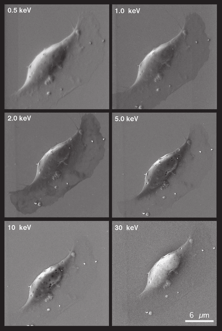
Figure 3–57. Secondary electron micrograph series of increasing electron energies from 0.5 to 30 keV
from a keratinocyte. The micrographs are recorded with an “in-lens” FESEM. The image contrast
varies signifi cantly with the electron energy. Inhomogeneities in the “leading edge” of the keratino-
cyte, which has a thickness of about 200–400 nm, are most clearly visible at 2 keV. (Micrographs kindly
provided by Dr. R. Wepf, Beiersdorf AG, Hamburg, Germany.)
Chapter 3 Scanning Electron Microscopy 237
characterization of polymers (Berry, 1988; Butler et al., 1995; Brown and
Butler, 1997; Sawyer and Grubb, 1996).
4 Scanning Electron Microscopy at Elevated Pressure
The scanning electron microscopic investigation of specimens must
meet several requirements, which were mentioned in previous sec-
tions. To sum it up, it can be said that specimens (1) have to be compat-
ible with the low pressure in the specimen chamber (∼10
−3
Pa in
conventional SEM and 10
−5
–10
−4
Pa in fi eld emission SEM), (2) have to
be clean, i.e., the region of interest has to give free access to the primary
beam, (3) need suffi cient electrical conductivity, (4) need to be resistant
to some extent to electron radiation, and (5) have to provide a suffi cient
contrast. In a narrower sense, only metals, alloys, and metallic com-
pounds fulfi ll those requirements. Numerous preparation procedures
mentioned in Section 2.4 were developed in the past and are still in the
process of improvement, to provide a suffi cient electrical conductivity
to nonconductive specimens, to remove the water in samples, and to
replace it or to rapidly freeze it in a structure-conserving manner.
Nevertheless, there was and still is enormous interest in investigating
specimens in their genuine state.
Thirty years ago Robinson (1975) proposed examining any uncoated
insulating specimen in the SEM at high accelerating voltages in the
specimen chamber, which had been modifi ed to contain a small residual
water vapor environment. It appeared that the presence of the water
vapor suffi ciently reduced the resistance of the insulator so that no
charging effects were detected in backscattered electron micrographs.
Danilatos (1980) developed an “atmospheric scanning electron micro-
scope” (ASEM), which later was called an “environmental scanning
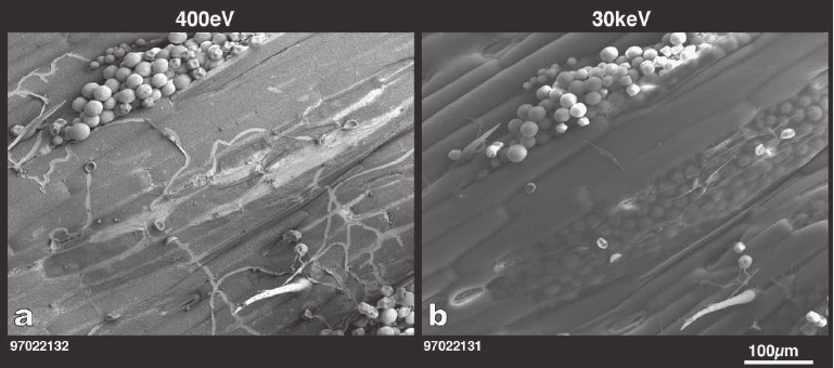
Figure 3–58. Secondary electron micrograph pair of the cuticula of a leaf recorded at electron energies
of 0.4 (a) and 30 keV (b) with an “in-lens” SEM. The low-energy image contains information only from
the surface whereas the 30-keV image also reveals information about structural features below the
surface, e.g., new spores, which are not visible in (a). (Micrographs kindly provided by Dr. R. Wepf,
Beiersdorf AG, Hamburg, Germany.)
238 R. Reichelt
electron microscope” (ESEM®) (Danilatos, 1981) and is now a registered
trademark. To enable the investigation of water and water-containing
specimens in their native state at stationary conditions a minimum pres-
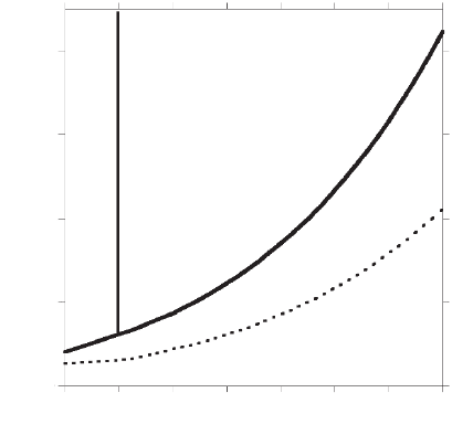
sure of water vapor of about 612 Pa is required at 0°C (cf. Figure 3–59).
Stationary conditions in the specimen chamber of the SEM can be
accomplished by controlling the water vapor pressure p in close vicinity
of the specimen as well as the specimen temperature T such that the p–T
values always correspond to points on the solid p–T graph in Figure 3–
59. For example, at 20°C a water vapor pressure as large as about 2330 Pa
is required for stationary conditions. p–T values below the solid graph,
e.g., 300 Pa at 0°C (Figure 3–59), corresponds to a relative humidity of
less than 100%, thus representing nonstationary conditions.
How can stationary conditions be reached during imaging of a wet
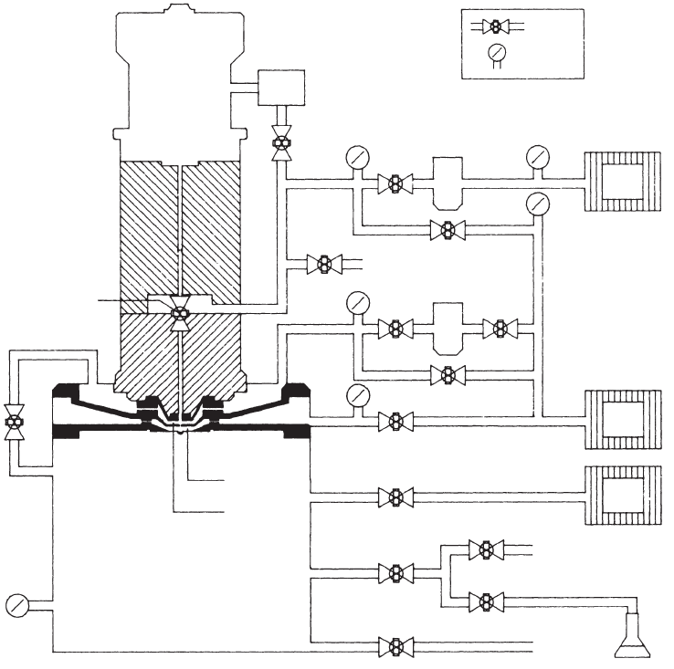
sample in the specimen chamber of an SEM? Figure 3–60 shows the
cross section of the ESEM, which permits investigations at pressures
suffi cient for stationary conditions. Basically, the electron beam pro-
pagates in the column as in a conventional SEM until it reaches the fi nal
aperture. Then, since the pressure increases gradually as the electrons
proceed toward the specimen, the electrons undergo signifi cant scatter-
ing on gas molecules until they reach the specimen surface.
The electron–gas interaction is discussed in detail by Danilatos
(1988). According to this study the average number of scattering events
per electron n can be approximated by
n = σ
g
p
g
L/kT (4.1)
where σ
g
represents the total scattering cross section of the gas molecule
for electrons, L is the electron path length in gas, and k is the Boltzmann
constant. These approximations hold for Λ >> L, where Λ represents the
4000
3000
Solid
Liquid
Vapor
2000
p [Pa]
1000
0
–5 10
T [°C]
15 20 25 3005
Figure 3–59. Phase diagram of water. Solid line, 100% relative humidity (satu-
rated vapor conditions); dashed line, 50% relative humidity. (Data from Lax,
1967.)
Chapter 3 Scanning Electron Microscopy 239
Figure 3–60. Schematic cross section of the fi rst commercial Electroscan Environmental SEM (ESEM
®
)
showing the vacuum and pumping system. Two pressure-limiting apertures separate the electron
optical column from the specimen chamber. Differential pumping of the stage above and between the
two pressure-limiting apertures ensures the separation of high vacuum in the column from low vacuum
in the specimen chamber. The differential pumping of two stages and optimum arrangement of the
pressure-limiting apertures can work successfully to achieve pressures up to 10
5
Pa in the specimen
chamber. [From Danilatos, 1991; with kind permission of Blackwell Publishing Ltd., Oxford, U.K.]
GUN
CHAMBER
ION
PUMP
manual valve
Valve
Gauge
RP1
RP2
RP3
G2
G4
G1
G3
G5
G7
SPECIMEN CHAMBER
EC2
EC1
V7
V8
V9
regulator
valve
V10
V11
VENT
AUX IL IARY GAS
WATER VAPOR
V13
V2
V3
V4V5
VENT
V12
V1
V6
DIF
1
DIF
2
mean free path of a beam electron in the gas. According to Eq. (4.1) the
average number of collisions increases linearly with the gas pressure p
g
and the path length in the specimen chamber. Furthermore, n depends
via the scattering cross section on the type of gas molecules and on the
temperature. When the beam electrons start to be scattered by the gas
molecules, the fraction of scattered electrons is removed from the focused
beam and hit the specimen somewhere in a large area around the point
of incidence of the focused beam. The scattered electrons form a “skirt”
around the focused beam, which has a radius of 100 µm for a pathlength
of 5 mm (conditions: E
0
= 10 keV, water vapor pressure = 10
3
Pa) (Danila-
tos, 1988). Using a phosphor imaging plate, the distribution of unscat-
240 R. Reichelt
tered beam electrons and the scattered “skirt” electrons was directly
imaged by exposure to the electron beam for a specifi ed time (Wight and
Zeissler, 2000). Related to the electron beam intensity within 25 µm, the
“skirt” intensity as a function of the distance from the center drops to
15% at 100 µm, 5% at 200 µm, and 1% at 500 µm (conditions: E
0
= 20 keV,
water vapor pressure = 266 Pa, l = 10 mm) (Wight and Zeissler, 2000). The
signals generated by the electrons of the skirt originate from a large area,
which contributes to the background, whereas the unscattered beam
remains focused to a small spot on the specimen surface, although its
intensity is reduced by the fraction of electrons removed by scattering.
The resolution obtainable depends on the beam diameter and the size of
the interaction volume in the specimen, which is analogous to the situa-
tion in conventional and high-resolution SEM, i.e., the resolving power
of ESEM can be maintained in the presence of gas.
The detection of BSE, CL, and X-rays is to a great extent analogous to
the detection in a conventional SEM, because these signals can pene-
trate the gas suffi ciently (Danilatos, 1985, 1986). However, the situation
is completely different for the detection of SE. The conventional Ever-
hart–Thornley detector would break down at elevated pressure in the
specimen chamber. However, the gas itself can be used as an amplifi er
in a fashion similar to that used in ionization chambers and gas propor-
tional counters. An attractive positive voltage on a detector will make
all the secondary electrons drift toward it. If the attractive fi eld is suffi -
ciently large, each drifting electron will be accelerated, thus gaining
enough energy to cause ionization of gas molecules, which can create
more than one electron. This process repeating itself results in a signifi -
cant avalanche amplifi cation of the secondary electron current, which
arrives at the central electrode of the environmental secondary electron
detector (ESD) (Danilatos, 1988). The avalanche amplifi cation works
best only in a limited pressure range and can amplify the SE signal up
to three orders of magnitude (Thiel et al., 1997). Too high pressure in the
specimen chamber makes the mean free path of the electrons very
small and a high electric fi eld between specimen and detector is required
to accelerate them suffi ciently. Too low pressure in the chamber results
in a large mean free electron path, i.e., only a few ionization events take
place along the electron path from the specimen to the detector, thus the
avalanche amplifi cation factor is low. The new generation of ESD, the
gaseous secondary electron detector (GSED), which consists of a 3-mm-
diameter metallic ring placed above the specimen, provides better dis-
crimination against parasitic electron signals. Both the ESD and GSED
are patented and are available only in the ESEM.
However, the ionization of gas molecules creates not only electrons
but also ions and gaseous scintillation. The latter can be used to make
images (Danilatos, 1986), i.e., in that case the imaging gas acts as a
detector. This principle is used in the patented variable pressure sec-
ondary electron (VPSE) detector. Nonconductive samples attract posi-
tive gas ions to their surface as negative charge accumulates from the
electron beam, thus effectively suppressing or at least strongly reduc-
ing charging artifacts (Cazaux, 2004; Ji et al., 2005; Tang and Joy, 2003;
Thiel et al., 2004; Robertson et al., 2004). The gas ions can affect or even
reverse the contrast in the GSED image under specifi c conditions, e.g.,
Chapter 3 Scanning Electron Microscopy 241
at specimen regions of enhanced electron emission, where the rate of
electron–ion pairs increases (Thiel et al., 1997). The highly mobile elec-
trons generated by electron–gas interaction are removed from the gas
by rapid sweeping to the GSED, which in turn causes an increased
concentration of positive ions during image acquisition due to different
electric fi eld-induced drift velocities of negative and positive charge
carriers in the imaging gas (Toth and Phillips, 2000).
However, imaging of wet, soft specimens can be hampered by the
effect of surface tension (Kellenberger and Kistler, 1979), which may
fl atten and hereby deform the specimen. Obviously, this is a mislead-
ing situation demonstrating that “environmental conditions” do not
necessarily guarantee structural preservation.
As mentioned above, about 612 Pa is the crucial minimum pressure for
wet specimens. In addition to the ESEM, which enables imaging with SE
at pressures up to about 6500 Pa, numerous variable pressure SEM
(VPSEM), high pressure SEM, and low vacuum SEM (sometimes the
abbreviation LVSEM is used, which cannot be distinguished from the
low-voltage SEM) became commercially available. The water vapor pres-
sure in the specimen chamber of those SEM is typically at maximum
300 Pa, i.e., below the crucial value of 612 Pa, which is not suffi cient for
imaging of wet specimens at stationary conditions. To separate the speci-
men pressure of maximum 300 Pa from the high vacuum in the column
only one pressure-limiting aperture is suffi cient. For imaging at pressures
in the range from 250 to 300 Pa backscattered electrons are utilized.
Very recently, Thiberge et al. (2004) demonstrated scanning electron
microscopy of cells and tissues under fully hydrated atmospheric condi-
tions using a small chamber with a polyimide membrane (145 nm in thick-
ness) that is transparent to beam and backscattered electrons. The
membrane protects the fully hydrated sample from the vacuum. BSE
imaging at acceleration voltages in the range of 12–30 kV revealed struc-
tures inside cultured cells and colloidal gold particles having diameters
of 20 and 40 nm, respectively. Another interesting experimental setup is
the habitat chamber designed to keep living cells under fully hydrated
atmospheric conditions as long as possible and to reduce the exposure time
to the lower pressure in the ESEM below 2 min (Cismak et al., 2003).
Scanning electron microscopy at elevated pressure is increasingly
used in very different fi elds. Apart from variations in the pressure and
chamber gas a heating stage (maximum temperature about 1500°C)
allows changes in the specimen temperature. For example, chemical
reactions such as corrosion of metals, electrolyte–solid interactions,
alloy formation, and the degradation of the space shuttle ceramic
shields by increasing oxygen partial pressures at high temperatures
are possible with micrometer resolution. The onset of chemical reac-
tions that depend on various parameters can by studied in detail.
Insulators, including oil and oily specimens, can be directly imaged.
Water can also be imaged directly in the ESEM, which allows studies
of wetting and drying surfaces (e.g., de la Parra, 1993; Stelmashenko et
al., 2001; Liukkonen, 1997) and direct visualization of the dynamic
behavior of a water meniscus (Schenk et al., 1998; Rossi et al., 2004).
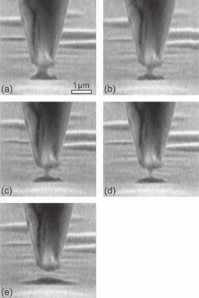
Figure 3–61 shows an example of dynamic studies of a water menis-
cus between the scanning tunneling tip and a support when the tip is
242 R. Reichelt
Figure 3–61. Time-resolved sequence of secondary electron images recorded with an ESEM®-E3
(ElectroScan Corp., Wilmington, MA). The water meniscus between the hydrophilic tungsten tip
(normal electron beam incidence) and the Pt/C-coated mica (incidence angle of 85°) is clearly visible
(a–d). Due to locally decreasing relative humidity the meniscus becomes gradually smaller until it
snaps off (e). The absence of the meniscus leads to a signifi cant change of shape of the water bead
below the tip [cf. (d) and (e)]. Some water drops are located on the sample in front of and behind the
tip. The sequence was recorded within 11 s and each image was acquired within about 2 s. Experimen-
tal conditions: E
0
= 30 keV, I
p
= 200 pA, p
g
= 1.2 kPa. (From Schenk et al., 1998; with kind permission of
the American Institute of Physics, Woodbury, NY.)
Chapter 3 Scanning Electron Microscopy 243
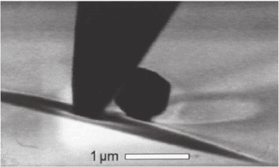
moved across the sample. The wetting of the tip indicates a hydrophilic
surface, whereas Figure 3–62 clearly indicates a hydrophobic tip
surface.
ESEM studies of the wettability alteration due to aging in crude
oil/brine/rock systems that are initially water wet are of signifi cant
importance in the petroleum industry in understanding the water
condensation behavior on freshly exposed core chips. Surface active
compounds are rapidly removed from the migrating petroleum, thus
changing the wettability and subsequently allowing larger hydropho-
bic molecules to sorb (Bennett et al., 2004; Kowalewski et al., 2003;
Robin, 2001). Furthermore, the ESEM is a powerful tool with which
study the infl uence of salt, alcohol, and alkali on the interfacial activity
of novel polymeric surfactants that exhibit excellent surface activity
due to their unique structure (Cao and Li, 2002).
Environmental scanning electron microscopy disseminates rapidly
among scientifi c and engineering disciplines. Applications range
widely over diverse technologies such as pharmaceutical formulations,
personal care and household products, paper fi bers and coatings,
cement-based materials, boron particle combustion, hydrogen sulfi de
corrosion of Ni–Fe, micromechanical fabrication, stone preservation,
and biodeterioration. In spite of the broad applications, numerous con-
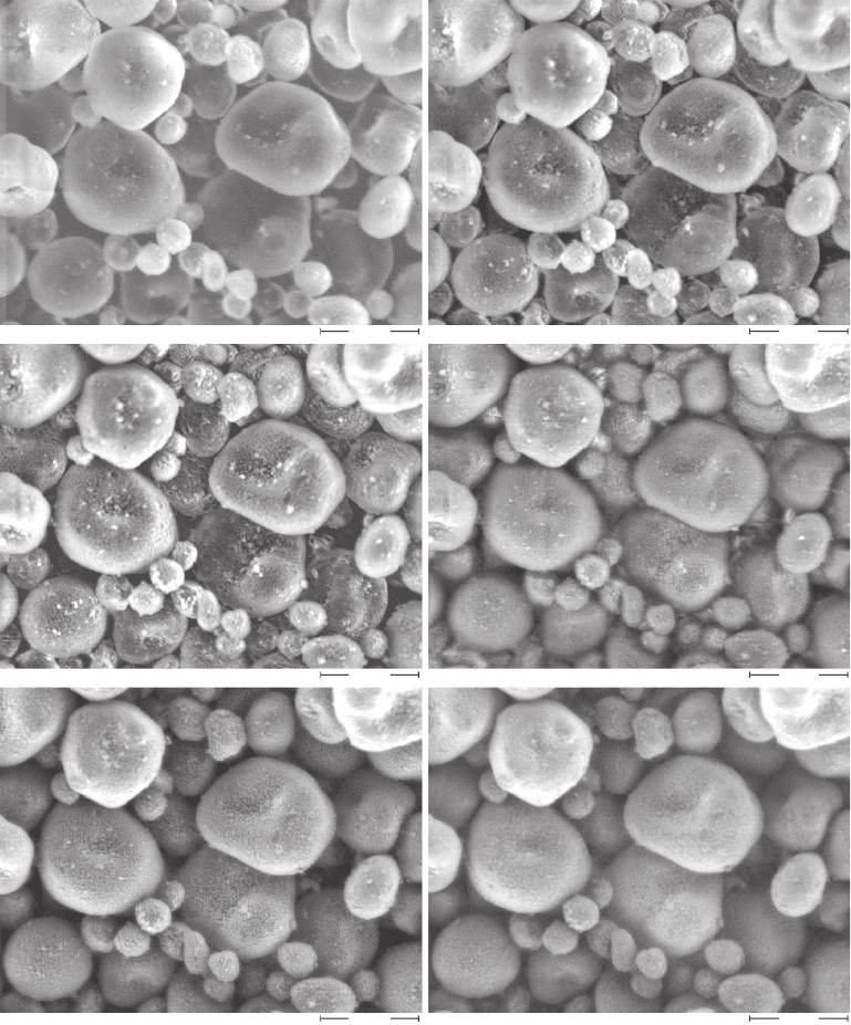
trast phenomena are not fully understood as yet. This is illustrated in
Figure 3–63 by a series of SE micrographs recorded at different electron
energies, but otherwise identical conditions. In addition, ESEM inves-
tigations of polymeric and bio logical specimens, which are known
from conventional electron microscopy to be highly irradiation sensi-
tive, are more diffi cult because water acts as a source of small, highly
mobile free radicals, which accelerate specimen degradation (Kitching
and Donald, 1996; Royall et al., 2001).
Figure 3–62. Secondary electron image recorded with an ESEM-E3 (Elec-
troScan Corp., Wilmington, MA) from a hydrophobic tungsten tip (normal
electron beam incidence) and a water bead on Pt/C-coated mica (incidence
angle of 85°). The shape of the deformed water surface in the sub micrometer
vicinity of the tip clearly indicates its hydrophobic surface. The spherical object
(black) at the right of the tip in the back is probably a polystyrene sphere and
any resemblance is purely coincidental. (From Schenk et al., 1998; with kind
permission of the American Institute of Physics, Woodbury, NY.)
244 R. Reichelt
Figure 3–63. Secondary electron micrograph series of the starch glycolys D recorded at electron ener-
gies from 30 keV down to 5 keV (see the individual legends below each micrograph) with an ESEM
demonstrating the effect of the electron energy on image contrast. (Micrographs kindly provided by
Fraunhofer-Institut für Werkstoffmechanik, Halle, Germany; the project was supported by the State
Sachsen-Anhalt, FKZ 3075A/0029B.)
Glycolys D 30 kV 500x 15 mm 3Torr
Glycolys D 20 kV 500x 15 mm 3Torr
60 µm
60 µm
60 µm
60 µm
60 µm
60 µm
Glycolys D 15 kV 500x 15 mm 3Torr
Glycolys D 10 kV 500x 15 mm 3Torr
Glycolys D 7.5 kV 500x 15 mm 3Torr
Glycolys D 5 kV 500x 15 mm 3Torr
