Czichos H., Saito T., Smith L.E. (Eds.) Handbook of Metrology and Testing
Подождите немного. Документ загружается.

918 Part D Materials Performance Testing
16.3.3 Detection of the Damage
of Composites on the Mesoscale:
Application Examples
Putting aside the mechanical interpretation of compos-
ite damage in polymers, is it possible to characterize the
damage of these materials based on a single method that
combines the physical and chemical interpretations?
The answer must evidently be no. The problems calls
for nondestructive testing methods, which include ul-
trasonics, x-rays, thermography, and others methods. At
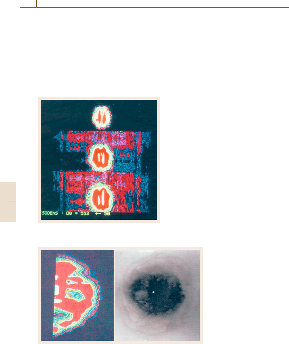
Fig. 16.58 Three cross sections of the specimen after im-
pact
Fig. 16.59 Comparison of radiogra-
phy (right) and tomography (left)of
delamination
the microscopic level, an electron or acoustic micro-
scope is the required nondestructive evaluation tool.
Between the macroscopic and microscopic levels,
there exists a very important middle domain, typical
of composite materials, which is treated at the meso-
scopic level: that of transverse cracking, distribution of
reinforcements and of porosity, that is, the domain of
sequential cells. It is very difficult today to characterize
defects at these different levels other than by radiogra-
phy.
The various applications of x-ray tomography al-
low for the study of composite materials at any level,
macroscopic, mesoscopic or microscopic. This instru-
ment is well adapted to reveal defects on a centimeter
scale, such as a delamination, but it is also capable
of revealing the distribution of the reinforcements on
a millimeter scale, and of detecting fibber cracking as
well as cracking along fibber matrix interfaces on a mi-
crometer scale. In addition, the attenuation of x-rays,
which depends on the physical and chemical nature of
the composites constituents, can be measured by x-rays
scanners.
Changes in the polymeric matrix due to aging, or
even the transformation of amorphous polymers into
crystallites, can be studied by x-ray tomography. This
astonishing tool, long used by the medical field, is
also well adapted to the study of composite mater-
ials and polymers due to its ability to detect defects
on a wide range of scales, as well as its ability of
physical and chemical analysis by measuring x-ray at-
tenuation.
X-ray scanning can thus provide information on the
physical and chemical aspects of the material and on
the geometry of flaws and damage on any scale. This
Part D 16.3
Performance Control 16.3 Computerized Tomography – Application to Organic Materials 919
tool provides solutions to many problems faced by the
materials scientist, as demonstrated in the following re-
sults.
Damage in a Carbon Epoxy Composite Plate
Specimen After an Impact
The specimen studied is a rectangular coupon whose di-
mensions are 255 × 172 × 8.5 mm. It is made up of 64
plies of carbon-epoxy fiber. The stacking sequence is
(45/90/ −45/0) 8 s.
The specimen was subjected to the impact of an
aluminum ball 12.7 mm in diameter moving at a speed
of 120 m/s. Then it was cycled in compression under
110 MPa to intensify the damage.
In this case, the scanner examination has been com-
plemented by radiography, zinc-iodide-enhanced x-ray,
and by optical micrography. This was done in collab-
oration with the NASA Langley Research Center in
Hampton, Virginia.
Global Examination of the Plain Specimen
A study on the axial slices and on the frontal slices
has been carried out. On the frontal views, we note
a strongly damaged area whose density is higher than
550 H, slightly elliptical shaped, and whose largest
dimension is orientated in the compression direc-
tion (Fig. 16.58).
Modeling of the Damage
The damage of this composite specimen is complex be-
cause of the great number of plies. The envelope of
the damaged area has the shape of a barrel, delamina-
tions being more numerous in the center of the plain
specimen than near the surface.
Synthesis of the Observations
Made on the Impacted Specimen
To facilitate the interpretation of observations made
with medical scanners, damage to the impact speci-
men is examined by radiography at a microscopic scale
and by scanning electronic microscopy at a microscopic
scale.
A comparison between radiography and tomogra-
phy is made (Fig. 16.59). We would like to note that the
scanner provided much better resolution of the outline
of the damaged area. Nevertheless, enhanced x-ray ra-
diography was required to reveal the very complicated
network of microcracks.
To conclude, we emphasize that the resolution of the
images of the microcracks with the medical scanner is
not satisfactory at the moment.
Damage in Rubber Submitted
to High Hydrostatic Pressure
It is well known that triaxiality of stresses including
a high hydrostatic pressure has a large effect on the me-
chanical behavior of rubbers and particularly on fatigue
mechanisms.
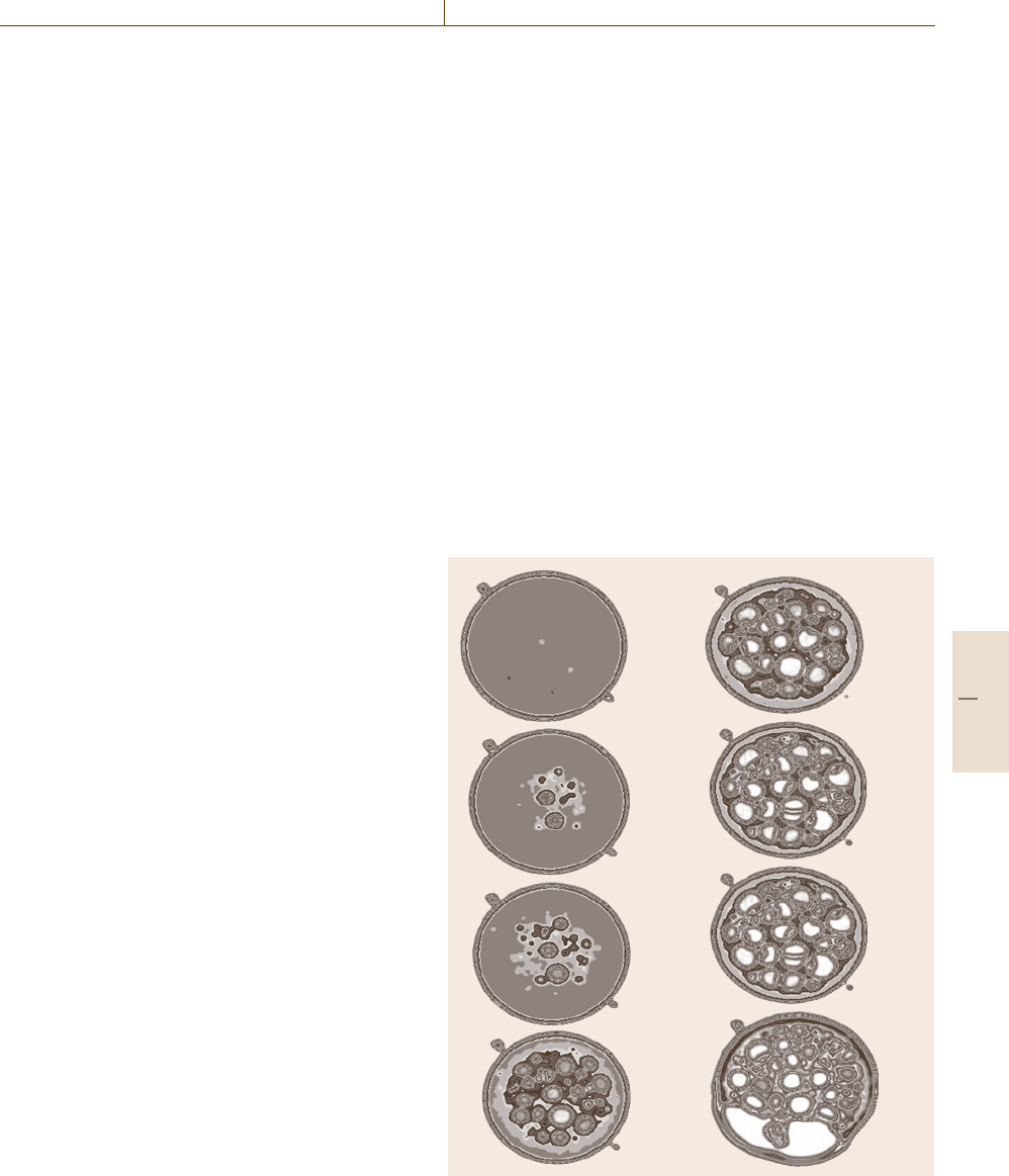
In a pancake specimen loaded in tension the hy-
drostatic pressure is about 2.5 × the shear modulus of
rubber for an elongation of 20% in NR. From observa-
tions by x-ray tomography, it seems that the first cavities
are formed in the center of the specimen at 20% of
elongation for a fracture at 380% (Fig. 16.60).
It must be pointed out that no other NDT method is
available to observe cavitation in rubber.
16.3.4 Observation of Elastomers
at the Nanoscale
The damage caused by fatigue and cracking in rubber
or in composites with elastomer matrices seems to be
considerably influenced by the transformation from an
15 %
20 %
28 %
60 %
140 %
200 %
290 %
320 %
Fig. 16.60 Cavitation growth due to high hydrostatic pressure (NR)
Part D 16.3
920 Part D Materials Performance Testing
amorphous to a crystalline phase, when this latter phase
exists. This hard-to-detect transformation is generally
studied by x-ray diffraction, a method by which we can-
not detect the gradients of transformation within the
rubber nor make local observations.
In contrast, x-ray tomography allows for the obser-
vation of the crystalline transformation of rubber and
its localization owing to the images generated by scan-
ner. The attenuation of the intensity of the x-ray beam,
similar to x-ray diffraction, reveals the existence of
crystallites.
In order to validate this hypothesis, test samples
of natural, crystallisable rubber NR and of synthetic,
noncrystallizable rubber (SBR) were studied at differ-
ent temperatures, knowing that low temperatures induce
a partial crystallization of NR.
a)
b)
DT mean value (pixel)
TD mean value (pixel)
Temperature (K)
Temperature (K)
3500 100 150 200 250 300
350
1180
1170
1175
1175
1175
1100
1200
1160
1150
1140
1130
1190
1120
50 125 200 275
NR_1
NR_2
SBR1
SBR2
Amorphous zone
Semicristal zone
1225
1212
1200
1187
1175
1162
1150
1137
1125
1112
1100
1087
1075
1062
1237
1050
Maximum crystallization
temperature
Transition zone
Glassy zone
z
y
x
Rubbery zone
Rubbery zone
T
g
Glassy zone
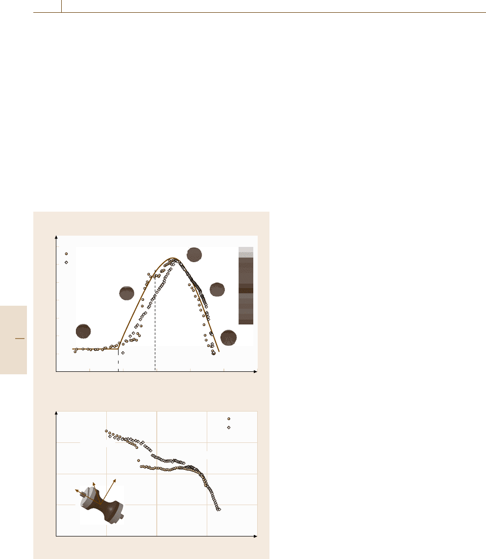
Fig. 16.61a,b Evolution of the TD depending on the temperature in
NR
(a) and SBR (b)
The crystallization of rubber (NR) may be ob-
served on the samples, which were cooled down to
−200
◦
C. This microscopic phenomenon can clearly
be evaluated by means of x-rays CT on the meso-
scopic scale, which is another benefit of medical
scanning. To show this effect, some of the tests were
carried out on the axisymmetric hourglass-shaped spec-
imens of NR and compared with SBR, which does
not show this phenomenon as it is always amor-
phous. Hourglass-shaped specimens are very suitable
for this test due to their large outer surface [16.32, 33].
So, the variation of mean values of the TD becomes
complex; the TD value is maximal at the level of
227 K (−46
◦
C) (Fig. 16.61), it exceeds 1180, whereas
at lower temperatures (83 K, −190
◦
C) the value is
1135.
This variation in TD displayed a crystallite state be-
tween 300 K (27
◦
C) and 227 K (−46
◦
C) above which
the amorphous state again becomes predominant be-
tween 227 K (−46
◦
C) and the transition temperature
(T
g
) of rubber. When there is no crystallization, varia-
tion in density certainly modifies the attenuation (TD).
In the case of SBR, which is always amorphous,
only the density varies with temperature, related to the
dilatation. An increasing in the temperature causes natu-
rally the increasing of the volume and the decreasing of
the density and finally the decreasing of the TD (pixel).
Figure 16.61b shows the evolution of the TD curve as
a function of the temperature for the SBR specimens. It
decreases from 1185 to 1120 in the temperature range
from 175 (−98
◦
C) to 300 K (27
◦
C). However, it shows
a transition zone at the level of 200 K (−73
◦
C), corre-
sponding to the transition temperature, T
g
, between the
glassy and rubbery phase. This zone is found between
1155 and 1160, corresponding to this glassy transfor-
mation.
16.3.5 Application Assessment of CT
with a Medical Scanner
The application examples show quite obviously that the
medical scanner is adapted to nondestructive evaluation
of composite materials from a technical point of view
and if we do not take into account the price of the de-
vice. The advantages of the method have been presented
with its excellent results. They confer on the medical
scanner a double role, which may form the subject of
two developments.
1. A nondestructive testing instrument with an indus-
trial purpose,
Part D 16.3

Performance Control 16.4 Computerized Tomography – Application to Inorganic Materials 921
2. An instrument comparable to the electronic micro-
scope with a more scientific purpose and able to give
information at a microscopic scale (several hun-
dreds of microns).
In the first case, the resolution power of the medical
scanner is good enough for a device of classical NDT.
We have found only one major inconvenience, namely
the lack of reliability of the scanner to make precise
measurements of the dimensions of the defects within
more than 25%.
In the second case, the resolution of the scan-
ner is not good enough to study the transverse cracks
in the matrix. We can estimate the spatial resolu-
tion of the scanner at some hundreds of micrometers,
whereas we would need 10 μm. We are dealing here
with an important need which needs more investi-
gation.
Nevertheless, we should point out that the reso-
lution in density of the scanner is of the order of
10
−3
, which makes it possible to resolve all the de-
fects contained by composites whether they are cracks,
delaminations, fiber bundles, or porosities. The varia-
tion of the Hounsfield density must be related at the
volumetric density but also to the modification of the
chemical microstructure of the materials. From this
point of view, the scanner enables studies of the crys-
tallization of polymeric matrix of rubbers, for example.
This means that x-ray tomography is a multiscale NDT
method.
16.4 Computerized Tomography – Application to Inorganic Materials
The study of volume properties as well as of dimen-
sional features with computed tomography requires
an optimized selection of source–detector combina-
tion depending on the material composition (energy-
dependent linear attenuation coefficient μ), the size
of the samples and the maximum thickness of ma-
terial that has to be irradiated, d. Additionally the
manipulator system and the mounting need to suf-
fice the required accuracy. The maximum SNR of
a CT measurement of a homogenous sample is given
for μd
∼
=
2, corresponding to a transmission of about
11% [16.40]. This value is valid only for line detec-
tors with a sufficient detector collimator and shielding
between single detector elements. Due to some lim-
itations, especially for flat detectors, the conditions
for optimum image quality differ from this theoretical
value.
For organic materials with low atomic numbers
and low density, medical scanners can be applied
in many cases. As an example fiber-reinforced heli-
copter blades have been investigated for many years
by computed tomography, using commercial medical
scanners [16.41]. Inorganic materials require radiation
sources with higher energy and appropriate detector
systems. The different kinds of computed tomography
equipment developed at Federal Institute for Materials
Research and Testing (BAM) represents the state of
the art on this field and are described in the following
knowing well that commercial solutions are in general
more efficient with regard to saving of time and user
guidance.
16.4.1 High-Energy CT
This field covers roughly the energy range given by
420 kV x-ray tubes over some radionuclide sources up
to electron linear accelerators with a maximum en-
ergy of 12 MeV. Most of the high-energy scanners used
have line detectors which can be shielded against the
scattered radiation. Starting with a translation/rotation
scanning principle and multidetector systems, today
line detectors are common with several hundred de-
tector elements and scanning times in the range of
some minutes and lower. At BAM a scanner for a 380-
kV x-ray tube and a
60
Co radionuclide source was
described as early as 1985 [16.42]. This universal scan-
ner was extended for measurements with a 12-MeV
electron linear accelerator (LINAC, Raytech 4000) in
combination with a multi-detector system with step-
motor-controlled collimator slits [16.43]. The actual
research covers the study of some effects of high-energy
cone beam CT with a LINAC and
60
Co using an a-
Si flat-panel detector (Perkin Elmer, 16-bit ADC, 256×
256 pixel, pixel size is 0.8mm
2
). Due to the focal spot
size of high-energy radiation sources, which is on the
order of about 1.5 mm, the spatial resolution is limited
to a few tenths of a mm.
16.4.2 High-Resolution CT
Using magnification techniques a much higher spa-
tial resolution can be reached. The condition hereby
is that the focal spot size can be lowered to the mi-
Part D 16.4
922 Part D Materials Performance Testing
crometer range, which is possible with microfocus x-ray
tubes. The usable energy range of commercial x-ray
tubes extends up to 225 kV. For many objects it would
be desirable to extend the energy range of microfocus
x-ray tubes. As an example most cellular metals are
made from aluminum and thus have a high penetration
depth for x-rays. However, they are often used to pro-
duce objects of irregular shapes (with an outer cover
of a different material or the foam skin itself). This
produces artefacts due to the high-attenuation parts. In
some applications foams of high-attenuation materials
(up to iron) are used. In these cases the choice of an
x-ray energy as high as possible reduces the artefacts
from beam hardening and exponential edge-gradient
effects.
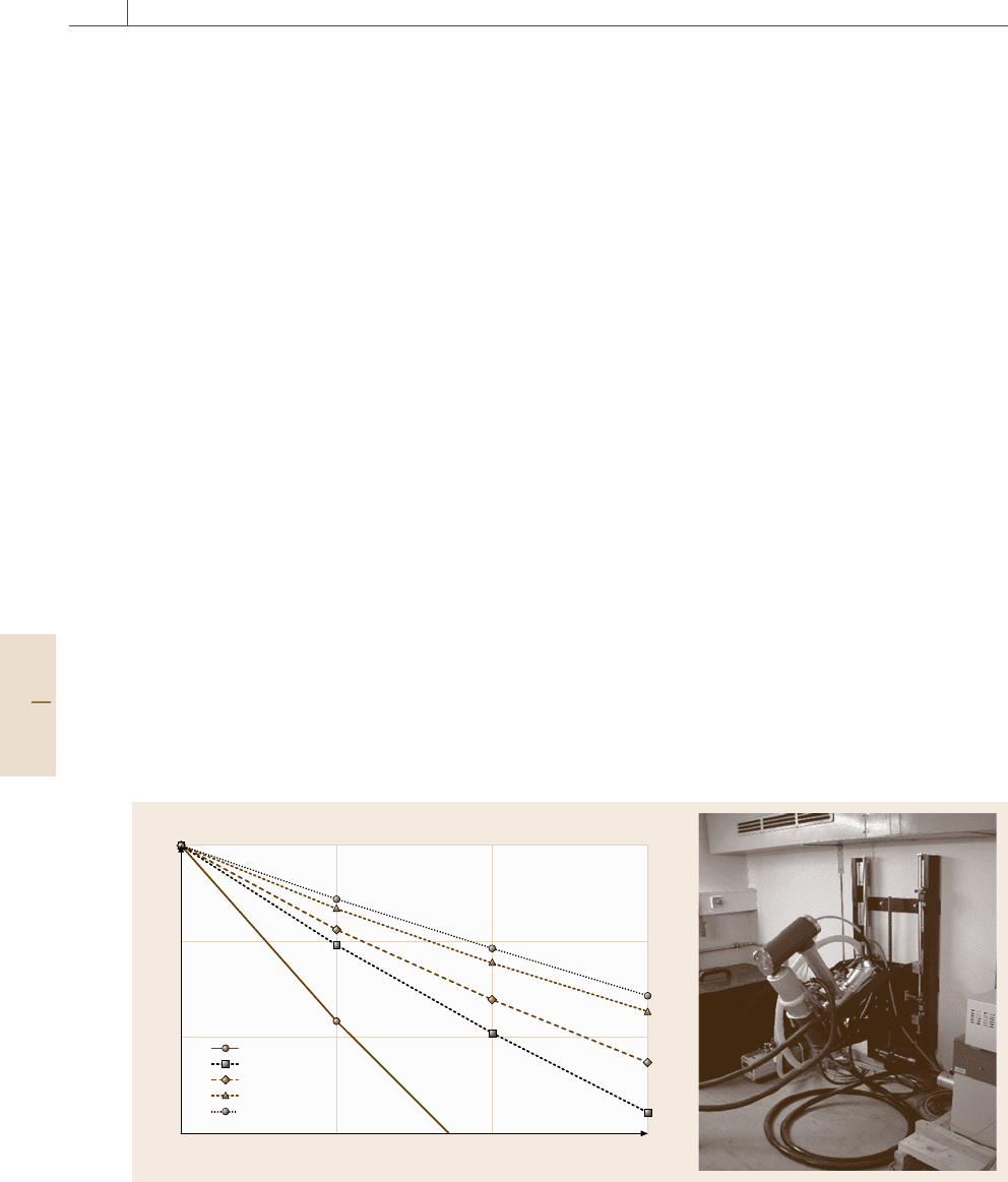
Figure 16.62a gives a graph that shows the gain
in attenuation possible with an extended range of kV.
For CT measurements the maximum absorption to free
beam ratio is about 20. From the graph it is seen that this
means an extension of measurable material in object
thickness from 20 to 30 mm of steel. At the end of 2002
a 320-kV microfocus x-tray tube (MX-5 tube, build by
YXLON, Halfdangsgade 8, 2300 S Copenhagen) was
integrated into a CT system (Fig. 16.62a). The bipolar
tube is build up from a standard −200 kV tube. The
original probe head is changed to the bipolar part which
has a build in anode with an additional high voltage sup-
plyofupto+120 kV. The distance between x-ray spot
to tube outside is enlarged to 25 mm because the target
position is no longer at ground voltage level. This means
that the tube in this modification has a lower maximal
magnification, but this corresponds to the bigger object
Attenuation
100 kV
160 kV
200 kV
320 kV
380 kV
Thickness (mm)
3001020
10
0
10
–1
10
–2
10
–3
a) b)
Fig. 16.62 (a) Attenuation curve for iron as function of x-ray energy (b) 320-kV micro-focus tube
size. For example with 320 kV and 0.1 mA the optimum
spatial resolution is about 35 μm.
In practical operation the second part is fixed to
+120 kV while the variation in energy is done with the
first. A range of 130 kV to 320 kV results. To the first
part a standard microfocus target can be attached and
the tube can thus be used as normal 200 kV microfocus
tube with high magnification. As detector an a-Si flat-
panel detector (Perkin Elmer, 16-bit ADC, 1024 × 1024
pixel, pixel size is 0.4mm
2
) is used [16.44].
16.4.3 Synchrotron CT
With transmission target x-ray tubes the focal spot size
can be lowered to 1–5 μm, corresponding to a spatial
resolution of less than 10 μm. Due to the limited inten-
sity given by electron scattering and the heat generated,
which can melt the target, measuring times are increas-
ing to some hours and more. Synchrotron radiation
sources offer photon flux densities, which are several or-
ders of magnitude higher compared to laboratory x-ray
tubes. The additional advantage is the fact that the high
photon flux density can be reached with monochromatic
radiation giving some advantages over tomography,
such as the absence of beam-hardening artefacts.
To extend the usable energy range at the Berlin Elec-
tron Storage Ring Company for Synchrotron Radiation
(BESSY) a 7-T wavelength shifter with a critical en-
ergy of 13.5 keV has been installed in the storage ring
to operate the first hard x-ray beam line at BESSY,
called BAMline. The main optical components of the
beam line are a double crystal monochromator (DCM)
Part D 16.4
Performance Control 16.4 Computerized Tomography – Application to Inorganic Materials 923
and, for the first time for imaging, a double multilayer
monochromator (DMM, 320 double layers of W and
Si, with thicknesses of 1.2and1.6 nm, respectively).
The latter is preferred for use in tomographic facilities,
giving a factor of 100 higher photon flux due to the
increased bandwidth of 2% compared with 0.01% of
the DCM [16.45–47]. The detector system consists of
a 2048× 2048 photometric camera system together with
a scintillator screen of GdOS. Depending on the lens
combination used nominal voxel sizes of 1.5–7.2 μm
can be used.
16.4.4 Dimensional Control
of Engine Components
A current research topic is the improvement of dimen-
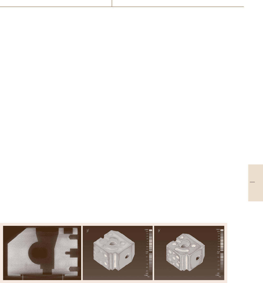
sional control with CT.AsanexampleFig.16.63 shows
a cross section of an aluminum test sample investigated
with the 320-kV equipment. This sample was measured
additionally with the LINAC CT. Standard operation
for a comparison is the conversion of the voxel data in
a point cloud and the analysis of files in the stereolitho-
graphic data format (STL).
The comparison performed using the STL data
shows principal deviations between the computer-aided
design (CAD) model and real sample, which are due to
the limited energy of the 320-kV equipment and espe-
cially the smoothed edges in the LINAC measurement
due to the limited detector pixel size.
Fault Detection
in Telecommunication Equipment
The transmission characteristics of glass-fiber cables
can be changed after installation or during lifetime.
Fig. 16.63 Test sample of aluminum (∅ about 100 mm). The image (left) shows a cross section of the measurement
with the 320-kV equipment (320-kV x-ray: 220 kV, 100 μA; prefilter: 1.5 mm Sn; projections: 900/360
◦
; exposure time:
2.28 s per projection; voxel: (0.12 mm
3
); matrix: 1023× 1023 ×605). The image (middle) shows the deviation against the
CAD data and the image (right) the deviation against the measurement with LINAC (10.5 MeV, 20 Gy; prefilter: 90 mm
Fe; projections: 720/360
◦
; exposure time: 0.8 s per projection; voxel: (0.65 mm
3
); matrix 255×255×201)
To localize such flaws optical time-delay reflection
(OTDR) methods are frequently used. The location of
flaws can be determined with an accuracy of a few
mm. An analysis of the type and size of flaws to-
gether with geometry control of the complex-shaped
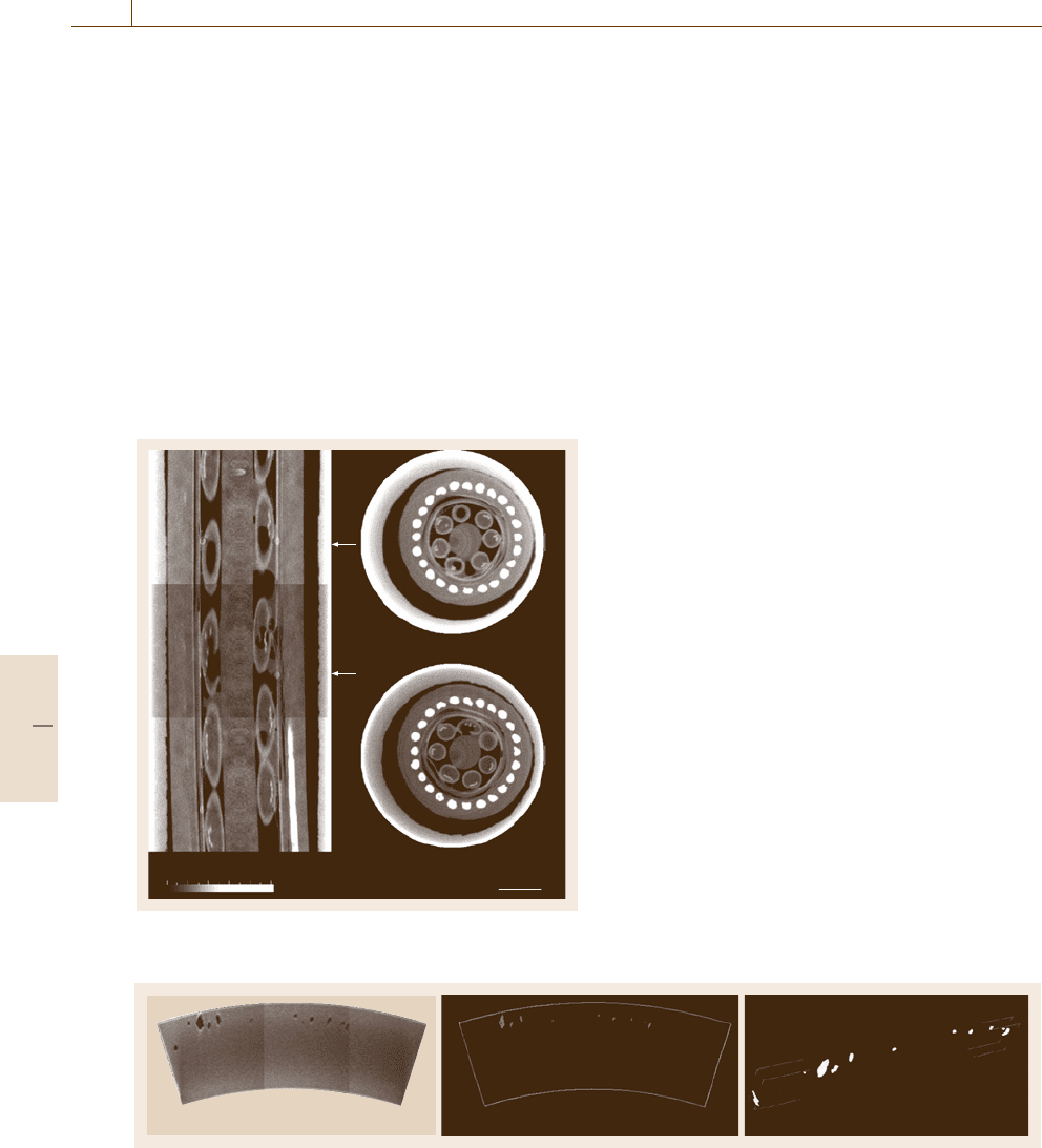
cable can be performed with CT. Figure 16.64 shows
a vertical slice of a section (length 48 mm) of the ca-
ble together with two horizontal slices. The bunches
of four glass fibers are embedded in a protective cov-
ering. One of the seven coverings contains no fibers.
The diameter of each glass fiber is 125 μm. The
outer diameter of the cable is 16 mm. Some protec-
tive coverings show flaws (Fig. 16.64 bottom right)
and an incomplete embedding of fiber bunches. To
avoid the influence of sample preparation the investi-
gated sample volume was part of a 15-m-long cable
segment.
Flaw Extraction in Cu Samples
As a first example results are shown for flaw extraction
in electron-beam-welded Cu samples (size about 40 ×
100 × 300 mm) using high-energy 3-D CT (Fig. 16.65).
For studies of probability of detection (POD) a 100%
inspection of volumetric flaws are performed. Due to
the limited size of the flat-panel detector the sample
was measured in three positions and the overlapping
data sets are joined after the image reconstruction. The
reconstructed images show some artefacts given by
scattered radiation, beam hardening and inherent detec-
tor effects. Therefore a filter was used to reduce these
artefacts. Figure 16.65 shows as result a cross section
trough the sample.
The flaws are evaluated twofold; by a local threshold
operation using some image-processing modules (e.g.
Part D 16.4
924 Part D Materials Performance Testing
an STL converter module) developed for the image-
processing system AVS and the Volume Graphics (VG
Studio Max) image-processing tool. The geometry of
the envelope and the voids are lead together and con-
verted to STL format for comparison with ultrasonic
results and for theoretical simulation of the irradia-
tion process. The volume of all flaws of the sample is
1100 voxels, determined with the AVS system, which is
in good agreement with the result from the VG system,
which gives a volume of a 1072voxel. The total volume
is 4 304 500 voxels.
Calibration of NDT Methods
The CT imaging method combined with the geomet-
rical correctness of the dimensions of the investigated
i240 k250
k550
5mm0616 m
0 50 100 150 200 250
Fig. 16.64 Vertical and two horizontal cross sections of
a glass-fiber cable
Fig. 16.65 Electron beam welding of a section (size 40 × 100 × 300 mm) of a Cu canister. The image (left)showsacross
section containing the flaws, flaws after segmentation (middle) projected into a plane and the flaws after conversion to the
STL data format (right). Source: LINAC 10.5 MeV, 15 Gy; prefilter: 90 mm Fe; projections: 720/360
◦
; exposure time:
0.2 s per projection; voxel: (0.653 mm
3
); matrix: 255×255×532
samples are the main advantages to the use of CT for
the calibration of other NDT methods, such as ultra-
sonic technique (UT) or eddy-current techniques. As an
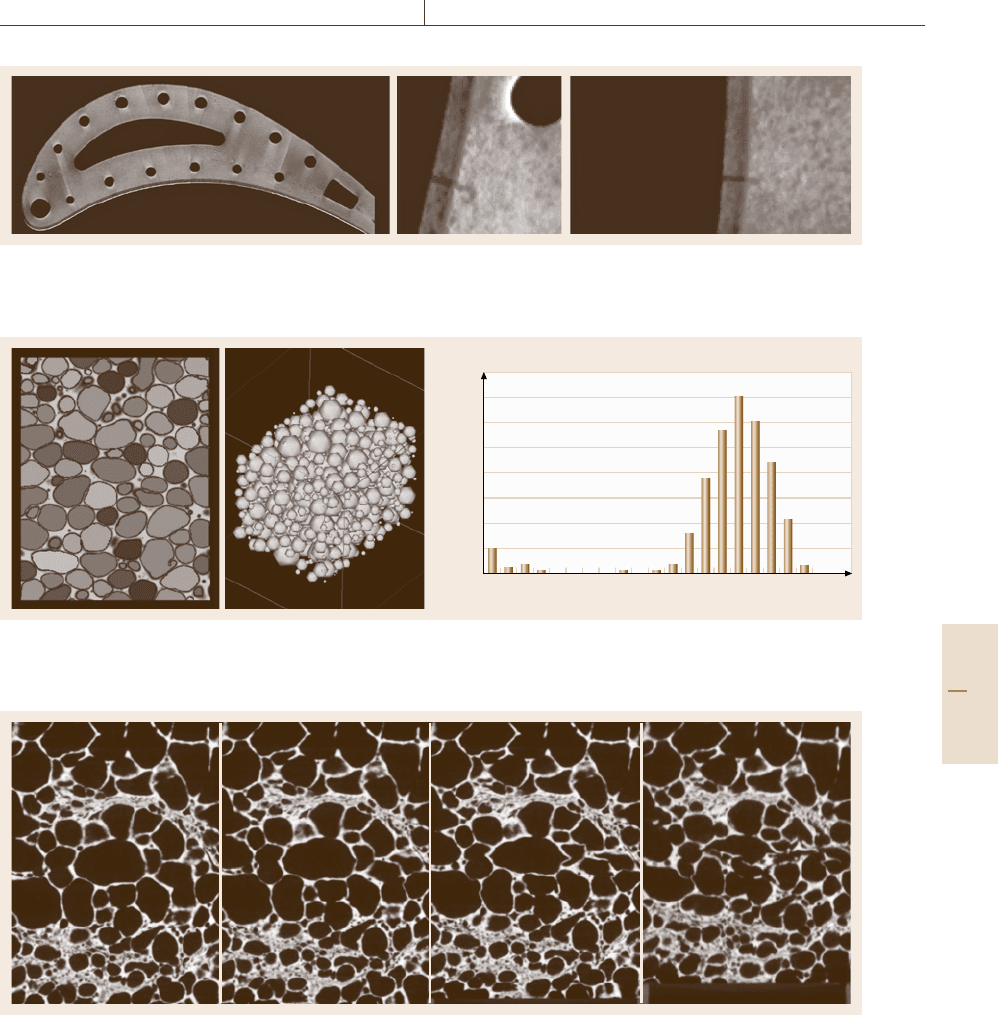
example Fig. 16.66 shows the investigation of flaws of
coated turbine blades. With optical methods only the
flaws in the coating layer can be visualized but not in
the matrix material. With high-resolution CT the crack
configuration was analyzed. The result was used for
calibration of other NDT techniques (the eddy-current
method).
Pore Detection and Pore-Size Calculation
in Al Foam
Depending on the type of foam there are two ways to
measure the size of the pores inside.
•
If the pores are closed, a threshold operation is first
used to obtain the binary area of all pores. Start-
ing on one edge, an algorithm then searches in 3-D
for the first marked voxel and colors all adjacent
as belonging to the same pore until no more con-
nected voxels are found. The searching algorithm
then goes on to the next pore. Figure 16.67 (left im-
age) shows the result; the image is a slice through
the searched foam, the different colors of each pore
are used to detect pores, which were not separated.
The size distribution is given in Fig. 16.67 (right im-
age). While the volume is searched for pores, the
volume and the center of gravity are also stored, and
further parameters can be calculated. From these
parameters a simplified model of the foam can be
generated and calculation of real foams with finite-
element method (FEM) programs becomes possible.
The center image of Fig. 16.67 shows the same
foam, representing the pores as spheres with the
same diameter.
•
If the pores are open, the inverted nonmetal part has
to be 3-D-eroded until single areas result for all the
Part D 16.4
Performance Control 16.4 Computerized Tomography – Application to Inorganic Materials 925
Fig. 16.66 Turbine blade with protective layer (layer thickness of about 400 μm). The CT cross sections show a crack
only in the protective layer (right image) and a crack in the matrix material
Number of spheres
Radius (mm)
1.120.910.70.490.28 1.330.07
140
120
100
80
60
40
20
160
0
Fig. 16.67 Pore detection (left). Spheres with a diameter as calculated from the volume of found pores (right) at position
of pore centers. The pore size distribution is shown in the diagram on the right
Fig. 16.68 A vertical slice through the foam before and after three compression states (from left to right). From the
different 3-D image data sets the same slice was extracted showing the internal deformation of the foam
pores. The pore radius can then be calculated from
the number of voxels counted per pore. Knowing
the number of erode steps, the pore radius has to
be enlarged by this value.
Internal Deformation
The failure mechanisms of strength-tested foams were
studied on samples before compression and after com-
pression. 3-D CT images of the samples were used
Part D 16.4
926 Part D Materials Performance Testing
100 mm
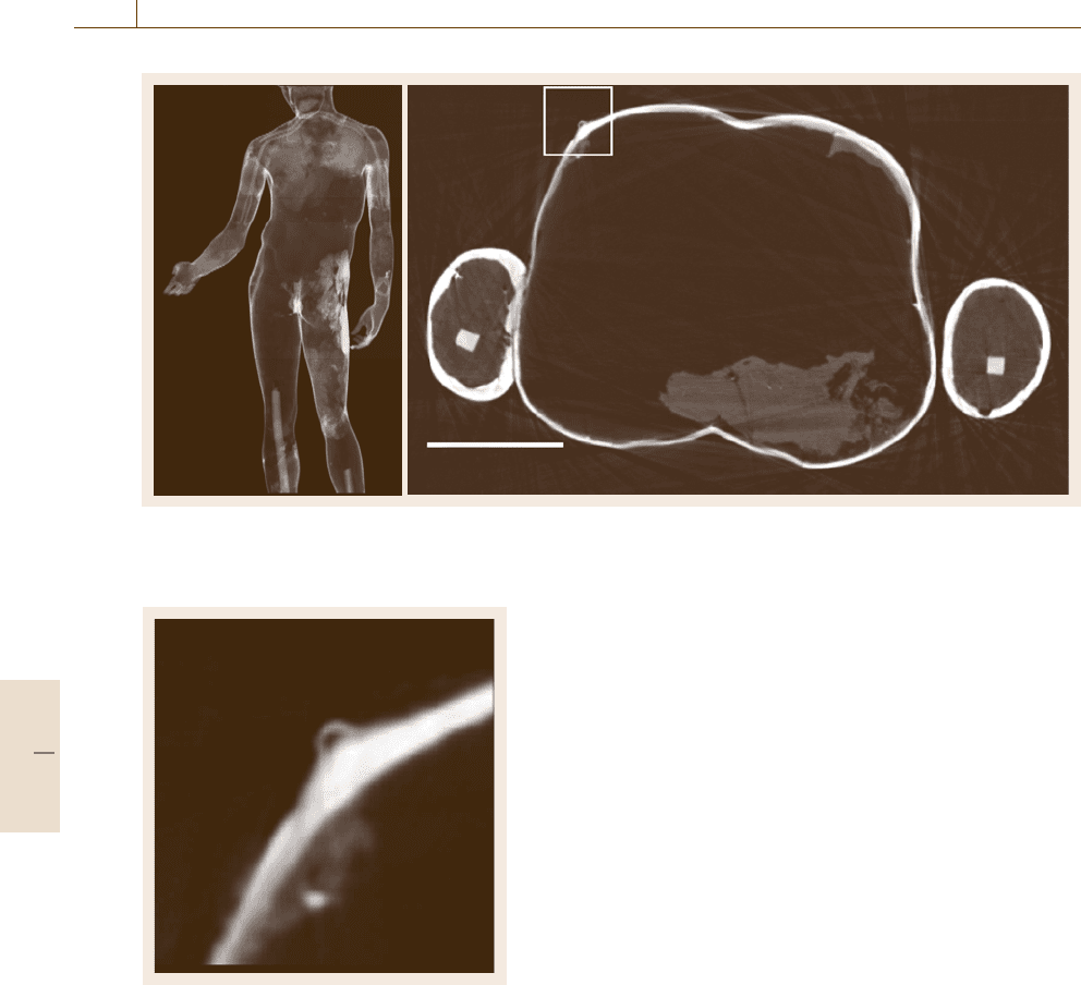
Fig. 16.69 Digital radiography of the antique bronze statue Idolino (left image). A cross section is shown on the right,
representing the inner structure as well as the technique to fit together the separate cast parts of the statue
Fig. 16.70 Enlarged detail (fivefold magnification), marked
in Fig. 16.69 by the white square
to find out where foam deformation has started, for
which a program for image comparison in 3-D was de-
veloped. First the size of the region to be compared
with the region of the sample after the compression
test has to be defined. This region has to be big-
ger than the mean pore size. The region is moved in
different directions in 3-D until it fits best with the
initial sample, and the shift of the parts of the strength-
tested sample is written to an array. In this way, for
all small regions of the probe we obtain shifts with re-
spect to the initial sample in three directions: Δx, Δy
and Δz.
The displacement after the compression test is
shown (unit mm) using different grey levels, which are
painted over the original foam structure, in order to
show the displacement of compressed foam compared
to original foam (Fig. 16.68).
Art Objects
Nondestructive methods such as x-ray techniques have
been focused on art objects since the discovery of
x-rays by W. C. Roentgen. In a long-standing col-
laboration with the Antikensammlung Berlin several
large Greek and Roman bronzes were examined, to
evaluate work traces and the interior of these archaeo-
logical samples. In 2000 the interior of the hellenistic
Getty Bronze and the early Augustan Idolino in Flo-
rence were investigated at BAM [16.48, 49]. From the
still being results Fig. 16.69 shows a digital radiogra-
phy (left image) which is used to localize the precise
location of cross sections. The tomogram (right im-
age of Fig. 16.69) gives a view of the interior of the
statue, with parts of the structure fitting the differ-
ent cast parts. A detail of the tomogram shown in
Fig. 16.69 is presented in Fig. 16.70 with a fivefold
enlargement.
Part D 16.4

Performance Control 16.5 Computed Tomography – Application to Composites and Microstructures 927
16.5 Computed Tomography – Application to Composites
and Microstructures
In computed tomography (CT) the interface contrast
of heterogeneous materials can be strongly enforced
by to x-ray refraction effects. This is especially de-
sirable for materials with low absorption or mixed
phases of similar absorption that result in low con-
trast. X-ray refraction [16.50, 51] is an ultrasmall-angle
scattering (USAXS) phenomenon. Refraction contrast
has also been applied for planar refraction topogra-
phy, a scanning technique for improved nondestruc-
tive characterization of high-performance composites,
ceramics and other low-density materials and compo-
nents [16.52].
X-ray refraction occurs when x-rays interact with
interfaces (cracks, pores, particles, phase boundaries),
preferably at low angles of incidence, similarly to the
behavior of visible light in transparent materials, e.g.
lenses or prismatic shapes. X-ray optical effects can be
observed at small scattering angles of between several
seconds and a few minutes of an arc, as the refractive
index n of x-rays is nearly unity (n
∼
=
1–10
−5
). In other
terms, due to the short x-ray wavelength below 0.1nm,
x-ray light scattering is sensitive to inner surfaces and
interfaces of nanometer dimensions.
16.5.1 Refraction Effect
In analogy to visible optics the interaction of x-rays
with small transparent structures with dimensions above
several nanometers results in coherent scattering gov-
erned by wavelength, structural dimensions and shape,
local phase shift and absorptive attenuation. However,
differently from optical conditions, the refractive index
of x-rays near 1 causes beam deflections into the same
small-angle region of several minutes of arc as diffrac-
tion. Thus the resulting interference is due to phase
modulation due to the refractive index and the absorp-
tive and Raleigh diffraction, both of which depend on
the path length through matter.
However if the dimensions of the scattering ob-
jects are much larger than several tens of nanometers,
as is common in classical small-angle scattering, the
interference fringes are no longer observable by clas-
sical small-angle cameras as they are too narrow.
The resulting smeared angular intensity distribution
is then simply described by a continuous decay ac-
cording to the rules for refraction by transparent
media, e.g. applying Snell’s law [16.51]. This purely
geometrical refraction approach is appropriate for
small-angle x-ray (and neutron) scattering effects by
micrometer-sized structures and is applied in the fol-
lowing.
If ε is the real part of the complex index of re-
fraction n, ρ is the electron density and λ is the x-ray
wavelength, then n is
n = 1 −ε, with ε ≈ρλ
2
and ε
∼
=
10
−5
(16.30)
for glass under 8 keV radiation. In contrast to optics
convex lenses cause divergence of x-rays as n < 1.
Figure 16.71 demonstrates the effect of small-angle
scattering by refraction of cylindrical lenses: a bundle
of 15-μm glass fibers (for composites) deflects a pin-
hole x-ray beam within several minutes of an arc. In
fibers and spherical particles the deflection of x-rays oc-
curs twice, when entering and when leaving the object
(insert Fig. 16.71). The oriented intensity distribution is
collected by an x-ray film or a CCD camera while the
straight (primary) beam is removed by a beam stop. The
shape of the intensity distribution of such cylindrical ob-
jects is a universal function independent of materials,
Oriented
small angle
scattering
by refraction
Detector/
film
Sample,
fibers
Collimation
Mo-K
α
, 20 keV
n =1–ε with ε ≈ρλ ≈ 10
–5
2θ
Refracted
beam I
R
*
(2θ)
Fig. 16.71 Effect of oriented small-angle scattering by re-
fraction of glass fibers; n index of refraction, ε real part of
n, λ wavelength
Part D 16.5
