Bongard Frederic , Darryl Sue. Diagnosis and Treatment Critical Care
Подождите немного. Документ загружается.


IMAGING PROCEDURES
147
pneumonia (bronchopneumonia), and interstitial pneumo-
nia. Lobar pneumonia is characterized on x-ray by relatively
homogeneous regions of increased lung opacity and air
bronchograms. The entire lobe need not be involved, and in
fact, with early therapy, consolidation does not usually affect
the entire lobe. Pathologically, the infecting organism reaches
the distal air spaces, resulting in edema filling the alveoli. The
infected edema fluid spreads centripetally throughout the
lobe via communicating channels to adjacent segments. Air
bronchograms are common. Streptococcus pneumoniae
(pneumococcal) pneumonia is the classic lobar pneumonia,
although other organisms, including Klebsiella pneumoniae
and Legionella pneumophila, may produce an identical pat-
tern. Since the airways are not primarily involved, volume
loss is not conspicuous. Indeed, expansion of the lobe may
occur in Klebsiella or pneumococcal pneumonia.
Bronchopneumonia (lobular pneumonia) results from
inflammation involving the terminal and respiratory bron-
chioles rather than the distal air spaces. Since the process
focuses in the airways, the distribution is more segmental
and patchy, affecting some lobules while sparing others.
Pathologically, there is less edema fluid and more inflamma-
tion of the mucosa of bronchi and bronchioles. Patchy con-
solidation is seen radiographyically. Mild associated volume
loss may also be present. Air bronchograms are not as com-
mon a feature in bronchopneumonia as in lobar pneumonia.
The most common organisms producing classic bronchop-
neumonia are Staphylococcus aureus and Pseudomonas
species.
Interstitial pneumonia is typically caused by viruses or
Mycoplasma pneumoniae. In the immunocompromised
patient, Pneumocystis carinii (now known as Pneumocystis
jerovicii) is an important cause of interstitial pneumonia.
The pathologic process is located primarily in the intersti-
tium, and the classic radiograph reflects the interstitial
process and demonstrates an increase in linear or reticular
markings in the lung parenchyma with peribronchial
thickening and occasionally septal lines (Kerley A and B
lines). Although the pathologic process is primarily
located in the interstitium, proteinaceous fluid is exuded
into the air spaces and consequently may progress to a
pneumonia that radiographically appears alveolar.
Radiographic Features
A. Plain Films—Although plain films cannot provide a spe-
cific microbial diagnosis in a patient with pneumonia, radi-
ology has a central role in both initial evaluation and
treatment. The chest radiograph documents the presence
and extent of disease. Associated parapneumonic effusions,
mediastinal or hilar adenopathy, cavitation, and abscess
formation—as well as predisposing conditions such as cen-
tral bronchogenic carcinoma—may be identified. Such
information can guide the clinician to a high-yield diagnos-
tic procedure such as thoracentesis or bronchoscopy, which
may be necessary in a patient who cannot produce adequate
sputum for bacteriologic culture. The chest radiograph is
also critical in evaluating the patient’s response to therapy.
Antibiotic therapy is frequently empirical, and the chest radi-
ograph may be the first indicator of failure of antibiotics and
a need for change in management. A pneumonia that does
not clear despite antibiotic therapy should raise the suspicion
of central airway obstruction by a mass or foreign body or
may represent a bronchoalveolar carcinoma mimicking
pneumonia.
Localization of the consolidation to a specific lobe is
important not only to correlate with the physical examina-
tion but also to guide the bronchoscopist when necessary.
In addition, different types of pneumonia may be more
likely to occur in specific regions. For example, reactivation
tuberculosis occurs most commonly in the apical and pos-
terior segments of the upper lobes and the superior seg-
ment of the lower lobes. The silhouette sign is useful in
determining the site of pneumonia. When consolidation is
adjacent to a structure of soft tissue density (eg, the heart or
the diaphragm), the margin of the soft tissue structure will
be obliterated by the opaque lung. For example, right mid-
dle lobe consolidation may cause loss of the margin of the
right heart border, lingular consolidation may cause loss of
the left heart border, and lower lobe pneumonia may oblit-
erate the diaphragmatic contour.
Intrathoracic nodal enlargement may be a useful diag-
nostic feature. Enlargement of the hilar or mediastinal lymph
nodes is uncommon in bacterial pneumonia and most viral
pneumonias. Tuberculosis, atypical mycobacterial infections,
fungal infections such as coccidioidomycosis and histoplas-
mosis, and viral infections such as measles and Epstein-Barr
virus may be associated with adenopathy.
Pleural effusions occur in up to 40% of patients with bac-
terial pneumonia. A parapneumonic effusion consists of
intrapleural fluid in association with pneumonia or lung
abscess. Empyema is defined as pus in the pleural space.
Thoracentesis is required for differentiation between a sim-
ple parapneumonic effusion and an empyema, and the deci-
sion to place a chest tube depends on the characteristics and
the quantity of the effusion. A pleural effusion usually is
identified radiographically on a plain film, although ultra-
sound or CT may be necessary in some cases.
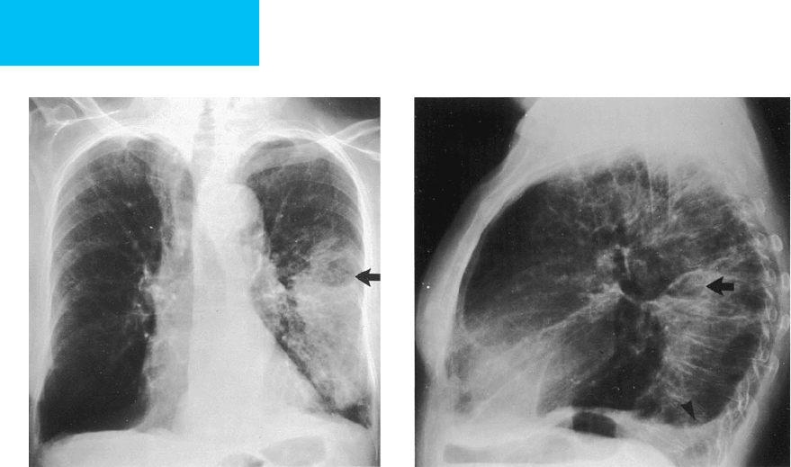
1. Lung abscess and cavitation—Cavitation of pneumo-
nia results from destruction of lung tissue by the inflamma-
tory process, leading to lung abscess formation (Figure 7–6).
Although often seen in pneumonias due to gram-negative
organisms such a Pseudomonas and Klebsiella, cavitation is
rare in pneumococcal pneumonia. Pneumonias due to
Mycobacterium tuberculosis, atypical mycobacteria, and fungi
and those due to anaerobes and staphylococci also frequently
cavitate. Cavitary lung abscesses must be distinguished from
bullae, pneumatoceles, cavitary lung cancers, and other
lucent lesions. Most abscesses have a wall thickness between
5 and 15 mm, allowing differentiation from bullae and pneu-
matoceles, which usually have thin, smooth walls. A lung
abscess is usually surrounded by adjacent parenchymal con-
solidation, which may serve to differentiate an abscess from a
CHAPTER 7
148
cavitary bronchogenic carcinoma. Complications of lung
abscess include sepsis, cerebral abscess, hemorrhage, and
spillage of contents of the cavity into uninfected lung or
pleural space.
In one review, 18% of lung abscesses were radiographi-
cally occult, with only nonspecific lung opacities or nod-
ules identified. In these patients, the diagnosis was made at
surgery or at postmortem examination. One reason lung
abscesses were not identified was probably failure to use a
horizontal beam in obtaining the chest radiographs. With
semierect or supine positioning, air-fluid levels within the
cavity were obscured. In cases where erect chest films are
unobtainable, decubitus or cross-table lateral views can be
obtained with a horizontal beam and may be diagnostic.
2. Nosocomial pneumonia—Definitive diagnosis of noso-
comial pneumonia is difficult because both the clinical fea-
tures and the chest radiographic findings may be present in
other disease processes and because abnormalities on chest
radiographs are often present prior to development of noso-
comial pneumonia. Clinical suspicion in patients with
underlying heart and lung disease is important. For example,
the incidence of nosocomial pneumonia is increased in
patients with ARDS as well as in other patients with respira-
tory failure.
Radiographically, nosocomial pneumonia is heralded
by the development of new or worsening parenchymal
opacities, usually multifocal. Since nosocomial pneumonias
are most often due to aerobic gram-negative organisms or
staphylococci, abscesses and pleural effusions may develop.
Development of cavitation helps to distinguish nosocomial
pneumonia from other causes of parenchymal opacification
such as atelectasis, lung contusion, or pulmonary edema.
B. Computed Tomography—The cross-sectional imaging
plane and superior contrast resolution make CT useful in
the evaluation of complicated inflammatory diseases.
Cavitation, which may be obscured on plain films, is easily
identified on CT. Localization of parenchymal diseases
facilitates the direction of invasive studies such as bron-
choscopy or open lung biopsy. Superimposed pleural and
parenchymal processes are more easily differentiated on CT
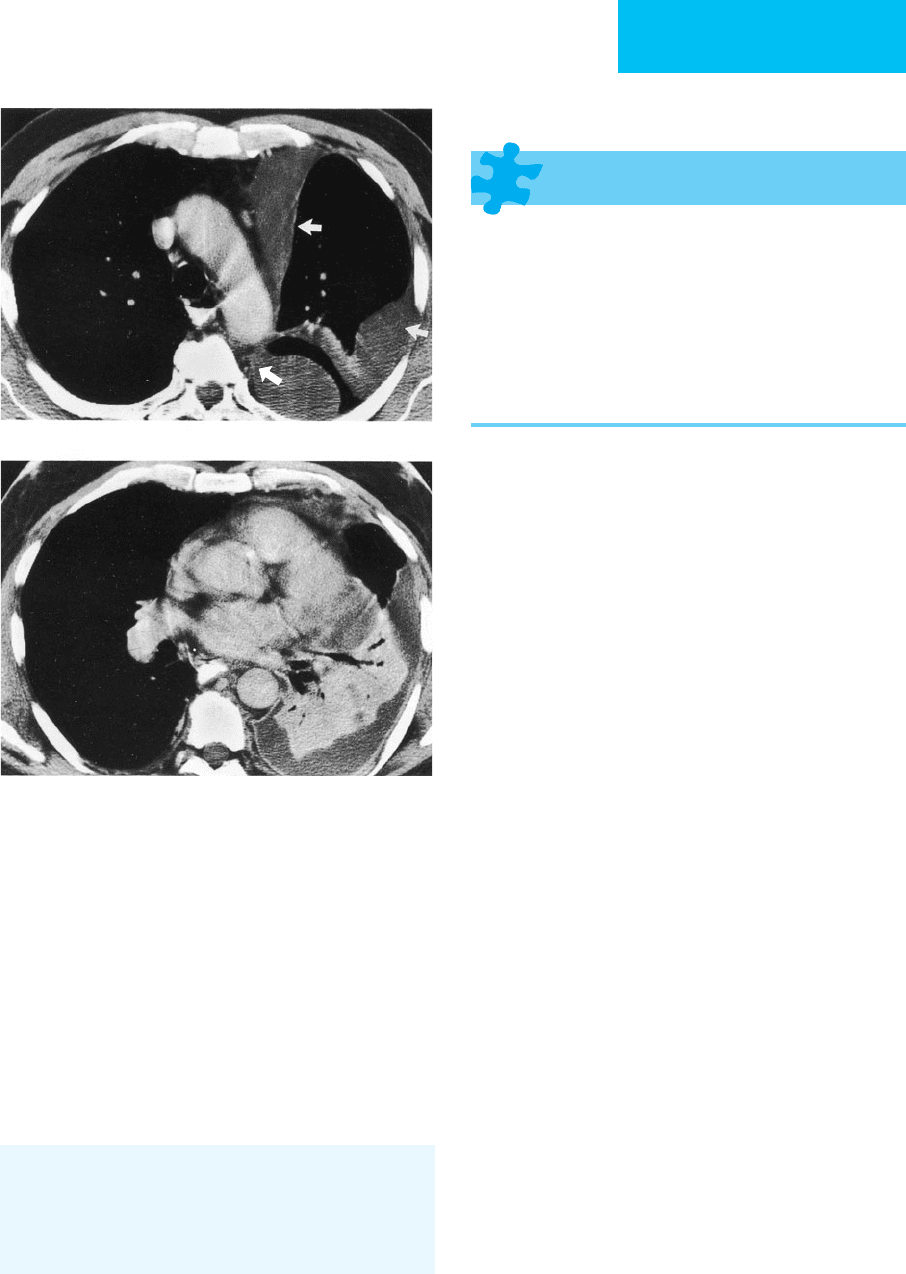
than on plain films (Figure 7–7). Loculated pleural effusion
or empyema associated with pneumonia may be difficult to
evacuate, and CT may serve to guide thoracentesis, chest
tube placement, or percutaneous drainage of large lung
abscesses.
Empyema and lung abscess are more easily distinguished
on CT than on conventional radiographs. Separation of
thickened visceral and parietal pleural surfaces (“split pleura
sign”) may be seen in empyema. Other useful findings
included wall characteristics, with smooth, uniform walls
seen in empyema and thick, irregular walls more commonly
seen in lung abscess. The size and shape of the lesion are less
helpful; lung abscesses generally tend to be round—as
opposed to lenticular in empyemas. The administration of
A
B
Figure 7–6. Cavitary pneumonia. Posteroanterior (A) and lateral (B) chest radiographs demonstrate consolidation
with cavitation (arrows) in the superior segment of the left lower lobe secondary to Pseudomonas aeruginosa. A small
left pleural effusion is present, best seen on the lateral view (arrowhead). Changes of chronic obstructive pulmonary
disease are also present.
IMAGING PROCEDURES
149
intravenous contrast material facilitates differentiation of
pleural and parenchymal disease because the lung
parenchyma will enhance with contrast, whereas the pleural
effusion will retain its low attenuation.
Franquest T: Imaging of pneumonia: Trends and algorithms. Eur
Respir J 2001;18:196–208. [PMID: 11510793]
Sharma S et al: Radiological imaging in pneumonia: recent innova-
tions. Curr Opin Pulm Med 2007;13:159–69. [PMID: 17414122]
Vilar J et al: Radiology of bacterial pneumonia. Eur J Radiol
2004;51:102–13. [PMID: 15246516]
Aspiration Pneumonia
ESSENTIALS OF RADIOLOGIC
DIAGNOSIS
Consolidation in dependent regions of the lung, varying
with position of patient at time of aspiration, but may
be multilobar and bilateral.
Cavitation and abscess formation may be seen, but
pleural effusions are infrequent.
May lead to necrotizing pneumonia and lung abscess.
Aspiration of gastric contents may result in noncardio-
genic pulmonary edema, cavitation, and atelectasis.
General Considerations
Aspiration pneumonia results from endotracheal aspiration
of oropharyngeal or gastric secretions. Aspiration is thought
to be a common occurrence in the healthy adult, with the
incidence during sleep estimated to be as high as 45%.
Small-volume aspirates are cleared by physical entrapment
and coughing along with the mucociliary elevator action of
the respiratory epithelium. Inactivation by IgA antibodies
and opsonization and ingestion of bacteria by phagocytic
cells play a role as well. Although organisms are present in
pathogenic numbers even in small-volume aspirates, nor-
mal individuals are able to clear these organisms without
sequelae.
Several clinical conditions predispose patients to aspira-
tion. Depressed levels of consciousness secondary to medica-
tions, alcohol intoxication, seizures, anesthesia, or neurologic
disease result in impaired upper airway reflexes.
Endotracheal intubation increases the rate of aspiration,
with both high-volume, low-pressure cuffs and uncuffed or
low-volume, high-pressure tubes implicated. The incidence
of aspiration is even higher in patients with tracheostomies
as compared with endotracheal tubes. Nasogastric and feed-
ing tubes, gastric distention, gastroesophageal reflux, hiatal
hernia, decreased esophageal mobility, and vomiting have all
been cited as predisposing factors for aspiration. Severe peri-
odontal disease is also a risk factor for aspiration pneumonia.
Bacterial colonization of gastric secretions also plays a role in
the development of aspiration pneumonia. Although gastric
acidity prevents significant bacterial colonization, antacid
therapy for prophylaxis for stress ulcers may change gastric
pH, resulting in increased bacterial colonization of gastric
contents.
Aspiration pneumonia occurs when a normal host aspi-
rates a large amount of contaminated matter, overwhelming
host defenses, or when smaller amounts are aspirated in a
patient with impaired defenses. Aspiration pneumonia is
caused by mixed anaerobic and aerobic organisms, with up
to 80% of cases caused by multiple strains of bacteria. The
A
B
Figure 7–7. Pneumonia with loculated empyema. A. CT
shows a loculated pleural effusion in the left hemithorax
(arrows). B. More caudally, dense consolidation with air bron-
chograms secondary to pneumonia is present in the left
lower lobe. The consolidated lung enhances with contrast and
is easily distinguished from the surrounding pleural effusion.
CHAPTER 7
150
organisms responsible for the pneumonia vary with the
clinical setting—community-acquired, nursing home, or
hospitalized patients—and reflect colonization of the upper
airway. Aerobic bacteria associated with community-
acquired aspiration pneumonia are mostly streptococci,
whereas gram-negative organisms, particularly Klebsiella and
Escherichia coli, are seen more often in nosocomial infection.
The major anaerobic organisms include Fusobacterium
nucleatum, Peptostreptococcus, Bacteroides melaninogenicus,
and Bacteroides intermedius.
There are three general clinical patterns that may be seen
following aspiration: (1) respiratory compromise followed
by rapid clinical and radiographic improvement, (2) rapid
clinical and radiographic progression, and (3) transient sta-
bilization followed by protracted worsening of clinical and
radiographic status, with bacterial superinfection or ARDS.
Aspiration of acidic gastric contents resulting in an acute
pulmonary reaction with pulmonary edema is sometimes
referred to as Mendelson’s syndrome. Manifestations depend
on the volume, pH, and distribution of the aspirate. The
absorption of acid by the pulmonary vasculature and subse-
quent pulmonary injury are almost immediate and lead to
consolidation, alveolar hemorrhage, and collapse with tran-
sudation of fibrin and plasma into the alveoli. Aspiration of
a combination of acid and gastric particulate material pro-
duces a more severe injury pattern than either acid or gastric
particulate matter alone.
Radiographic Features
Aspiration pneumonia results in consolidation in dependent
regions of the lung. The location of the consolidation will
vary according to the patient’s position at the time of aspira-
tion. In the supine patient, the superior segments of the
lower lobes, the posterior segment of the right upper lobe,
and the posterior subsegment of the left upper lobe are
involved—whereas in the upright patient, the basal segments
of the lower lobes are more often affected, particularly on the
right. The more obtuse angle between the trachea and the
right main stem bronchus compared with the angle of the
trachea and the left main stem bronchus results in a higher
percentage of right-sided abnormalities in the supine patient.
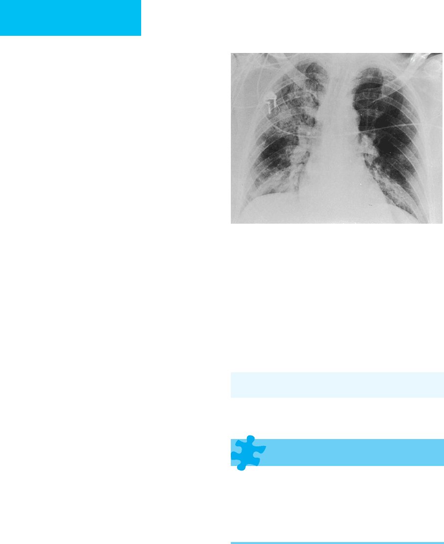
Consolidation is usually multilobar and bilateral (Figure 7–8).
Because of frequent infection with anaerobes, cavitation and
abscess formation may be seen. Effusions are infrequent.
CT is useful in the evaluation of aspiration disease and to
differentiate aspiration from other parenchymal diseases. CT
is also more sensitive than chest radiographs for the detec-
tion of aspirated foreign bodies.
Complications of simple aspiration pneumonia include
necrotizing pneumonitis and lung abscess. Necrotizing pneu-
monia results in multiple small cavities within the involved
lung and may extend into the pleural space, leading to
empyema formation. Lung abscess radiographically appears
as a cavitary lesion within a focus of consolidation, usually
solitary. Empyema is less likely in lung abscess since extension
of infection into the pleural space is usually impeded by the
barrier effect of the fibrous wall of the abscess cavity.
Patients who aspirate gastric contents may develop a
chemical pneumonitis that shows characteristics consistent
with noncardiogenic pulmonary edema. ARDS and features
of secondary bacterial infection may follow, including lung
necrosis and cavitation. Atelectasis may be a feature of airway
obstruction with food particles.
Franquet T et al: Aspiration diseases: Findings, pitfalls, and differen-
tial diagnosis. Radiographics 2000;20:673–85. [PMID: 10835120]
Chronic Obstructive Pulmonary Disease
ESSENTIALS OF RADIOLOGIC
DIAGNOSIS
Hyperinflation.
Bullae or blebs.
Pulmonary arterial deficiency pattern (areas of decreased
pulmonary vasculature).
Features of pulmonary hypertension.
General Considerations
Chronic obstructive pulmonary disease (COPD) is any pul-
monary disorder characterized by airflow obstruction.
Emphysema and chronic bronchitis are the most com-
mon examples. Emphysema is defined as a lung condition
characterized by enlargement of the air spaces distal to the
Figure 7–8. Aspiration pneumonia. Multiple areas of
pulmonary opacification are present bilaterally—secondary
to aspiration pneumonia following drug overdose.
IMAGING PROCEDURES
151
terminal bronchiole, accompanied by destruction of the
walls without obvious fibrosis. Four principal types of
emphysema are described: centrilobular, panlobular,
paraseptal, and paracicatricial. Chronic bronchitis is usually
defined in clinical terms, manifested by chronic productive
cough for at least 3 months for a minimum of 2 consecutive
years and characterized by excessive secretion of mucus in
the bronchi. Emphysema and chronic bronchitis frequently
coexist.
Radiographic Features
There is considerable controversy regarding the utility of the
chest radiograph in the evaluation of emphysema. Although
moderate to severe emphysema is usually apparent on the
chest radiograph, mild disease is difficult to appreciate.
Hyperinflation results from obstruction of small airways,
resulting in air trapping. Radiographic features include an
increase in size of the retrosternal clear space, flattening of
the hemidiaphragms, increased height of the lung, and
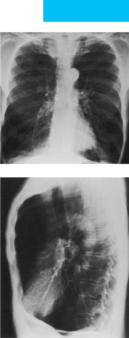
increased radiolucency (Figure 7–9). Measurements
obtained from chest x-rays have shown that the height of the
lung and the height of the arc of the right hemidiaphragm
correlate best with spirometric measures such as the forced
expiratory volume in 1 second (FEV
1
) and forced vital capac-
ity (FVC). A lung height of 29.9 cm or greater, as measured
from the tubercle of the first rib to the dome of the right
hemidiaphragm, will identify 70% of patients with abnormal
pulmonary function tests. A height of the right hemidi-
aphragm of less than 2.6 cm on the lateral projection identi-
fies 68% of patients with abnormal pulmonary function tests.
Bullae and blebs appear as focal regions of hyperlucency.
Although good indicators of emphysema, they also may be
seen in patients without COPD. Bullae are recognized as
hyperlucent or avascular regions and occasionally are demar-
cated peripherally by a fine curvilinear wall. The lung adja-
cent to large bullae may be compressed, and redistribution of
pulmonary blood flow away from areas of extensive bullous
disease may occur. The arterial deficiency pattern refers to
regions of radiolucent, hypovascular pulmonary
parenchyma characterized by a decrease in the size and num-
ber of vessels. This appearance may be due to multiple bul-
lae. Emphysema eventually can lead to pulmonary arterial
hypertension, manifested radiographically by disproportion-
ate enlargement of the central pulmonary arteries and right
heart chambers.
The radiographic appearance of the lungs in chronic
bronchitis is even less specific. Unlike that of emphysema, the
diagnosis of chronic bronchitis is based on clinical symp-
toms and not morphologic appearance. In addition, chronic
bronchitis and emphysema frequently coexist, making pure
chronic bronchitis difficult to characterize. Radiographic
findings suggesting chronic bronchitis include thickening of
bronchial walls and increased linear markings (“dirty
lungs”). Hyperinflation and hypovascularity have been
described but are probably due to concomitant emphysema.
A
B
Figure 7–9. Chronic obstructive pulmonary disease.
Posteroanterior (A) and lateral (B) chest radiographs show
hyperinflated lungs with increased anteroposterior diame-
ters, flattening of the diaphragm, and increased retroster-
nal clear space.

CHAPTER 7
152
High-resolution CT (HRCT) is more sensitive than plain
radiographs in the detection of emphysema. On HRCT,
emphysema appears as regions of low attenuation, lung
destruction, or simplification of the pulmonary vasculature.
The type of emphysema can often be defined by its pattern
and distribution on CT, with centrilobular CT predominantly
upper zone in distribution and panlobular emphysema more
diffuse or more severe within the lower lobes. The CT
appearance of chronic bronchitis may be overshadowed by
coexisting emphysema. Bronchial wall thickening and cen-
trilobular abnormalities have been described.
Cleverley JR, Muller NL: Advances in radiologic assessment of
chronic obstructive pulmonary disease. Clin Chest Med
2000;21:653–63. [PMID: 11194777]
Goldin JG: Quantitative CT of emphysema and the airways.
J Thorac Imaging 2004;19:235–40. [PMID: 15502610]
Shaker SB et al: Imaging in chronic obstructive pulmonary disease.
COPD 2007;4:143–61. [PMID 17530508]
Webb WR: Radiology of obstructive pulmonary disease. AJR
1997;169:637–47. [PMID: 9275869]
Asthma
ESSENTIALS OF RADIOLOGIC
DIAGNOSIS
Hyperinflation.
Peribronchial thickening.
Increased lung markings centrally.
Subsegmental atelectasis.
General Considerations
Asthma is a disease characterized by widespread narrowing
of the airways that fluctuates in severity over short periods of
time either spontaneously or following therapy.
Hyperactivity of airways may be induced by a variety of stim-
uli, and asthma is usually divided into two types: intrinsic
and extrinsic. Pathologic changes include smooth muscle
hypertrophy, mucosal edema, mucous hypersecretion, and
plugging of airways by thick, viscid mucus. The result is nar-
rowing of the airway diameter.
Radiographic Features
The radiographic manifestations of asthma vary from a normal
radiograph to hyperinflation, atelectasis, or barotrauma.
Radiographic findings may be categorized as (1) those common
features of asthma that do not affect management and are there-
fore not unanimously considered abnormalities and (2) find-
ings that influence patient management. The incidence of
radiographic abnormalities depends on the age of the patient
and the definition of abnormal by the investigator.
A. Uncomplicated Asthma—Hyperinflation, bronchial wall
thickening, and prominent perihilar vascular markings are all
features commonly seen in uncomplicated asthma that do not
alter patient management. Hyperinflation, characterized by
flattening of the hemidiaphragms and an increase in the ret-
rosternal clear space, results from air trapping. Bowing of the
sternum, another sign of hyperinflation, is seen more fre-
quently in the pediatric population, probably secondary to
more pliable osseous structures. Hyperinflation is not specific
for asthma and occurs in other pulmonary diseases associated
with air trapping, including emphysema and cystic fibrosis.
Bronchial wall thickening results from edema of the
bronchial wall and can be diagnosed when the walls of sec-
ondary bronchi peripheral to the central bronchi appear
abnormally thickened. Identification of bronchial wall thick-
ening may be difficult and is best made when serial films are
compared. Mucous plugs may be identified as tubular or
branching soft tissue densities; plugging of large airways may
result in atelectasis. Prominent perihilar vascular shadows
and prominence of the main pulmonary artery segment are
probably due to transient pulmonary arterial hypertension
and are more often seen in children.
B. Complications of Asthma—Radiographic findings that
alter medical management and therefore are considered
manifestations of complicated asthma consist of pneumonia,
segmental or lobar atelectasis, and barotrauma, including
pneumomediastinum and pneumothorax. Exacerbation of
asthma secondary to pneumonia is usually secondary to viral
infection. Although subsegmental atelectasis from mucous
plugging is common in uncomplicated asthma, plugging of
large airways may result in lobar collapse (see Figure 7–4).
Lobar atelectasis occurs more often in children, with an inci-
dence between 5% and 10%.
Pneumomediastinum complicating asthma is uncom-
mon but has been reported in 1–5% of cases of acute asthma.
This complication occurs primarily in children; the pre-
sumed mechanism is an increase in intraalveolar pressure
and subsequent alveolar rupture secondary to mucous plug-
ging, giving rise to pulmonary interstitial emphysema.
Central dissection of air along the perivascular sheaths
results in pneumomediastinum and may eventuate in subcu-
taneous emphysema and pneumothorax. In aerated lung,
pulmonary interstitial emphysema is usually not identifiable,
but the sequelae of pneumomediastinum and pneumotho-
rax may be recognized.
C. Assessment of Asthma Severity—Several studies have
addressed the usefulness of chest radiography in acute asthma.
Although the findings of hyperinflation, increased perihilar
markings, bronchial wall thickening, and subsegmental atelec-
tasis are seen frequently, identification of these abnormalities
does not change medical management. Most investigators
agree that a chest radiograph should be obtained when asthma
is diagnosed initially to rule out other causes of wheezing such
as airway obstruction by tumor or foreign body, congestive
heart failure, bronchiectasis, or pulmonary embolism.

IMAGING PROCEDURES
153
D. High-Resolution CT—HRCT is rarely used to evaluate
patients with asthma. Bronchial wall thickening with narrowing
of the bronchial lumen is identified. Mild bronchiectasis also
may be seen with mucous plugging of small centrilobular
bronchioles, resulting in a tree-in-bud appearance. Air trap-
ping may be identified with focal or diffuse hyperlucency,
accentuated on expiratory images.
Grenier PA et al: New frontiers in CT imaging of airway disease.
Eur Radiol 2002;12:1022–44. [PMID: 11976844]
Lynch DA: Imaging of asthma and allergic bronchopulmonary
mycoses. Radiol Clin North Am 1998;36:129–42. [PMID: 9465871]
Mitsunobu F, Tanizaki Y: The use of computed tomography to
assess asthma severity. Curr Opin Allergy Clin Immunol.
2005;5:85–90. [PMID: 15643349]
Silva CI et al: Asthma and associated conditions: High-resolution
CT and pathologic findings. AJR 2004;183:817–24. [PMID:
15333375]
Sung A et al: The role of chest radiography and computed tomog-
raphy in the diagnosis and management of asthma. Curr Opin
Pulm Med 2007;13:31–6. [PMID: 17133122]
Epiglottitis
ESSENTIALS OF RADIOLOGIC
DIAGNOSIS
Enlargement of the epiglottis and thickening of the
aryepiglottic folds on lateral radiographs of the neck.
Ballooned hypopharynx, narrowed tracheal air column,
prevertebral soft tissue swelling, and obliteration of the
vallecula and piriform sinuses.
General Considerations
Epiglottitis is a potentially lethal infection of the epiglottis and
larynx resulting in supraglottic airway obstruction. Although
usually a disorder of children aged 3–6 years, epiglottitis can
occur in adults as well. In the pediatric patient, the causative
organism is usually Haemophilus influenzae, whereas in adults
the etiologic agents also include H. parainfluenzae, pneumo-
cocci, group A streptococci, and S. aureus. Epiglottitis results
in edema of the epiglottis, aryepiglottic folds, false cords, and
subglottic region and may involve the entire pharyngeal wall.
The clinical presentation differs somewhat in children and
adults, with fever more common in the pediatric patient.
Radiographic Features
The radiologic examination may be diagnostic. However, sud-
den death from airway obstruction is known to occur, and
patients should be accompanied by a physician during the
examination in the event that emergency endotracheal intuba-
tion or tracheostomy is necessary. Films should be obtained in
the erect position to minimize respiratory distress; manipula-
tion of the neck should be avoided. A single lateral radiograph
of the neck should be confirmatory. In the patient with obvi-
ous (classic) epiglottitis, roentenographic diagnosis is not nec-
essary, and airway management is started immediately.
In acute epiglottitis, enlargement of the epiglottis and
thickening of the aryepiglottic folds are noted in 80–100% of
patients. The normal epiglottis has a shape like a little finger,
whereas the enlarged epiglottis has been likened to a thumb
(“thumb sign”). Other radiographic features of acute epiglot-
titis include a ballooned hypopharynx, narrowed tracheal air
column, prevertebral soft tissue swelling, and obliteration of
the vallecula and the piriform sinuses. In one report of an
affected adult, CT examination demonstrated enlargement of
the epiglottis and aryepiglottic folds as well as induration of
preepiglottic fat. CT is not appropriate in children with sus-
pected epiglottitis and is rarely required in an adult.
Radiography may be useful in distinguishing epiglottitis
from other causes of upper airway obstruction in the pedi-
atric patient such as croup, retropharyngeal abscess, or for-
eign body aspiration.
Pulmonary Embolism
ESSENTIALS OF RADIOLOGIC
DIAGNOSIS
Chest radiograph usually abnormal but nonspecific, showing
atelectasis. Useful to exclude other causes of symptoms
such as pneumonia, pneumothorax, and pulmonary edema.
In pulmonary embolism, chest radiograph may show
focal oligemia and radiolucency. In pulmonary infarc-
tion, may show peripheral parenchymal opacities.
Pleural effusions occur frequently.
Ventilation-perfusion lung scan can be used to assess
probability of pulmonary embolism in a given patient.
Spiral or multidetector CT allows for direct visualization
of thrombus and parenchymal and pleural changes sec-
ondary to pulmonary embolism.
Pulmonary angiography considered the “gold standard”
for the diagnosis of pulmonary embolism, but is rarely
performed. If clinical suspicion of pulmonary embolism is
high but the patient has an indeterminate, intermediate,
or low-probability ventilation-perfusion scan or an inde-
terminate CT angiogram, pulmonary angiography is nec-
essary for diagnosis.
General Considerations
Pulmonary embolism is a common life-threatening disorder
that results from venous thrombosis, usually arising in the
deep veins of the lower extremities. In situ pulmonary arterial

CHAPTER 7
154
thrombosis is exceedingly rare. The signs and symptoms of
pulmonary embolism are nonspecific, and can be seen in a
variety of pulmonary and cardiovascular diseases. The clini-
cian must stay alert to the possibility of pulmonary embolism
in any patient at risk for Virchow’s triad of venous stasis, inti-
mal injury, and hypercoagulable state. The high morbidity
and mortality rates of pulmonary embolism and the not
inconsequential risk of anticoagulant therapy make accurate
diagnosis of venous thromboembolism crucial. A variety of
imaging resources, including chest radiography, ventilation-
perfusion scans, pulmonary angiography, and spiral or helical
CT, play a role in the diagnosis of pulmonary embolism.
Radiographic Features
A. Chest Radiograph—Although the chest x-ray is abnor-
mal in 80–90% of cases, findings are nonspecific. Despite its
low sensitivity and specificity, the chest radiograph may
exclude other diseases that can mimic pulmonary embolism,
such as pneumonia, pneumothorax, or pulmonary edema. In
addition, the chest radiograph is necessary for proper inter-
pretation of the ventilation-perfusion radionuclide scan.
Radiographic findings include atelectasis, pleural effusion,
alterations in the pulmonary vasculature, or consolidation.
Linear opacities (discoid or plate atelectasis) occur commonly
in pulmonary embolism as well as in several other disorders in
which ventilation is impaired. These linear shadows are most
prevalent in the lung bases and are presumed to be secondary
to regions of peripheral atelectasis from small mucous plugs.
Some investigators have suggested that these linear opacities
are caused by infolding of subpleural lung in low-volume
states with hypoventilation, distal airway closure, and
decreased surfactant production. Linear shadows also may
occur secondary to regions of fibrosis due to pulmonary
infarction or prior inflammatory disease. Pleural effusions are
a frequent finding, occurring in up to 50% of patients. The
effusions are usually small and unilateral. Effusions may be
present with or without pulmonary infarction, although
patients with lung infarction tend to have larger, more slowly
resolving effusions that are often hemorrhagic. Alterations in
the pulmonary vasculature are manifested radiographically by
focal oligemia and radiolucency (Westermark’s sign). These
findings result from obstruction of pulmonary vessels either
by thrombus or by reflex vasoconstriction. Focal oligemia usu-
ally requires occlusion of a large portion of the vascular bed
and is uncommonly observed. Associated enlargement of the
central pulmonary artery may be seen secondary to a large
central embolus or acute pulmonary hypertension.
It is estimated that approximately 10–15% of pulmonary
thromboemboli cause pulmonary infarction. By virtue of
dual blood supply via the pulmonary and bronchial arterial
circulations, infarcts are relatively uncommon, occurring
more often peripherally, where collateral flow via bronchial
arteries is reduced. The incidence of pulmonary infarction is
also greater in patients with left ventricular failure, in whom
there is compromise of the bronchial circulation. Infarcts are
more common in the lower lobes and vary in size from less
than 1 cm to an entire lobe. Radiographically, they appear as
regions of parenchymal opacity adjacent to the pleura, typi-
cally developing 12–24 hours following the onset of symp-
toms. Initially ill-defined, the lesion becomes more discrete
and well-demarcated over several days. Air bronchograms
are uncommon, presumably because the bronchi are filled
with blood. Hampton and Castleman described the classic
appearance of a pulmonary infarct as a wedge-shaped, well-
defined opacity abutting the pleura (Hampton’s hump), but
this is observed in a minority of cases.
Infarcts may resolve entirely or may clear with residual lin-
ear scars or pleural thickening. The appearance of a resolving
infarct has been likened to a melting ice cube in that the
infarct shrinks in size while maintaining its basic configura-
tion. This is in contrast to infectious processes, which show
gradual resolution or fading of the entire involved area.
B. Ventilation-Perfusion Lung Scan—The ventilation-
perfusion (
.
V/
.
Q) scintigraphic lung scan was previously fre-
quently performed in the patient with suspected pulmonary
embolism before the advent of CT pulmonary angiography
and still has a role in the diagnosis of this disease today. A nor-
mal perfusion scan virtually excludes pulmonary embolism.
Interpretation of
.
V/
.
Q scans is complex, and an abnormal
.
V/
.
Q
scan does not make a definitive diagnosis of pulmonary
embolism. Instead, the
.
V/
.
Q scan in conjunction with the chest
radiograph may be used to determine the probability of pul-
monary embolism in a given patient. The results of a
.
V/
.
Q scan
in an individual patient then must be evaluated in conjunction
with the clinical data to determine the course of action for that
specific patient. Based on these combined data, the decision to
treat the patient or not or to perform additional diagnostic
procedures is made.
Ventilation-perfusion scans are based on the premise
that pulmonary thromboembolism results in a region of
lung that is ventilated but not perfused. The study consists
of two scans—the perfusion scan and the ventilation scan—
that are compared for interpretation. The perfusion scan
involves injection of an agent such as macroaggregated albu-
min labeled with technetium-99m (
99m
Tc). This agent is
trapped via the precapillary arterioles and identifies areas of
normal lung perfusion. Following injection, the patient is
immediately scanned in multiple projections. Regions of the
lung with absent perfusion will appear photon-deficient.
The ventilation scan is performed by having the patient
inhale a radionuclide, usually xenon (
133
Xe), krypton
(
81m
Kr), or
99m
Tc. Images are obtained during an initial
breath-hold of approximately 15 seconds while breathing in
a closed system (equilibrium) and during a “washout”
phase. Most images are obtained in a posterior projection,
allowing for evaluation of the largest lung volume.
Ventilation scans can also be performed using a radionu-
clide aerosol. This has the advantage of allowing multiple
images to be acquired with the patient in the same positions
as during the perfusion scan.
Although the concept behind
.
V/
.
Q scanning is simple,
image interpretation is quite complex. Perfusion scans are
IMAGING PROCEDURES
155
quite sensitive in the detection of perfusion abnormalities.
However, several disorders other than pulmonary
thromboembolism may cause perfusion defects, including
COPD, pulmonary edema, lung cancer, pneumonia, atelecta-
sis, and vasculitis. In an attempt to increase the specificity of
radionuclide lung scans, ventilation scans were added to per-
fusion scans. Whereas pulmonary embolism results in a
region of nonperfused lung, ventilation to this region is
maintained, resulting in a perfusion defect without an asso-
ciated ventilation defect (mismatch). In obstructive pul-
monary disease, both perfusion and ventilation are impaired,
resulting in a matched perfusion and ventilation defect.
There has been considerable controversy regarding the effi-
cacy, reliability, and interpretation of
.
V/
.
Q scans. The majority of
these studies were retrospective, resulting in bias secondary to
patient selection. Standardized criteria have been established
that are used most often in the interpretation of the
.
V/
.
Q scan.
The chest radiograph, the size and number of perfusion
defects, and the match or mismatch of ventilation defects are all
taken into consideration in assigning probability categories for
pulmonary embolism. There are four probability categories:
normal, low, indeterminate or intermediate, and high. Fewer
than 8% of patients in the low-probability category had pul-
monary embolism documented by angiography, whereas those
in the high-probability category had pulmonary embolism
documented in approximately 90% of cases. Of the
intermediate-probability group, 20–33% had pulmonary
embolism documented angiographically. In a multicenter
prospective study (PIOPED) of the value of the ventilation-
perfusion study in acute pulmonary embolism, 88% of patients
with high-probability scans had pulmonary embolism,
whereas 33% of those with intermediate-probability scans and
12% of those with low-probability scans had pulmonary
embolism. However, only a minority of patients with pul-
monary embolism had high-probability scans. Angiography
was required for a substantial number of patients to make a
definitive diagnosis of pulmonary embolism in this study.
C. CT Pulmonary Angiography—The search for a noninva-
sive study that can detect thrombus rather than the second-
ary effects of thrombi has lead to the use of CT scanning for
the evaluation of pulmonary embolism. Contrast-enhanced
helical (spiral) or electron beam CT has sensitivities and
specificities of approximately 90% in the diagnosis of pul-
monary embolism involving segmental or larger pulmonary
arteries. Although subsegmental thrombi may be missed, the
clinical significance as well as the incidence of an isolated
subsegmental clot remains uncertain. Multidetector CT
(MDCT) demonstrates subsegmental pulmonary artery
embolism with greater frequency. Given the relatively nonin-
vasive nature of the technique and its high sensitivity and
specificity for central clot, many institutions have chosen to
perform CT pulmonary angiography as the initial study in
the investigation of suspected pulmonary embolism, bypass-
ing the ventilation-perfusion scan. Using CT venography, the
deep veins of the pelvis and lower extremities also may be
evaluated. Scanning of the lower extremities may be performed
3–4 minutes after scanning the pulmonary arteries, without
additional contrast material.
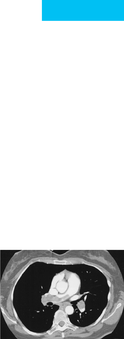
CT findings of pulmonary embolism include partial or
complete filling defects within the pulmonary artery due to
nonocclusive or occlusive thrombi, contrast material stream-
ing around a central thrombus, complete cutoff of vascular
enhancement, enlargement of an occluded vessel, and mural
defects (Figure 7–10). Parenchymal and pleural changes that
occur with pulmonary emboli are also easily detected on CT.
Oligemia of lung parenchyma distal to the occluded vessel
may be present. Pulmonary embolism may result in hemor-
rhage that is visible as ground-glass opacification or consoli-
dation on CT. An infarct may appear as a peripheral region of
consolidation, typically wedge-shaped with a central region of
lower attenuation due to uninfarcted lobules. Pleural effu-
sions are seen commonly. Acute right-sided heart failure may
occur secondary to pulmonary embolism and is suggested on
CT by right ventricular dilatation and deviation of the inter-
ventricular septum toward the left ventricle. On non-
contrast-enhanced CT, a region of increased attenuation
within the pulmonary artery may suggest acute central pul-
monary embolism. CT also may provide an alternative diag-
nosis in patients with suspected pulmonary embolism and
may demonstrate pulmonary edema, pneumonia, pericardial
disease, aortic dissection, or pneumothorax.
Pitfalls in the interpretation of CT pulmonary angiogra-
phy include breathing artifacts in patients unable to breath-
hold, inadequate contrast opacification of the pulmonary
arteries, and suboptimal visualization of vessels that are
obliquely oriented relative to the transverse imaging plane
(eg, the segmental branches of the right middle lobe and
lingula). Partially opacified veins may be confused with
thrombosed arteries, and hilar lymph nodes and mucus-
filled bronchi may be misinterpreted as thrombi.
Figure 7–10. Acute pulmonary embolism. CT pul-
monary angiogram demonstrates low-attenuation filling
defects within the right pulmonary artery and within the
left lower lobe pulmonary artery. There is distention of
the left lower lobe pulmonary artery.

CHAPTER 7
156
D. Pulmonary Angiography—Pulmonary angiography is gen-
erally considered the most sensitive and specific imaging method
for the diagnosis of pulmonary embolism. Angiography is indi-
cated when there is disagreement between the results of the CT
angiogram or
.
V/
.
Q scan and the clinical suspicion of pulmonary
embolism; when the CT angiography is indeterminate or the
.
V/
.
Q
scan is indeterminate or is of intermediate probability, when
there is a contraindication to anticoagulant therapy, or when
other studies are indeterminate, therapy involves more compli-
cated treatment such as an inferior vena cava filter, surgical
embolectomy, or thrombolytic therapy. Complications of pul-
monary angiography are related to the catheter and its manipu-
lation through the heart and to reactions to intravenous contrast
material. Dysrhythmias, heart block, cardiac perforation, cor
pulmonale, and cardiac arrest may occur. Relative contraindica-
tions to pulmonary angiography include elevated right ventricu-
lar and pulmonary arterial pressures, bleeding diathesis, renal
insufficiency or failure, left-sided heart block, and a history of
contrast material allergy. Pulmonary angiography can be per-
formed in all these settings if appropriate measures are taken to
reduce the risk of the procedure.
At angiography, the diagnosis of pulmonary embolus is
made when an intraluminal filling defect or an occluded pul-
monary artery is identified. Secondary findings include
decreased perfusion, delayed venous return, abnormal
parenchymal stain, and crowded vessels, which, though sug-
gestive, may be seen in other pulmonary disorders.
E. MRI—The role of MRI and MR angiography (MRA) in the
diagnosis of pulmonary embolism remains unclear.
Although central and peripheral emboli have been detected
on MRA, and physiologic information on ventilation and
perfusion may be provided, CT is more readily accessible and
suitable for imaging of the critically ill patient.
F. Imaging Techniques in Chronic Pulmonary Embolism—
Chronic pulmonary embolism may lead to right ventricular
failure and pulmonary arterial hypertension. Radiographic
findings include enlargement of the right side of the heart and
of the main and proximal pulmonary arteries and decreased
peripheral vascularity. Bronchial arteries distal to the occluded
pulmonary artery may become dilated. As in patients with
acute pulmonary embolism, evaluation of the patient with sus-
pected chronic pulmonary embolism includes
.
V/
.
Q scanning,
CT pulmonary angiography, and pulmonary angiography. In
addition to direct visualization of clot, other signs of chronic
pulmonary embolism seen on CT angiography include abrupt
narrowing of the vessel diameter, cutoff of distal lobar or seg-
mental arterial branches, webs and bands, and an irregular or
nodular arterial wall. Calcification within the vessel is uncommon
but may be present. Recanalization and eccentric location of
thrombi also suggest chronicity. Direct pulmonary angiography
may demonstrate similar findings. Findings indicative of pul-
monary arterial hypertension, such as enlargement of the main
pulmonary artery, pericardial fluid, and right ventricular enlarge-
ment, also may be seen on CT. Abnormalities of the lung
parenchyma may include local regions of decreased lung atten-
uation and perfusion.
Han D et al: Thrombotic and nonthrombotic pulmonary arterial
embolism: Spectrum of imaging findings. Radiographics
2003;23:1521–39. [PMID: 14615562]
The PIOPED Investigators: Value of the ventilation-perfusion scan
in acute pulmonary embolism. JAMA 1990;263:2753–9.
[PMID: 2332918]
Quiroz R et al: Clinical validity of a negative computed tomogra-
phy scan in patients with suspected pulmonary embolism: A
systematic review. JAMA 2005;293:2012–7. [PMID: 15855435]
Stein PD et al: Diagnostic pathways in acute pulmonary embolism:
Recommendations of the PIOPED II investigators. Radiology
2007;242:15–21. [PMID: 17185658]
Stein PD et al: Multidetector computed tomography for acute pul-
monary embolism. N Engl J Med 2006;354:2317–27. [PMID:
16738268]
Swensen SJ et al: Outcomes after withholding anticoagulation
from patients with suspected acute pulmonary embolism and
negative computed tomographic findings: A cohort study. Mayo
Clin Proc 2002;77:130–8. [PMID: 11838646]
Winer-Muram HT et al: Suspected acute pulmonary embolism:
Evaluation with multi-detector row CT versus digital subtrac-
tion pulmonary arteriography. Radiology 2004;233:806–15.
[PMID: 15564410]
Wittram C et al: CT angiography of pulmonary embolism:
Diagnostic criteria and causes of misdiagnosis. Radiographics
2004;24:1219–38. [PMID: 15371604]
Septic Pulmonary Emboli
ESSENTIALS OF RADIOLOGIC
DIAGNOSIS
Wedge-shaped or rounded peripheral opacities of vary-
ing size, usually multiple and more numerous in the
lower lobes.
Thin-walled cavities, sometimes with necrotic debris,
are common.
On CT scan, peripheral nodules, wedge-shaped periph-
eral opacities, and cavitation.
General Considerations
Infections of the right side of the heart or of the peripheral
veins may give rise to septic pulmonary emboli. Risk factors
include intravenous drug use, indwelling catheters, pelvic
inflammatory disease, organ transplantation, and immuno-
logic deficiencies such as lymphoma or AIDS. Infectious
thrombophlebitis also may result from infection of the phar-
ynx extending to the parapharyngeal space and internal jugular
venous system (Lemierre’s syndrome or postanginal sepsis).
Tricuspid valve endocarditis is the most common source of
septic emboli in the intravenous drug user. S. aureus is the most
commonly isolated organism, followed by streptococci.
