Bongard Frederic , Darryl Sue. Diagnosis and Treatment Critical Care
Подождите немного. Документ загружается.


137
007
Imaging Procedures
Kathleen Brown, MD
Steven S. Raman, MD
Nam C. Yu, MD
An unprecedented array of imaging options is now avail-
able to the physician in the ICU. The choice of a particular
imaging modality is occasionally difficult and should be
based on recommendations in the literature, local expert-
ise, type of equipment available, and the experience of the
radiologists. Given the increasing emphasis on cost-
effective practice, clinicians and radiologists must maxi-
mize the diagnostic and therapeutic yield of procedures
while minimizing costs. Optimal management of critically
ill patients also requires close communication between the
critical care team and the diagnostic and interventional
radiologist. An established practice of daily ICU radiology
rounds with the participation of the radiologist facilitates
this level of communication.
In a traditional model, all ICU films would be placed on
a designated mechanical film alternator within either the
ICU or the radiology department. With rapid advances in
imaging options and telecommunications feasibility, new
models for ICU imaging are being developed. In one model,
films are acquired electronically and displayed in a patient
archival and communications system (PACS) or on Web-
based servers. The PACS unit is able to display plain radi-
ographs, ultrasound and nuclear medicine studies, computed
tomography (CT), and magnetic resonance images (MRI).
Suboptimal exposures may be corrected in part by adjusting
contrast and window levels. High-resolution monitors may
be placed at designated sites in the ICU and throughout the
hospital. An ideal system integrates PACS with the hospital
information system (HIS) and the radiology information
system (RIS) to display clinical and radiologic information.
These systems may greatly improve the efficiency of clini-
cians, nurses, and support staff.
Although neurologic and musculoskeletal imaging stud-
ies play an important role in the care of the critically ill
patient, this chapter will limit discussion to imaging of the
chest and abdomen, with a focus on adult ICU patients.
IMAGING TECHNIQUES
Most radiographic examinations in the ICU are obtained at
the bedside utilizing conventional analog or digital equip-
ment. In most facilities, ultrasound and portable gamma
cameras for planar nuclear medicine studies are useful and
critical adjuncts for bedside examinations in the ICU. Other
imaging methods, including high-quality ultrasound, CT,
nuclear medicine techniques, and MRI, are used selectively
due to cost and transport issues. Interventional procedures,
either at the bedside or in the radiology suite, are also fre-
quently performed under imaging guidance.
Plain Radiography
Digital systems are being used increasingly in the ICU for
portable radiography. With these systems, images are
obtained using a photo-stimulable phosphor imaging plate
instead of film. The exposed imaging plate is scanned, read,
and processed by computer, and the image can be transmit-
ted to an ICU console or viewed as a hard copy on a conven-
tional view box. Chest radiographs are the most common
imaging examination, accounting for approximately 40% of
the volume in a radiology department. As many as one-third
of these chest radiographs may be obtained at the bedside
(portable radiographs), and in the ICU almost all chest radi-
ographs are taken using the portable technique. The utility
and effectiveness of routine daily portable chest radiographs
have been studied, and—despite limitations of the technique—
these films play an important role in identifying and follow-
ing pulmonary and cardiac disorders in ICU patients. Chest
radiographs are also used to evaluate the positions of and
complications from catheters and support devices used in
the care of critically ill patients.
Likewise, imaging of the abdomen generally should begin
with plain radiographs, which provide a readily accessible
Copyright © 2008 by The McGraw-Hill Companies, Inc. Click here for terms of use.

CHAPTER 7
138
means of diagnosing perforation, bowel obstruction, and
ileus. However, because the overall sensitivity of plain radi-
ographs remains low, further imaging with CT may be nec-
essary to confirm suspected perforation and related
complications (eg, abscess) and to inspect the features of the
bowel walls and surrounding fat. Supine radiographs are most
appropriate for verifying nasogastric or feeding tube place-
ment and for investigation of renal stones and possible ileus
or bowel obstruction. Additional views (ie, semiupright, left
lateral decubitus, and cross-table lateral) may be helpful in
cases of bowel perforation, ileus, or obstruction.
Ultrasound
Ultrasound examination at the bedside in the ICU is relatively
inexpensive and does not use ionizing radiation. In the thorax,
ultrasound is used most often to evaluate and localize pleural
fluid collections, to determine whether such collections are
free or loculated, and as a guide to thoracentesis. Ultrasound is
also helpful in clarifying peridiaphragmatic processes because
the diaphragm is easily visualized, allowing differentiation of
supradiaphragmatic and infradiaphragmatic fluid collections.
The greatest utility of ultrasound, however, is in the evalua-
tion of abdominal disease. Ultrasound provides rapid assess-
ment of hepatobiliary and genitourinary disease and may be
used to guide percutaneous drainage of intraabdominal
abscesses. It allows rapid evaluation of the hepatobiliary sys-
tem, gallbladder, kidneys, pelvic organs, and scrotal disorders.
Visualization of vascular perfusion and parenchymal flow is
a useful feature, especially in transplanted organs.
Ultrasound is also indispensable for guidance of bedside pro-
cedures such as central line placement, cholecystostomies,
biopsies, and drainage of fluid collections.
Computed Tomography
By virtue of multiplanar imaging capabilities and improved
contrast resolution, multidetector CT (MDCT) has been
shown to be very valuable in increasing diagnostic accuracy
and guiding therapeutic procedures for critically ill patients.
MDCT allows for more rapid scanning of patients, with
imaging of the entire chest, abdomen, and pelvis with thin
sections during a single breath-hold. Such short acquisition
times have facilitated the use of CT for evaluation of vascu-
lar disorders such as aortic dissection and pulmonary
embolism. CT also allows for improved characterization of
pulmonary diseases, particularly acute respiratory distress
syndrome (ARDS), and is a critical diagnostic tool for the
evaluation of an acute abdomen.
Transportation of the ICU patient to the CT scanner
requires a coordinated effort from hospital personnel, includ-
ing ICU physicians and nurses, respiratory therapists, radiol-
ogy technologists, and radiologists. Careful monitoring
during transport and during the procedure is essential and
must include arrhythmia monitoring and pulse oximetry.
Nuclear Scintigraphy
Nuclear scintigraphy has a number of applications in the
critically ill patient. Myocardial perfusion and infarct scan-
ning in cardiac disease, ventilation-perfusion scanning in
patients with suspected pulmonary embolism, evaluation of
gastrointestinal hemorrhage and acute cholecystitis, and
localization of occult infection are among the most common
indications for radionuclide imaging in the ICU patient.
Magnetic Resonance Imaging
MRI has supplanted CT in the evaluation of many disorders
because it does not employ ionizing radiation, because it pro-
vides excellent differentiation of vascular and nonvascular
structures without the use of intravenous contrast material, and
because it provides cross-sectional images in multiple planes. It
is generally considered the single best imaging method for eval-
uation of the CNS, head and neck, liver, and musculoskeletal
system. However, in many cases, MRI is not feasible in the eval-
uation of the critically ill patient because of interference caused
by ferromagnetic monitoring devices, the difficulty of ade-
quately ventilating and monitoring patients within the narrow
MRI gantry, and long scan times. MRI may be appropriate in
selected diagnostic dilemmas if MR-compatible equipment and
coordinated effort among caregivers can be arranged.
Mayo PH, Doelken P: Pleural ultrasonography. Clin Chest Med
2006;27:215–27. [PMID:16716814]
Nicolaou S et al: Ultrasound-guided interventional radiology in crit-
ical care. Crit Care Med 2007;35:S186–97. [PMID: 17446778]
Redfern RO et al: A picture archival and communication system
shortens delays in obtaining radiographic information in a
medical intensive care unit. Crit Care Med 2000;28:1006–13.
[PMID: 10809274]
Trotman-Dickenson B: Radiology in the intensive care unit (part 1).
J Intensive Care Med 2003;18:198–210. [PMID: 15035766]
Trotman-Dickenson B: Radiology in the intensive care unit (part 2).
J Intensive Care Med 2003;18:239–52. [PMID: 15035758]
IODINATED CONTRAST AGENTS
Adverse reactions to iodinated contrast agents occur at low
rates but are encountered not infrequently given their wide-
spread use. Older ionic agents, newer nonionic agents, and
the newest nonionic isoosmolar agents are available, with
the oldest agents having the highest incidence of adverse
reactions and the newest agents having a significantly lower
incidence. Idiosyncratic reactions range from benign
urticaria to, very rarely, life-threatening hypotension, laryn-
geal edema, and bronchospasm. These events are not consid-
ered truly allergic in nature because they are not
antibody-mediated and are inconsistently reproducible with
subsequent administrations. Pretreatment with corticos-
teroids appears to be effective for mild events, but corticos-
teroids should not be used in patients with a history of severe
reaction. In the latter situation, an alternative such as MRI with

IMAGING PROCEDURES
139
gadolinium contrast or carbon dioxide angiography should
be considered. Contrary to popular belief, allergy to shellfish
is not predictive of reactions to iodinated contrast agents.
Contrast nephropathy is another important complication
of intravascular iodinated contrast use and occurs in the setting
of preexisting renal compromise, most often due to dehydra-
tion, surgery, nephrotoxic drugs, or long-standing diabetes.
Again, the incidence is highest with the oldest agents and low-
est with the isoosmolar nonionic agents. Although the serum
creatinine level is a convenient measure of renal function, cre-
atinine clearance should be calculated for a more reliable
estimation—less than 25 mL/min or 25–50 mL/min with risk
factors identifying high-risk patients. Potentially effective pre-
ventive strategies include adequate intravenous hydration with
normal saline or sodium bicarbonate solution and administra-
tion of N-acetylcysteine. Metformin should be stopped until
48 hours following contrast use to avoid possible lactic acidosis
in the event of contrast nephrotoxicity. Rather than using a uni-
versal creatinine level cutoff, the decision to use contrast agents
should be made on a case-by-case basis, carefully weighing the
need for the study in high-risk patients. In affected patients, the
serum creatinine level peaks at 4–7 days and gradually normal-
izes. Progression to end-stage renal disease is exceptionally rare.
Bettmann MA: Frequently asked questions: Iodinated contrast
agents. Radiographics 2004;24:S3–10. [PMID: 15486247]
Merten GJ et al: Prevention of contrast-induced nephropathy with
sodium bicarbonate: A randomized, controlled trial. JAMA
2004;291:2328–34. [PMID: 15150204]
Meschi M et al: Facts and fallacies concerning the prevention of
contrast medium-induced nephropathy. Crit Care Med
2006;34:2060–8. [PMID: 16763513]
Tepel M et al: Prevention of radiographic-contrast-agent-induced
reductions in renal function by acetylcysteine. N Engl J Med
2000;343:180–4. [PMID: 10900277]
USE OF CENTRAL VENOUS CATHETERS
FOR CONTRAST INJECTION
Peripheral veins are the preferred routes of contrast agent
administration in imaging. When peripheral access is diffi-
cult, existing central venous catheters (CVCs) may be consid-
ered, with a few caveats. Intraluminal pressure limitations
may result in low contrast flow rates, producing a suboptimal
study, or catheter rupture may occur during rapid power
injection of the relatively viscous contrast material. While
most catheter manufacturers do not provide specific instruc-
tions in this regard and practice standards have not been
established, the following general precautions may be useful:
(1) High flow rates (>2 mL/s) should be avoided in most
temporary or tunneled CVCs, (2) silicone-type peripherally
inserted central catheters (PICCs) should not be used, (3) for
multilumen catheters, the largest-caliber port should be used
when possible, (4) Groshong-valve lines should not be used,
(5) pulmonary or systemic arterial lines should not be used,
and (6) catheter integrity and patency should be checked
before and after injection. Since no established guidelines are
available, hospital personnel should be knowledgeable about
the specific catheters used at their institution.
Funaki B: Central venous access: A primer for the diagnostic radi-
ologist. AJR 2002;179:309–18. [PMID: 12130425]
Salis AI et al: Maximal flow rates possible during power injection
through currently available PICCs: An in vitro study. J Vasc
Interv Radiol 2004;15:275–81. [PMID: 15028813]
Reynolds NJ, Grosvenor LJ: Problems with the rapid powered
injection of radiology contrast through multilumen catheters.
Anaesthesia 2003;58:923–4. [PMID: 12911383]
IMAGING OF SUPPORT & MONITORING
DEVICES IN THE ICU
Endotracheal & Tracheostomy Tubes
Both endotracheal intubation and tracheostomy may cause
potentially serious complications. Malpositioning of the
endotracheal tube into the right main stem bronchus occurs
in approximately 9% of endotracheal intubations. Such mal-
positioning may lead to atelectasis of the left lung, hyperin-
flation of the right lung, and possible pneumothorax. The
clinical assessment of tube location is frequently inaccurate,
and a chest radiograph should be obtained immediately fol-
lowing intubation. Tubes currently in use are usually radi-
ographically visible by virtue of a metallic wire or barium
marker in the wall of the tube. Periodic radiographs are
required to exclude inadvertent displacement of the tube by
cough, suctioning, or the weight of the respiratory apparatus.
Since endotracheal tubes are typically fixed in position at
the nose or mouth, flexion and extension of the neck may result
in motion of the tube relative to the carina, with the tube
descending during flexion and ascending during extension.
With the neck in neutral position, the ideal position of the tube
tip is 5–7 cm above the carina, which allows for a tolerable
change in tube position during flexion and extension. In 90%
of patients, the carina projects between the fifth and seventh
thoracic vertebrae on the portable radiograph; when the carina
cannot be clearly seen, the ideal positioning of the endotracheal
tube is at the T2–T4 level. The aortic arch also may be used to
estimate tube location because the carina is typically at the level
of the undersurface of the aortic arch. The balloon cuff should
not be greater in diameter than the trachea because cuff over-
inflation can cause pressure necrosis of the tracheal wall.
Inadvertent placement of the endotracheal tube into the
esophagus is uncommon but may be catastrophic when it
does occur. Esophageal intubation may be difficult to diag-
nose on the portable chest film because the esophagus fre-
quently projects over the tracheal air column. Gastric or
distal esophageal distention, location of the tube lateral to
the tracheal air column, and deviation of the trachea sec-
ondary to an overinflated intraesophageal balloon cuff are
radiographic signs of esophageal intubation. The right posterior
oblique view with the patient’s head turned to the right
CHAPTER 7
allows ease of separation of the esophagus and trachea and
should be obtained in equivocal cases.
Intubation may result in injury to the trachea, with tra-
cheal stenosis developing in approximately 19% of patients
following endotracheal intubation and approximately 65%
of patients with tracheostomy. In patients with translaryn-
geal intubation, the most frequent sites of stenosis are the
cuff site and the subglottic region.
Tracheostomy is typically performed in the patient who
requires relatively long-term ventilatory support. Although
the surgical mortality rate is less than 2%, the long-term
complication rate may be as high as 60%. Pneumothorax,
pneumomediastinum, subcutaneous emphysema, hemor-
rhage, and tube malposition may occur as early complica-
tions, whereas late complications include tracheal stenosis,
tracheo-innominate artery fistula, tracheoesophageal fistula,
stomal infection, aspiration, and tube occlusion. In addition,
the incidence of nosocomial pneumonia is increased second-
ary to airway bacterial colonization.
Central Venous Catheters
Central venous catheters are used frequently in the ICU patient
for venous access, especially for purposes of parenteral alimen-
tation, monitoring central venous pressure, and hemodialysis.
Such catheters are visible on the chest radiograph, and knowl-
edge of normal thoracic venous anatomy is required to assess
catheter location. The subclavian vein, the internal jugular vein,
and the femoral veins are the sites of venous access used most
commonly. Central venous lines inserted via a thoracic vein are
optimally positioned when the tip is past the valves in the sub-
clavian or brachiocephalic veins within the superior vena cava.
The preferred location for hemodialysis or pheresis catheters is
subject to debate, however, because some physicians believe
that catheter durability and performance are improved by
placement of the catheter tip within the upper right atrium.
Union of the subclavian and internal jugular veins to form
the brachiocephalic vein usually occurs behind the sternal end
of the corresponding clavicle. Whereas the right brachio-
cephalic vein has a vertical course as it forms the superior vena
cava, the left brachiocephalic vein crosses the mediastinum
from left to right in a retrosternal position to enter the superior
vena cava. The radiographic location of the superior vena cava
may be assessed relative to the tracheobronchial angle, with the
upper border of the superior vena cava usually just superior to
the angle of the right main stem bronchus and the trachea. The
junction of the superior vena cava and right atrium is at the
approximate level of the lower aspect of the bronchus inter-
medius. Changes in catheter location may occur with change in
patient position and changes in respiration.
Approximately one-third of catheters are incorrectly
positioned at the time of the initial chest radiograph. The
malpositioned catheter tip may result in venous thrombosis
or perforation as well as inaccurate venous pressure readings.
Positioning of the catheter tip within the right atrium may
result in cardiac perforation and tamponade, whereas a right
ventricular location may result in arrhythmias secondary to
irritation of the endocardium or interventricular septum.
Complications of central venous catheterization include
pneumothorax, hemothorax, and perforation, which may
result in pericardial effusion, hydrothorax, mediastinal
hemorrhage, or ectopic infusion of intravenous solutions
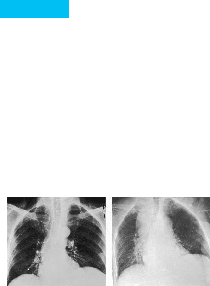
(Figure 7–1). Less common complications include air
A
B
Figure 7–1. Mediastinal hematoma following attempted central venous catheterization. A. Mediastinum appears
unremarkable prior to catheter placement. B. Following attempted central line placement, there is widening of the
superior mediastinum secondary to mediastinal hemorrhage due to a lacerated subclavian artery.
140

IMAGING PROCEDURES
141
embolism and catheter fracture or embolism. The incidence of
pneumothorax ranges between 1% and 12% and is higher with
a subclavian approach than with an internal jugular approach.
Pneumothorax may be clinically occult, and a chest radiograph
should be obtained to exclude a pneumothorax following line
placement. A radiograph should be obtained even following an
unsuccessful attempted line placement and is more critical
when contralateral venous cannulation is anticipated to avoid
the development of bilateral pneumothoraces. Although sel-
dom obtained in ICU patients, the cross-table lateral view may
be helpful to localize catheters malpositioned in the internal
mammary or azygos vein or in extravascular positions.
Venous air embolism is an uncommon complication of
central venous catheterization. Radiographically, air in the
main pulmonary artery is diagnostic, but other features include
focal oligemia, pulmonary edema, and atelectasis. Intracardiac
air or air within the pulmonary artery is seen easily on CT.
Long-term complications of venous access devices include
delayed perforation, pinch-off syndrome, thrombosis,
catheter knotting, and catheter fragmentation. Left-sided
catheters have a greater risk for perforation, with increased
risk in catheters abutting the right lateral wall of the superior
vena cava. In pinch-off syndrome, the catheter lumen is com-
promised by compression between the clavicle and the first
rib, leading to catheter malfunction and possible catheter
fracture. This is frequently first observed as subtle focal nar-
rowing of the catheter as it crosses the intersection of clavicle
and rib. As increasing numbers of chronically ill patients with
long-term venous catheters—including liver and bone mar-
row transplant recipients—are transferred to the ICU during
their hospital course, more such complications may be seen.
Access to the central venous system may be achieved
through a peripherally inserted central catheter (PICC)
placed via the antecubital fossa. These smaller catheters
course to the superior vena cava and may be associated with
fewer complications than catheters inserted via the internal
jugular or subclavian approach.
Pulmonary Artery Catheters
The pulmonary artery catheter has enhanced the manage-
ment of the ICU patient, allowing monitoring of left atrial
and left ventricular end-diastolic pressures and calculation of
vital data such as cardiac output and vascular resistance. The
catheter tip should lie within a large central pulmonary
artery; the ideal position for the pulmonary artery catheter is
within the right or left main pulmonary artery, below the
level of the left atrium. The catheter tip when deflated should
not be peripheral to the proximal interlobar arteries.
Complications associated with their use include arrhyth-
mias, pneumothorax, vascular perforation, venous air
embolism, and catheter-related sepsis. Knotting, kinking,
and coiling of the catheter also occur.
Pulmonary infarction, thrombosis, pulmonary artery rup-
ture, and infection represent other major complications asso-
ciated with indwelling pulmonary artery catheters. There is a
7% incidence of pulmonary ischemic lesions due to direct
injury from the use of pulmonary artery catheters. The major-
ity of these lesions are thought to be due to vascular occlusion
by the catheter itself. Continuous wedging of the catheter tip
in a peripheral pulmonary artery and central pulmonary
artery obstruction by the inflated balloon have been cited as
precipitating causes. In a smaller number of cases, emboli
arose from peripheral thrombosis around the catheter.
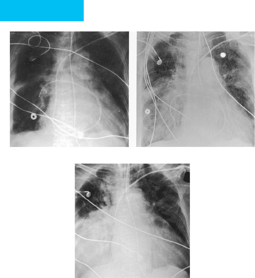
Pulmonary infarction secondary to a pulmonary artery
catheter has a radiographic appearance like that of infarction
from other causes. Typically, a wedge-shaped parenchymal
opacity is seen in the distribution of the vessel distal to the
catheter (Figure 7–2). The presence of a pleural effusion is
variable. Management consists of removal of the catheter;
anticoagulation is generally not required. Resolution of con-
solidation usually occurs in 2–4 weeks.
Pulmonary artery rupture is a catastrophic complication
of pulmonary artery catheterization, with a reported mortal-
ity rate of 46%. The incidence is low—no more than 0.2% of
catheter placements. Risk factors include pulmonary hyper-
tension, advanced age, and improper balloon location or
inflation. The mortality rate increases in anticoagulated
patients. Pseudoaneurysm formation has been reported sec-
ondary to rupture or dissection by the balloon catheter tip.
This appears radiographically as a well-defined nodule at the
site of the aneurysm, but it may be obscured initially by
extravasation of blood into the adjacent air spaces. Chest
radiographic findings often precede clinical manifestations,
and death due to rupture of pseudoaneurysm may occur
weeks following catheterization. The CT appearance of a
pulmonary artery pseudoaneurysm has been described as a
sharply defined nodule with a surrounding halo of faint
parenchymal density. Pulmonary artery pseudoaneurysm
now may be treated in some patients with transcatheter
embolization rather than surgical resection.
Location of the catheter tip should be monitored with
serial radiographs. Softening of the catheter over time may
result in migration of the catheter tip peripherally.
Redundancy of the catheter within the right heart favors
peripheral migration, and the intracardiac loop gradually
becomes smaller (see Figure 7–2).
Intraaortic Balloon Counterpulsation
Intraaortic balloon counterpulsation is used to improve car-
diac function in patients with cardiogenic shock and in the
perioperative period in cardiac surgery patients. The device
consists of a fusiform inflatable balloon surrounding the
distal portion of a catheter that is placed percutaneously
from a femoral artery into the proximal descending thoracic
aorta. The balloon is inflated during diastole, thereby
increasing diastolic pressure in the proximal aorta and
increasing coronary artery perfusion. During systole, the
balloon is forcibly deflated, allowing aortic blood to move
distally and decreasing the afterload against which the left
ventricle must contract, thus decreasing left ventricular
workload. The timing of inflation and deflation is controlled
by the ECG.
CHAPTER 7
142
The tip of the balloon ideally should be positioned just dis-
tal to the origin of the left subclavian artery at the level of the
aortic knob, maximizing the effect on the coronary arteries
while reducing the possibility of occlusion of the left subclavian
artery, embolization to cerebral vessels, or occlusion of the
abdominal vessels by the balloon. Complications associated
with the device are most often secondary to malpositioning of
the catheter and include obstruction of the subclavian artery
and cerebral embolism. Aortic dissection has been described,
and an indistinct aorta on chest radiographs has been suggested
as an early clue to intramural location, requiring confirmation
by angiography. Balloon leak or rupture also has been described.
A
B
C
Figure 7–2. Lung infarction secondary to pulmonary artery catheterization. A. Initial radiograph after catheterization
shows the tip of the catheter at the level of the right interlobar pulmonary artery. Mild redundancy of the catheter is present
within the dilated heart. B. At 24 hours, the patient developed hemoptysis. Radiograph now shows migration of the catheter
into a segmental arterial branch with increased density in the right lower lobe. C. Follow-up film demonstrates dense consol-
idation of the right middle and lower lobes secondary to infarction. (Reproduced, with permission, from Aberle DA, Brown K:
Radiologic considerations in the adult respiratory distress syndrome. Clin Chest Med 1990;2:737–54. Copyright 1990 Elsevier.)
IMAGING PROCEDURES
143
Cardiac Pacemakers and Automatic
Implantable Cardioverter Defibrillators
Cardiac pacemakers can be inserted by three approaches:
transvenous, epicardial, and subxiphoid. Most often the
transvenous approach is used, whereby wires are introduced
via the subclavian or jugular vein and fluoroscopically
guided into the right atrium and ventricle.
When viewed on a chest radiograph, the pacemaker lead
should curve gently throughout its course; regions of sharp
angulation will have increased mechanical stress and
enhance the likelihood of lead fracture. Excessive lead length
may predispose to fracture secondary to sharp angulation or
may perforate the myocardium, and a short lead can become
dislodged and enter the right atrium. Leads also may
become displaced and enter the pulmonary artery, coronary
sinus, or inferior vena cava. When possible, a lateral chest
radiograph is recommended to confirm pacemaker lead
location, with the electrodes located at least 3 mm deep to
the epicardial fat stripe. Other complications include venous
thrombosis or infection, either at the pulse generator pocket
or within the vein. Myocardium perforation may result in
hemopericardium and cardiac tamponade.
Biventricular pacing or cardiac resynchronization therapy is
a relatively new treatment for severe chronic heart failure. In
patients with dilated cardiomyopathy and intraventricular con-
duction delay, biventricular or left ventricular pacing can syn-
chronize contraction and increase cardiac output and exercise
tolerance. Percutaneous lead placement into a coronary vein via
the coronary sinus allows for left ventricular pacing. Many of
these patients also will have intravascular defibrillators because
of the risk of ventricular arrhythmias. The automatic
implantable cardioverter defibrillator (AICD) is used for treat-
ment of ventricular tachyarrhythmias unresponsive to conven-
tional antiarrhythmic drugs. Earlier devices consisted of a fine
titanium mesh placed on the cardiac surface and attached to a
generator source that provided an electrical output in the event
of ventricular arrhythmia. Devices currently in use typically are
combined with a cardiac pacemaker. Radiographs are used to
assess the location of wires.
Nasogastric Tubes
Nasogastric tubes are used frequently to provide nutrition
and administer oral medications as well as for suctioning gas-
tric contents. Ideally, the tip of the tube should be positioned
at least 10 cm beyond the gastroesophageal junction. This
ensures that all sideholes are located within the stomach and
decreases the risk of aspiration. Complications of nasogastric
intubation include esophagitis, stricture, and perforation.
Small-bore flexible feeding tubes have been developed
to facilitate insertion and improve patient comfort.
However, inadvertent passage of the nasogastric tube into
the tracheobronchial tree is not uncommon, most often
occurring in the sedated or neurologically impaired
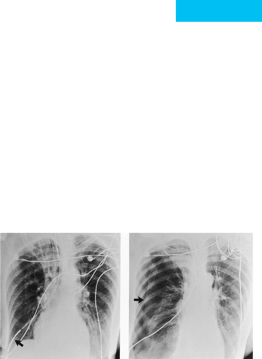
patient. In patients with endotracheal tubes in place, low-
pressure, high-volume balloon cuffs do not prevent passage
of a feeding tube into the lower airway. If sufficient feeding
tube length is inserted, the tube actually may traverse the
lung and penetrate the visceral pleura (Figure 7–3).
Figure 7–3. Malpositioned feeding tube. A. Feeding tube courses via the right main stem bronchus with the tip
(arrow) overlying the right costophrenic angle. An endotracheal tube is present. B. Following removal of the feeding
tube, a pneumothorax is seen (arrow).
A
B

CHAPTER 7
144
Removal of the tube from an intrapleural location may
result in tension pneumothorax, and preparations should
be made for potential emergent thoracostomy tube place-
ment at the time of removal.
In addition to feeding tubes, balloon tamponade tubes
occasionally are used for nasogastric intubation in the treat-
ment of bleeding esophageal and gastric varices. The balloon
can be easily recognized when distended, and correct posi-
tioning can be evaluated radiographically. Esophageal rup-
ture complicates approximately 5% of cases in which balloon
tamponade tubes are used.
Chest Tubes
Thoracostomy tubes (“chest tubes”) are used for the evacua-
tion of air or fluid from the pleural space. When chest tubes
are used for relief of pneumothorax, apical location of the tip
of the tube is most effective, whereas a tube inserted to drain
free-flowing effusions should be placed in the dependent
portion of the thorax. Chest radiographs, ultrasound, or CT
should be used to guide correct placement of the tube for
adequate drainage of a loculated effusion. Failure of the chest
tube to decrease the pneumothorax or the effusion within
several hours should arouse suspicion of a malpositioned
tube. Tubes located within the pleural fissures are usually less
effective in evacuating air or fluid collections. An interfis-
sural location is suggested by orientation of the tube along
the plane of the fissure on frontal radiographs and by lack of
a gentle curvature near the site of penetration of the pleura,
indicating failure of the tube to be deflected anteriorly or
posteriorly in the pleural space. The lateral view may be con-
firmatory. Uncommonly, thoracostomy tubes may penetrate
the lung, resulting in pulmonary laceration and bron-
chopleural fistula. Unilateral pulmonary edema may occur
following rapid evacuation of a pneumothorax or pleural
effusion that is of long standing or has produced significant
compression atelectasis of lung.
Cascade PN et al: Radiographic appearance of biventricular pacing
for the treatment of heart failure. AJR 2001;177:1447–50.
[PMID: 11717105]
Funaki B: Central venous access: A primer for the diagnostic radi-
ologist. AJR 2002;179:309–18. [PMID: 12130425]
Gayer G et al: CT diagnosis of malpositioned chest tubes. Br J
Radiol 2000;73:786–90. [PMID: 11089474]
Hunter TB et al: Medical devices of the chest. Radiographics
2004;24:1725–46. [PMID: 15537981]
Maecken T, Grau T: Ultrasound imaging in vascular access. Crit
Care Med. 2007;35:S178–85. [PMID: 17446777]
Salem MR: Verification of endotracheal tube position. Anesthesiol
Clin North Am 2001;19:813–39. [PMID: 11778382]
Vesely TM: Central venous catheter tip position: A continuing con-
troversy. J Vasc Intervent Radiol 2003;14:527–34. [PMID:
12761305]
IMAGING IN PULMONARY DISEASES
Routine Daily Chest Radiographs:
Technical Considerations & Utility
Portable chest radiographs are frequently obtained on a daily
basis on ICU patients and as indicated by changes in their
clinical situation. Several factors related to portable radiog-
raphy may lead to difficulty in evaluation of radiographs in
a critically ill patient. The equipment used for portable
radiographs requires longer exposure time than standard
radiographs obtained in the radiology department, some-
times resulting in artifacts due to respiratory, cardiac, and
gross patient motion. Inadequate exposure may result
from the limited power output of portable equipment.
Special attention must be paid to the multiple monitoring
devices required by the ICU patient, and considerable physical
effort by the technologists is required to transport portable
equipment.
Limitations imposed by the portable technique often
complicate image interpretation. Almost all portable chest
radiographs are taken with the patient supine and with the
film placed behind the back of the patient (anteroposterior)
rather than in the conventional upright, posteroanterior
position used in the radiology department. Supine chest
radiographs result in decreases in lung volume and can alter
the size and appearance of the lungs, the pulmonary vascula-
ture, and the mediastinum. Anteroposterior chest radi-
ographs cause cardiac magnification, making evaluation of
true cardiac size more difficult. Inspiratory films may be dif-
ficult to obtain because of respiratory distress, pain, sedation,
or alterations in mental status. These technical limitations
complicate diagnostic interpretation. Nonetheless, portable
radiography continues to be a primary method of imaging
critically ill patients.
The utility of daily radiographs may depend on the
underlying disease process. Routine daily radiographs are
of greatest utility in patients with pulmonary or compli-
cated cardiac disease. The American College of Radiology
Thoracic Expert Panel concluded that daily chest radi-
ographs are indicated for patients with acute cardiopul-
monary problems and those receiving mechanical
ventilation. In patients requiring cardiac monitoring or
stable patients admitted for extrathoracic disease, an ini-
tial admission film is recommended. Additional radi-
ographs are indicated when new support devices are
placed or a specific question arises regarding cardiopul-
monary status.
Krivopal M et al: Utility of daily routine portable chest radiographs
in mechanically ventilated patients in the medical ICU. Chest
2003;123:1607–14. [PMID: 12740281]
Tocino I et al: Routine daily portable x-ray. American College of
Radiology. ACR Appropriateness Criteria. Radiology 2000;215:
S621–6. [PMID: 11037473]
IMAGING PROCEDURES
145
Atelectasis
ESSENTIALS OF RADIOLOGIC
DIAGNOSIS
Shift in position of a fissure or change in position of hila
or mediastinum.
Elevation of hemidiaphragm.
Compensatory hyperexpansion of uninvolved lobes.
Increased opacity of the atelectatic lung.
Air bronchograms.
Narrowing of rib interspaces.
General Considerations
Atelectasis is the most common pulmonary parenchymal
abnormality seen in ICU patients. Signs and symptoms of
atelectasis are nonspecific, and atelectasis may coexist with
other pulmonary diseases. Multiple factors contribute to the
development of atelectasis. In the bedridden patient,
hypoventilation results in atelectasis of the dependent lung.
Central neurogenic depression, anesthesia, or splinting may
decrease alveolar volume, reducing surfactant and promot-
ing diffuse microatelectasis. Bronchial obstruction from
retained secretions and mucous plugging may lead to post-
obstructive collapse of the distal lung, particularly in patients
with pulmonary infection or chronic airway disorders. In the
intubated or postoperative patient, other factors are contrib-
utory. A malpositioned endotracheal tube with right main
stem bronchial intubation can cause atelectasis of the non-
ventilated left lung. Following cardiac surgery, left lower lobe
collapse occurs frequently due in part to the weight of the
heart unsupported by pericardium, which compresses the
left lower lobe bronchus. Phrenic nerve paresis secondary to
intraoperative cold cardioplegia results in diaphragmatic ele-
vation and is also thought to contribute to lower lobe atelec-
tasis. Pleural processes, including pneumothorax and pleural
effusion, may also result in atelectasis.
Radiographic Features
The radiographic appearance of atelectasis depends largely on
the degree and cause of lung collapse. Findings noted on the
chest radiograph in atelectasis range from subtle diminution in
lung volume without visible opacification to complete opacifi-
cation of a segment, lobe, or lung. Dependent atelectasis occur-
ring in supine patients may be demonstrated on thoracic CT
even in healthy individuals but is usually not appreciated on
plain chest radiography. Linear bands of opacity may be seen in
“discoid” or “platelike” atelectasis, whereas a patchy opacity is
seen with atelectasis of lung subtended by a segmental or sub-
segmental bronchus. With more extensive volume loss such as
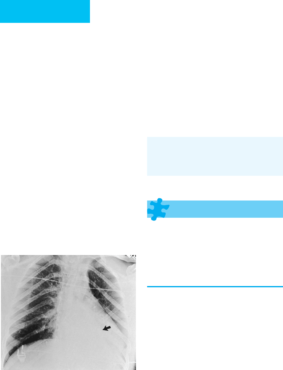
collapse of an entire lobe or lung, radiographic signs include an
increase in opacity of the atelectatic lung; shift in the position
of a fissure; change in the position of the mediastinum, hila, or
diaphragm; and hyperexpansion of the uninvolved lung
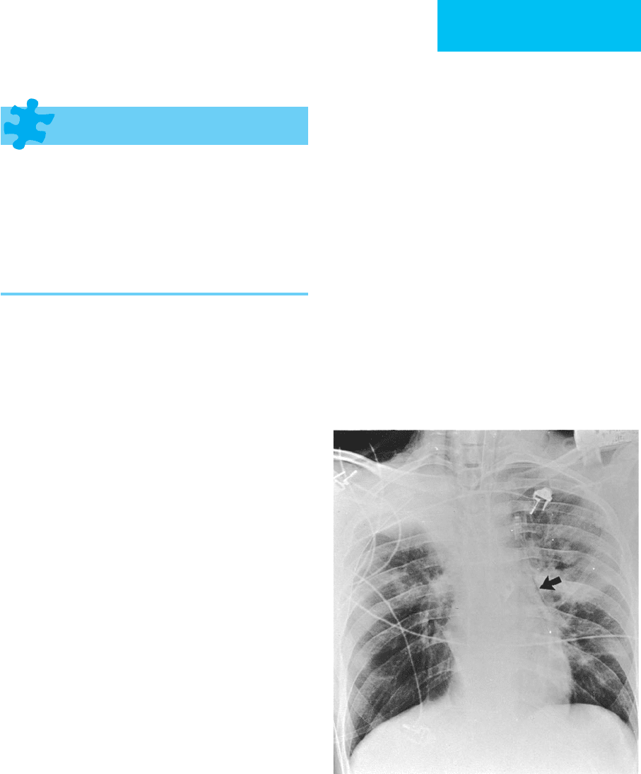
(Figure 7–4). In some cases, signs of volume loss may be absent
because of exudation of fluid into the atelectatic lung.
Air bronchograms are linear lucencies coursing through
opacified lung and represent patent bronchi and bronchi-
oles surrounded by opacified air spaces. Air bronchograms
are radiographically nonspecific and occur in any disorder
in which patent air-containing bronchi are situated within
consolidated lung, including atelectasis, pulmonary edema,
pneumonia, and hemorrhage. The presence of air bron-
chograms is also variable in atelectasis and depends on the
patency of the major airways and the cause of atelectasis.
Air bronchograms may be useful predictors of the effective-
ness of bronchoscopy in patients with lobar collapse.
Patients without air bronchograms are more likely to
demonstrate improvement following fiberoptic bron-
choscopy than those with air bronchograms. The absence of
air bronchograms in lobar collapse suggests that central
Figure 7–4. Atelectasis in a 22-year-old man with
status asthmaticus. The right upper lobe is opaque, and
there is elevation of the minor fissure consistent with
right upper lobe collapse. Areas of increased density in
the left lung are also due to atelectasis. Lucency adjacent
to the left heart border secondary to pneumomedi-
astinum is present (arrow), and there is subcutaneous
emphysema in the right supraclavicular region.
CHAPTER 7
146
airways may be plugged by secretions which by virtue of
their proximal location are amenable to bronchoscopic
removal. In contrast, the presence of air bronchograms sug-
gests that the collapse is more apt to be due to small airway
collapse or peripheral mucous plugs that are not effectively
treated by therapeutic fiberoptic bronchoscopy.
The left lower lobe is the most frequent location of lobar
atelectasis, with collapse occurring two to three times more
often in the left lower than in the right lower lobe. The cause
is uncertain, although many of the factors cited earlier are
contributory. The radiographic features of left lower lobe col-
lapse include a triangular opacity in the retrocardiac region
and loss of definition of the descending aorta and left hemidi-
aphragm—as well as other signs of volume loss outlined ear-
lier (Figure 7–5). Adequate penetration and patient
positioning are important in assessing left lower lobe disease.
Left lower lobe collapse may be falsely diagnosed secondary
to faulty radiologic technique. Cephalic angulation of the
radiographic beam by 10–15 degrees (lordotic positioning)
may cause projection of extrapleural fat onto the base of the
left lung and result in loss of tangential imaging of the apex of
the hemidiaphragm and subsequent loss of definition of the
diaphragm in the absence of left lower lobe disease. In
instances in which patients are examined radiographically
with even a small degree of lordosis, loss of definition of the
diaphragm therefore cannot be assumed to be secondary to
left lower lobe collapse. Ancillary findings, including depres-
sion of the hilum, crowding of vessels, and air bronchograms,
must be used to diagnose true left lower lobe disease.
Unusual appearances of lobar atelectasis may occur and
make diagnosis difficult. Atelectasis with marked volume
loss may be caused by peripheral airway obstruction and is
frequently chronic and easily missed. Atelectasis also may
present as a mass and be confused with tumor. Recognition
of the anatomic alterations described earlier is required for
differentiation.
Many other causes of parenchymal opacification may be
confused with atelectasis, including pneumonia and pul-
monary infarction. In addition to other features previously
discussed, temporal sequence may be helpful in distinguish-
ing atelectasis from other causes of focal parenchymal opaci-
fication. Whereas atelectasis may appear within minutes to
hours and also may clear rapidly, pneumonia and infarction
typically resolve over days to weeks.
Ashizawa K et al: Lobar atelectasis: Diagnostic pitfalls on chest
radiography. Br J Radiol 2001;74:89–97. [PMID: 11227785]
Tsai KL, Gupta E, Haramati LB: Pulmonary atelectasis: A frequent
alternative diagnosis in patients undergoing CT-PA for sus-
pected pulmonary embolism. Emerg Radiol 2004;10:282–6.
[PMID: 15290480]
Pneumonia
ESSENTIALS OF RADIOLOGIC
DIAGNOSIS
May present as lobar pneumonia, bronchopneumonia,
or interstitial pneumonia.
Parapneumonic effusions and cavitation may be present.
Hilar or mediastinal densities may lead to suspicion of
obstruction secondary to underlying malignancy.
In ICU patients, development of new or worsening
parenchymal pulmonary infiltrates may indicate nosoco-
mial pneumonia, especially if accompanied by cavitation.
General Considerations
Patients with severe pneumonia complicated by sepsis, respira-
tory failure, hypotension, or shock are seen frequently in the
ICU. Some patients will have acquired pneumonia outside of
the hospital (community-acquired), but an important problem
is that of nosocomial pneumonia, defined as lower respiratory
tract infection occurring more than 72 hours after admission.
Nosocomial pneumonia is the most common infection leading
to death among hospitalized patients. Factors contributing to
the high incidence of hospital-acquired pneumonias include
endotracheal intubation or tracheostomy, aspiration, and
impaired host defenses. Prior antibiotic therapy promotes col-
onization of the tracheobronchial tree.
Most radiologists sort the radiographic appearance of
pneumonias into three categories that may aid in differenti-
ation: lobar (alveolar or air space) pneumonia, lobular
Figure 7–5. Left lower lobe collapse in a 20-year-old
man with head trauma sustained in a motor vehicle acci-
dent. A triangular region of increased opacity is present
in the retrocardiac region secondary to left lower lobe
collapse. The major fissure is displaced inferiorly (arrow).
