Aughey E., Frye F.L. Comparative veterinary histology with clinical correlates
Подождите немного. Документ загружается.

8.97
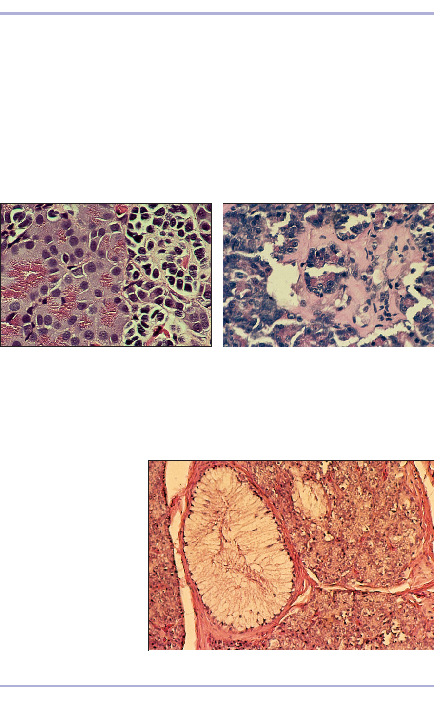
8.97 The pancreas of some snakes,
particularly many pythons, is
characterized by possessing
endocrine cells formed into giant
islets (often found at the edge of the
lobule) rather than into many small
nests of cells scattered randomly
throughout the parenchyma.
Illustrated is a section of the pancreas
from a regal (ball) python (Python
regius). H & E. ×125.
Reptilian, amphibian
and fish pancreas
Morphologically, the pancreas of teleost fish,
amphibians and reptiles is similar to that found in
mammals (8.95), but two major differences are
observed in some species. Many fish, and some
amphibians and reptiles, possess a pancreas that
has an intimate association, and admixture of cells,
with the spleen or liver (8.96). This combined
organ is termed a ‘splenopancreas’ or ‘hepato-
pancreas’, respectively, and the cells and tissues of
each organ receive blood from their respective
splenic, pancreatic or hepatic branches of the
splanchnic arteries and veins. Whereas the islet tis-
sue in most reptiles tends to be conventionally
arranged and evenly distributed, ‘giant’ islets of
Langerhans are characteristic in the pancreatic tis-
sues of some snakes, particularly members of the
family Boidae (pythons and boas); rather than
being scattered in a more or less random manner
throughout the pancreatic exocrine tissue, these
huge islets of endocrine cells tend to be localized in
specific areas of pancreatic tissue (8.97).
131
Digestive System
8.95 Pancreas of a salamander (Amphiuma tridactyla).
Exocrine pancreatic cells characterized by their fine
granular eosinophilic cytoplasm (on the left) extend a
finger-like isthmus into the islet of paler staining
endocrine cells bearing dense nuclei (on the right). The
islet cells are arranged into nest-like lobules that are
separated from each other and from the exocrine tissue
by thin strands of connective tissue that support small
blood vessels. H & E. ×250.
8.95
8.96 The splenopancreas of a milksnake (Lampropeltis
triangulum). The spleen is on the right, a large aggregate
of islet tissue is in the middle and a portion of the
exocrine pancreatic tissue is on the left. The islet displays
hyalinization. H & E. ×125.
8.96
Avian digestive system
The horny beak replaces functionally the lips and
teeth of mammals.
Oral cavity and oesophagus
The oral cavity is lined with stratified squamous
epithelium. The tongue is also lined with this type
of epithelium, with some keratinized areas. The
main mass of the tongue consists of striated muscle
and a small bar of cartilage or bone: the entoglos-
sal bone. There are no teeth. The glands in the lam-
ina propria of the oral cavity, tongue and pharynx
are simple-branched and mucus secreting.
The oesophagus is lined with stratified squamous
non-keratinized epithelium, with simple mucous
glands in the connective tissue lamina propria
(8.98). The muscularis externa consists of a thick
inner layer of circular and a thin outer layer of lon-
gitudinal smooth muscle. Lymphoid tissue accu-
mulates in the caudal oesophagus as the
oesophageal tonsil. The crop is an aglandular cau-
dal diverticulum situated two-thirds of the way
down the oesophagus (8.99). In the pigeon two lat-
eral glomerular sacs secrete crop milk.
Clinical correlates
Reptilian pancreas
Certain species of reptiles appear to have a
higher than expected incidence of some
tumours. In captivity, some lizards, especially
savanna monitors (Varanus exanthematicus),
seem to show a high incidence of adult-onset
diabetes mellitus and exocrine deficiency. No
evidence suggests that diabetes mellitus or
exocrine deficiency are as prevalent in wild
savanna monitors. Therefore, these disorders
seem to be artefacts of captive husbandry
(caused particularly by overfeeding and lack
of adequate exercise) resulting in spontaneous
acute and chronic pancreatitis with subse-
quent autodigestion of the pancreatic
parenchyma.
The migration of helminth larvae can also
induce pancreatitis with secondary fibrosis and
loss of secretory function both of exocrine and
of endocrine components.
132
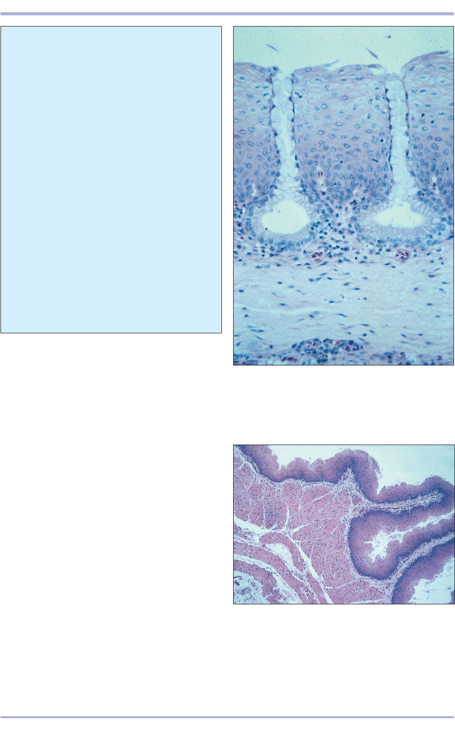
8.98 Oesophagus (bird). (1) Stratified squamous non-
keratinizing epithelium. (2) Simple tubular mucosal
glands. (3) Lamina propria. H & E. ×62.5.
8.98
8.99 Crop (bird). (1) Stratified squamous non-keratinized
epithelium. (2) Lamina propria. (3) Muscularis mucosae.
(4) Muscularis externa. H & E. ×12.5.
8.99
1
2
3
2
3
4
1
Comparative Veterinary Histology with Clinical Correlates
8.101
8.100
Stomach
The stomach consists of the glandular proventricu-
lus and a muscular ventriculus. The gastric epithe-
lium of the proventriculus is simple columnar and
mucus secreting. A thin lamina propria separates
it from the lobules of the submucosal glands. These
glands form an almost continuous mass of tissue,
with adjacent lobules separated by fine strands of
connective tissue. Each gland lobule contains a cen-
tral cavity with straight secretory tubules radiating
to the interlobular connective tissue. An excretory
133
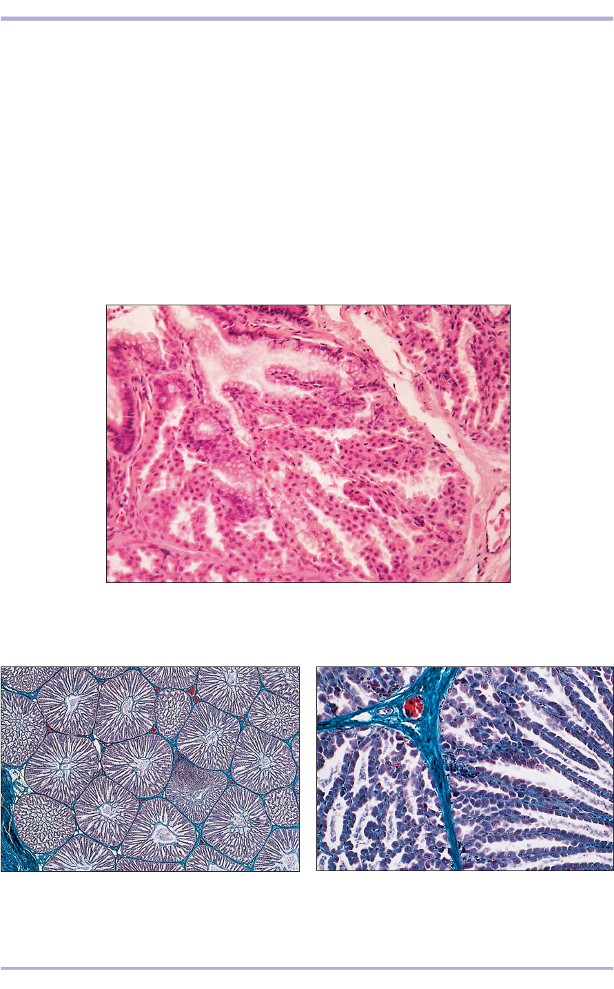
8.100 Proventriculus (bird). (1) Simple columnar mucus-secreting epithelium.
(2) Lamina propria. (3) Submucosal glands. (4) Muscularis externa. H & E. ×62.5.
8.101 Proventriculus (bird). (1) Simple columnar mucus-
secreting epithelium. (2) Lamina propria. (3) Submucosal
glands. (4) Muscularis externa. Masson’s trichrome. ×25.
8.102 Proventriculus (bird). The submucosal gland
lobules are separated by thin strands of connective tissue.
Masson’s trichrome. ×250.
8.102
1
2
2
3
4
4
3
1
duct drains onto the gastric mucosal surface. The
glands contain only one type of cell, which secretes
acid and pepsinogen, thus combining the functions
of both the chief and parietal cells of the mammal.
The muscularis externa is arranged as inner circu-
lar and outer longitudinal layers of smooth muscle
(8.100–8.102).
The ventriculus is the aglandular stomach or giz-
zard. The luminal surface is lined with secretory
product of the mucosal glands, which solidifies at the
surface to form a hard cuticle of koilin. The epithe-
lium is low columnar and continues within the
Digestive System
1
2
134
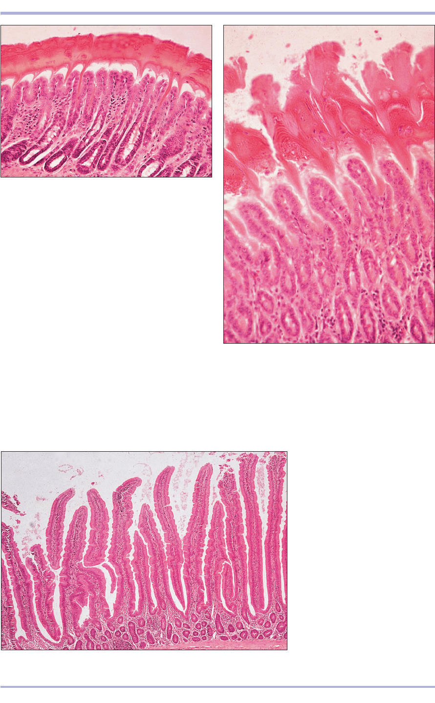
8.103 Ventriculus, gizzard (bird). (1) Cornified lining.
(2) Epithelium lining the gizzard. (3) Mucosal glands.
(4) Lamina propria. H & E. ×25.
8.103
8.104 Ventriculus, gizzard (bird). (1) Cornified lining.
(2) Lining epithelium. (3) Mucosal glands. H & E. ×125.
8.104
8.105 Duodenum (bird). (1) Simple
columnar epithelium. (2) Intestinal
mucosal glands. (3) Connective tissue
core of the villus. (4) Muscularis
mucosae. H & E. ×62.5.
8.105
1
3
2
4
1
2
3
3
4
1
2
simple straight tubular mucosal glands in the lamina
propria. A submucosa is present, and the muscularis
externa is a thick layer of smooth muscle (8.103 and
8.104). There is no muscularis mucosae.
Intestine
The small intestine is similar to that of mammals
but is more uniform throughout its length. Diffuse
lymphatic tissue is present in the lamina propria and
the submucosa, and a third layer of circular smooth
muscle may be present in the muscularis externa
(8.105–8.107).
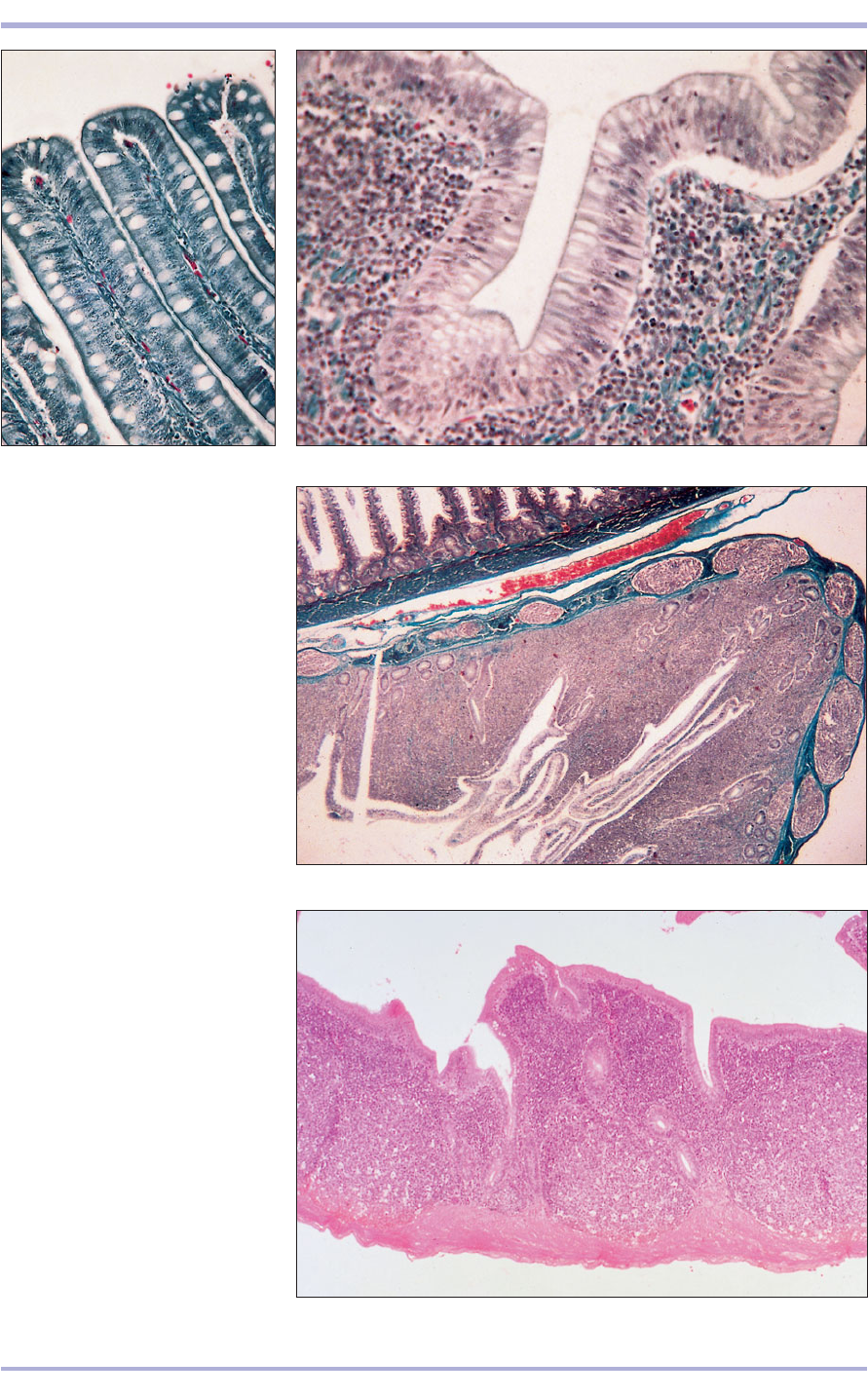
The caeca are two blind sacs at the junction of the
small and large intestine and are of considerable size
in domestic birds. The epithelium is simple colum-
nar with mucous cells. Lymphatic tissue is particu-
larly abundant, forming the caecal tonsil in the nar-
row proximal part of the caecum (8.108 and 8.109).
Comparative Veterinary Histology with Clinical Correlates
8.108
135
Digestive System
8.106 Duodenum (bird). The simple
columnar epithelium lining the
duodenum has a striated border to
allow absorption and goblet cells.
Masson’s trichrome. ×125.
8.107 Duodenum (bird). The simple
columnar epithelium rests on a
lamina propria with numerous
lymphatic cells. Masson’s trichrome.
×250.
8.108 Caecum (bird). (1) Lumen of
the caecum. (2) Mucosa consists of a
columnar epithelium and a lamina
propria with extensive deposits of
lymphatic tissue. (3) Muscularis.
(4) Lumen of the duodenum.
Masson’s trichrome. ×12.5.
8.109 Caecum (bird). Dense masses
of lymphatic tissue fills the lamina
propria. H & E. ×12.5.
8.106
8.107
4
1
3
2
8.109
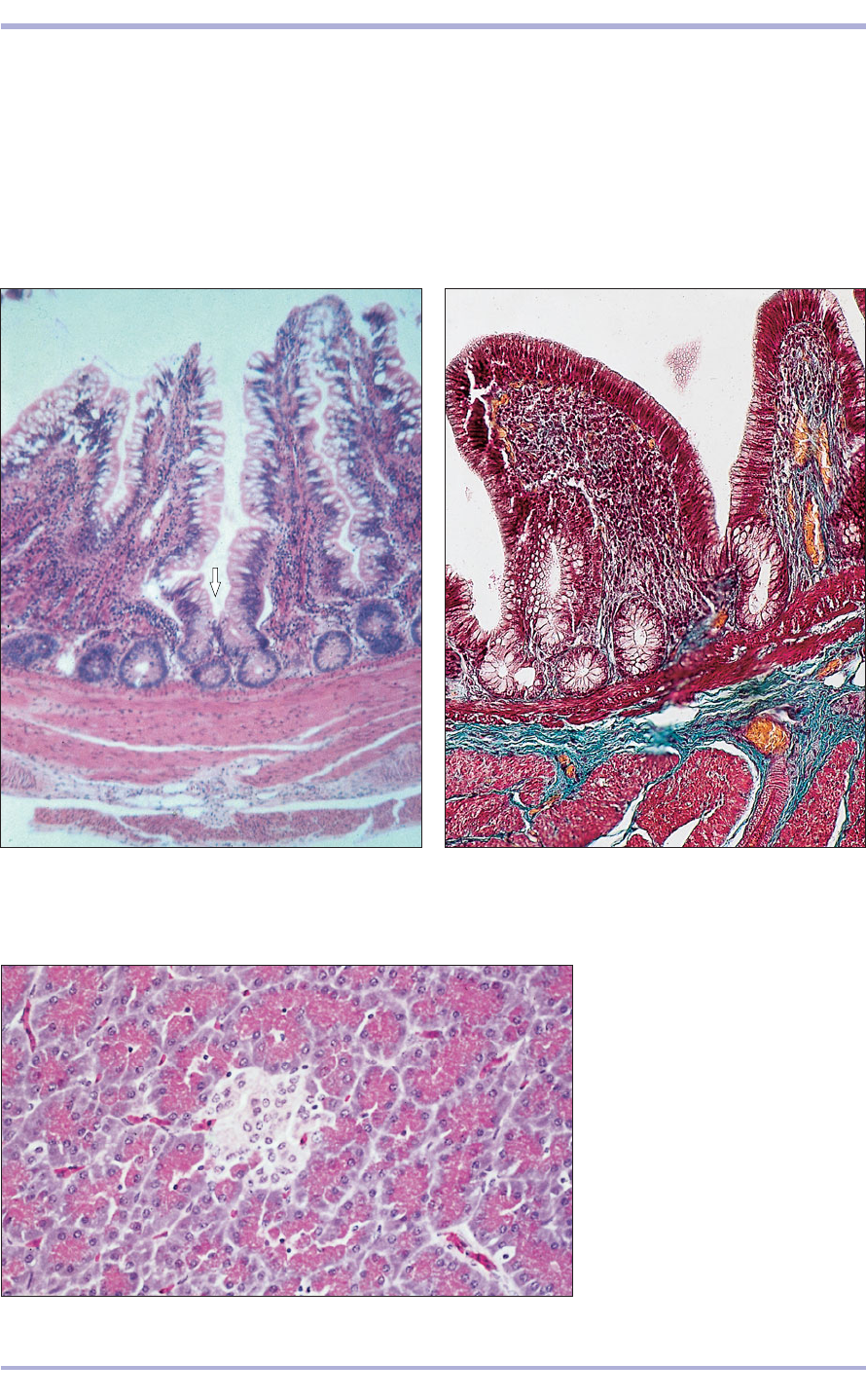
8.111 Cloaca (bird). (1) Simple columnar lining
epithelium. (2) Simple tubular glands. (3) Lamina
propria with lymphatic tissue. (4) Muscularis mucosae.
(5) Muscularis externa. Alcian blue/PAS. ×62.5.
8.111
The large intestine has the same histological
appearance as the caeca (8.110). The cloaca is lined
with tall columnar epithelium with a variable num-
ber of mucus-secreting cells. A vascular lamina pro-
pria separates it from the muscularis mucosae and
externa (8.111). (See Chapter 15 for the cloacal
bursa.)
The avian liver is very similar to the mammalian;
136
8.110 Rectum (bird). The lining epithelium is simple
columnar with goblet cells extending into the simple
tubular mucosal glands (arrowed). H & E. ×62.5.
8.110
8.112 Pancreas (bird). (1) Serous
units of the exocrine gland.
(2) Pancreatic islet, the endocrine
gland. H & E. ×125.
8.112
the connective tissue capsule extends into the gland
and divides it into lobes and lobules. The hepato-
cytes are arranged in rows, often two cells thick,
separated by sinusoids.
The avian exocrine pancreas is similar to the mam-
mallian, but has less interlobular connective tissue
(8.112). The endocrine pancreatic islets are of three
types: light (beta) islets, dark (alpha) islets and mixed.
2
1
2
1
3
4
5
Comparative Veterinary Histology with Clinical Correlates
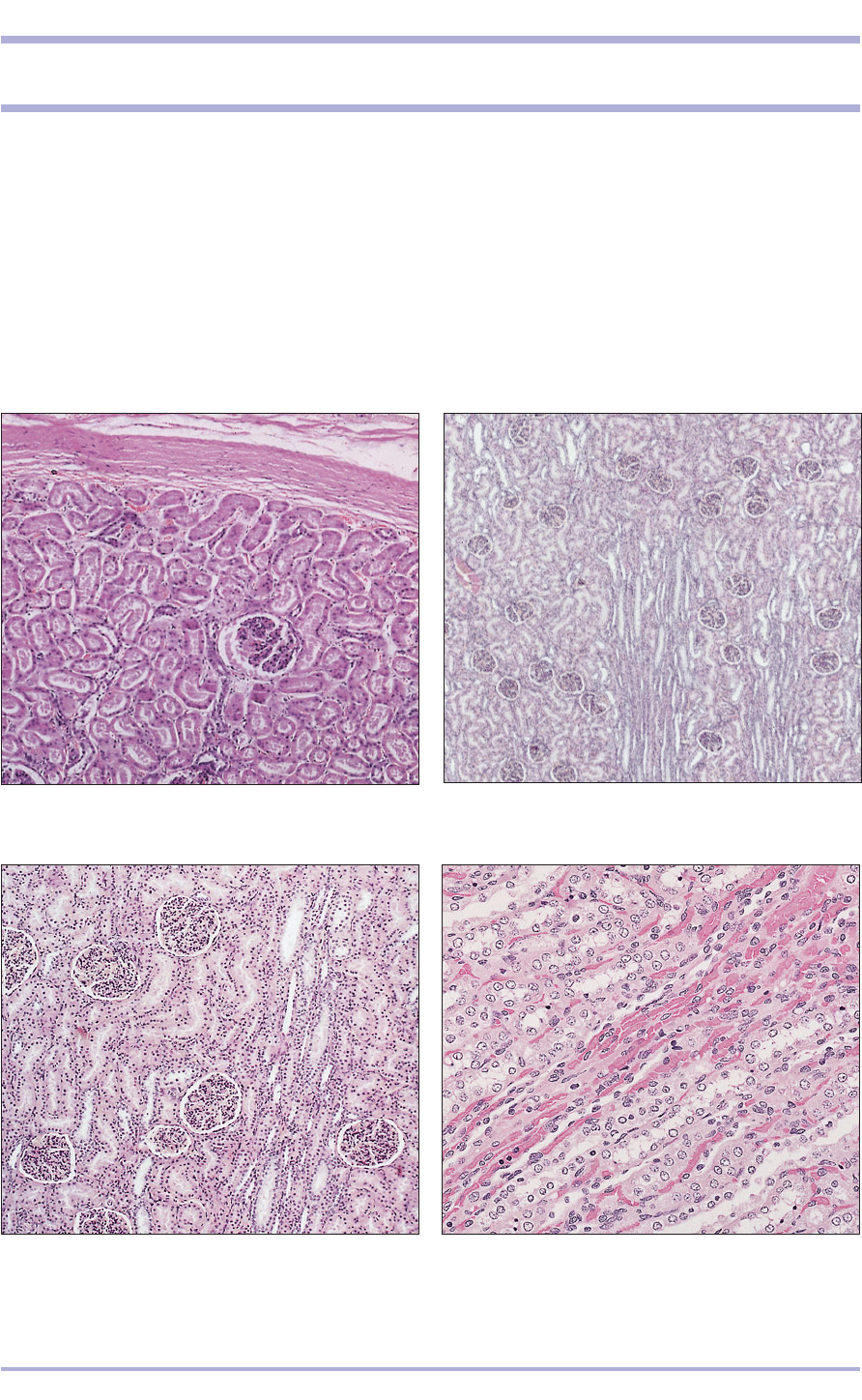
9.3 Kidney cortex (dog). (1) Renal corpuscle.
(2) Uriniferous tubules. (3) Medullary ray. H & E. ×125.
9.3
9.2 Kidney cortex (dog). (1) Renal corpuscle.
(2) Uriniferous tubules. (3) Medullary ray. H & E. ×12.
9.2
The urinary system of mammals comprises the kid-
neys, renal pelvis, ureters, bladder and urethra.
Kidneys
The kidneys are highly vascularized organs that fil-
ter the blood and excrete waste materials, excess
water and electrolytes via the ureters to the bladder
as urine (see Appendix Figures A1–A7). The kidneys
have an endocrine function: they secrete erythropoi-
etin, which stimulates erythrocyte production in the
bone marrow, and renin, which helps to regulate
blood pressure. Each kidney is enclosed in a tough
connective tissue capsule extending into the
parenchyma and has two regions – the cortex and
the medulla (9.1–9.4). Smooth muscle may be pre-
sent in ruminants. The hilus is a deep fissure on the
medial border of the kidney and contains the renal
artery, the renal vein and the ureter.
137
9. URINARY SYSTEM
9.1 Kidney (horse). (1) Capsule. (2) Outer area of the cortex.
(3) Renal corpuscle. (4) Uriniferous tubules. H & E. ×62.5.
9.1
2
3
1
2
3
1
1
2
3
4
9.4 Kidney medulla (dog). (1) Blood vessels. (2) Collecting
tubules. (3) Ascending thin limb. (4) Descending thin limb.
H & E. ×125.
9.4
2
3
4
1
1
1
138
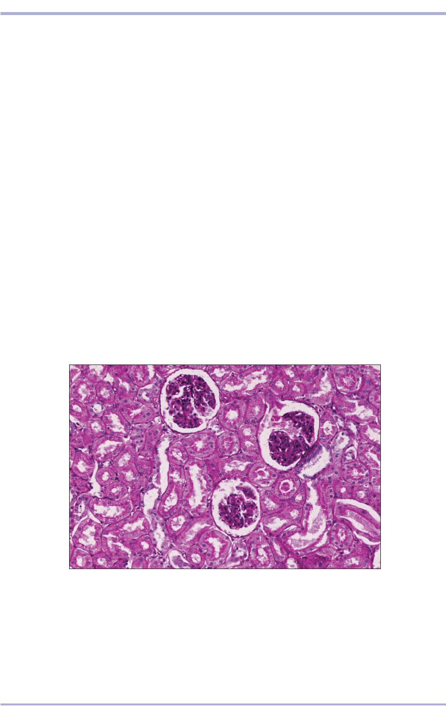
9.5 Kidney (horse). (1) Renal corpuscle with (2) capsular space (3) the urinary pole.
(4) Proximal convoluted tubule. (5) Distal convoluted tubule with (6) the macula
densa. H/PAS. ×250.
9.5
Nephron
The nephron is the functional unit of the kidney.
The major subdivisions are the renal corpuscle and
the uriniferous tubule.
Renal corpuscle
The blind end of the proximal tubule is indented
with a network of capillaries and supporting cells
to form a filtering system: the renal corpuscle. Each
renal corpuscle consists of a glomerulus and a
glomerular (Bowman’s) capsule. The outer layer of
the glomerular capsule is the capsular (parietal)
wall, which is separated from the glomerular (vis-
ceral) layer by the capsular (urinary) space (9.5).
The capillaries are lined with a fenestrated endothe-
lium resting on a basal lamina. The visceral epithe-
lial cells, or podocytes, closely invest the capillary
endothelium of the glomerulus and develop pri-
mary processes wrapped around each capillary.
These processes develop secondary foot processes
called pedicles. The foot processes of adjacent
podocytes interdigitate, resulting in the formation
of small gaps called slit pores (9.6). The podocyte
basal lamina is fused with the endothelial basal
lamina and blood passing through the capillary is
filtered through this common basal lamina into the
capsular space (9.6 and 9.7). Mesangial perivas-
cular cells are present between the endothelium
and the basal lamina. The capillaries of the
glomerulus are served by an afferent and an effer-
ent arteriole, entering and leaving the renal cor-
puscle at the vascular pole (9.8). At the opposite
pole is the capsular space, where the filtrate passes
into the proximal tubule at the urinary pole of the
renal corpuscle (9.8 and 9.9).
2
3
4
5
1
1
1
6
6
Comparative Veterinary Histology with Clinical Correlates
139
Urinary System
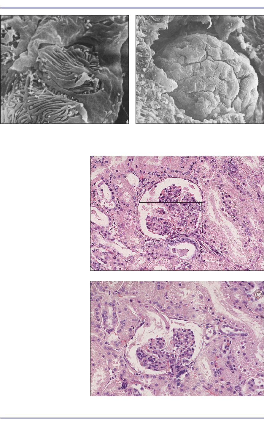
9.7 Kidney (dog). The renal corpuscle projects from the
surrounding tissue. Scanning electron micrograph. ×500.
9.7
9.6 Kidney (dog). (1) Part of the podocyte. (2) Secondary
foot processes, the pedicles – the filtration slit pores are
the spaces between the solid pedicles. Scanning electron
micrograph. ×1500.
9.6
9.8 Kidney (horse). The width of the
glomerular capsule is shown by the
line. (1) Renal corpuscle with
epithelial cells and mesangium.
(2) Vascular pole. (3) Capsular space.
(4) Distal convoluted tubule. (5) The
macula densa. (6) Proximal
convoluted tubule. H & E. ×250.
9.8
9.9 Kidney (horse). (1) Renal
corpuscle with epithelial cells and
mesangium. (2) Urinary pole opening
into (3) the proximal convoluted
tubule. (4) Distal convoluted tubule
and macula densa. H & E. ×250.
9.9
3
2
1
4
4
6
3
1
2
5
1
2
extends towards the medulla where the epithelium
changes abruptly to simple squamous. This part of
the tubule descends into the medulla as the thin
descending limb and bends sharply to return to the
cortex as the thick ascending limb, which was pre-
viously known as the loop of Henle. In the cortex
the epithelium becomes cuboidal or columnar and
forms the distal straight tubule and coils near the
glomerulus to become the distal convoluted tubule
(DCT). The DCT is shorter than the PCT, the
epithelium is cuboidal, the cytoplasm is paler and
there is no brush border.
Renal tubule
This nomenclature is based on the functional areas
of the renal tubule. The proximal convoluted tubule
(PCT) is long and lined with low columnar cells
with a basal nucleus. The cytoplasm is deeply
stained with eosin and the apical surface is a con-
tinuous brush border. The basal plasma membrane
is folded, with mitochondria in the cytoplasm giv-
ing a striated effect, and functions to increase the
surface for transport (cf. salivary glands striated
duct). The PCT is continued with the proximal
straight tubule. It is similar in appearance and
140
Comparative Veterinary Histology with Clinical Correlates
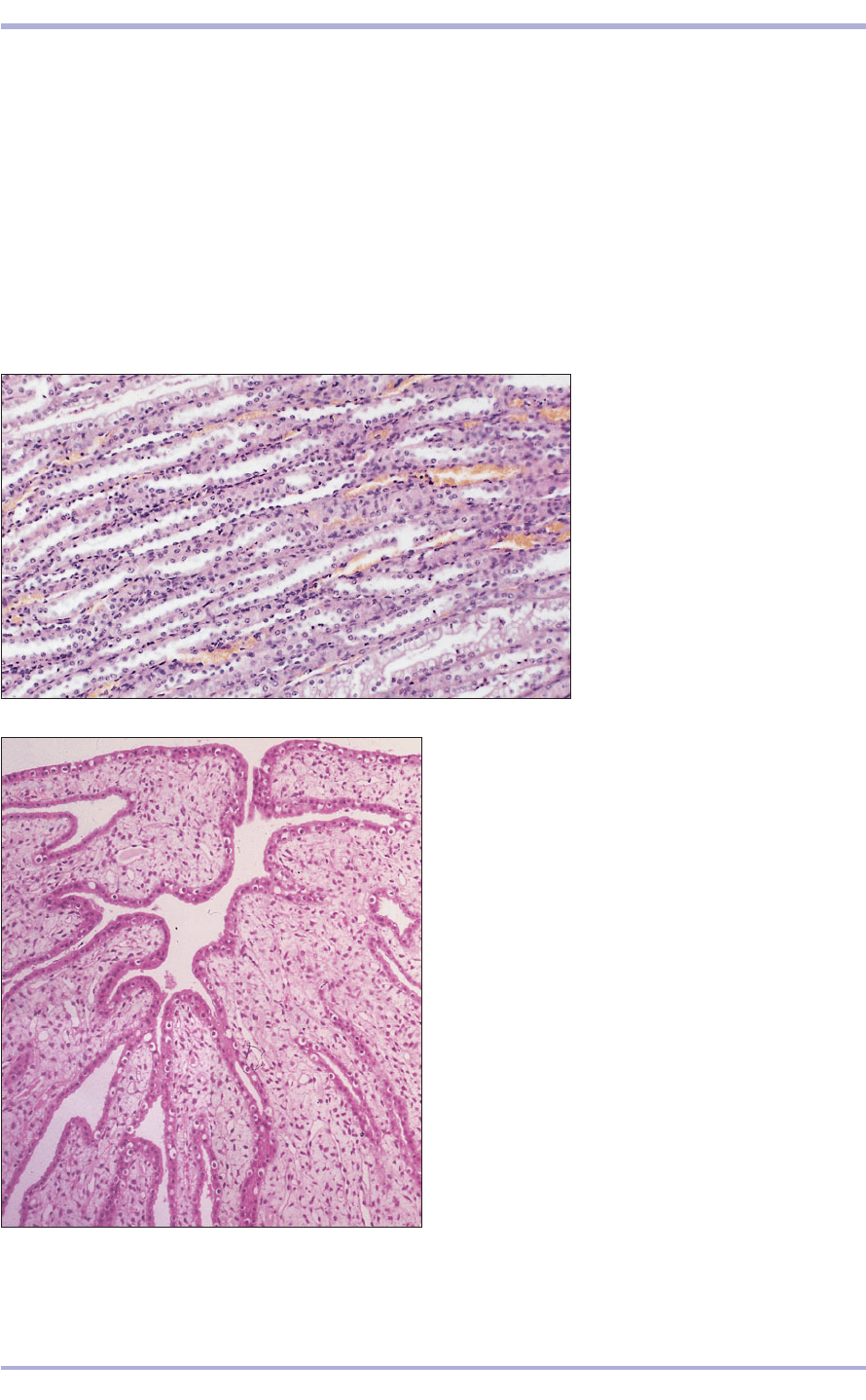
9.10 Kidney medulla (dog).
(1) Capillaries lined by endothelium.
(2) Collecting tubules lined by
cuboidal cells. (3) Ascending limb
lined by cuboidal epithelium.
(4) Descending limb lined by
squamous epithelium. H & E. ×125.
9.10
9.11 Kidney. Renal pelvis (sheep). The renal pelvis is lined
by urethelium resting on a vascular lamina propria. H & E.
×62.5.
9.11
The DCT approaches the glomerulus at the vas-
cular pole, where it thickens, and the cell nuclei of
the tubule wall become crowded together to form
the macula densa, part of the juxtaglomerular appa-
ratus. Juxtaglomerular cells are modified smooth
muscle cells in the walls of afferent arterioles close
to the glomerulus. The cells are epithelioid, contain
granules and produce renin (which plays a role in
the regulation of blood pressure) and angiotensin
(a vasoconstrictor and stimulus of aldosterone
secretion) (9.1–9.3 and 9.9).
Collecting tubule
The collecting tubule or duct (lined with poorly
staining cuboidal epithelium) is the terminal seg-
ment of the nephron, a continuation of the DCT
within the medulla (9.4 and 9.10), joining with oth-
ers to form straight ducts: the papillary ducts (of
Bellini). Here the epithelium becomes columnar, and
then becomes urethelium towards the opening into
the renal pelvis (9.11).
1
2
4
3
