Aughey E., Frye F.L. Comparative veterinary histology with clinical correlates
Подождите немного. Документ загружается.

8.68
8.69
121
Digestive System
Clinical correlates
Alimentary system of reptiles
and amphibians
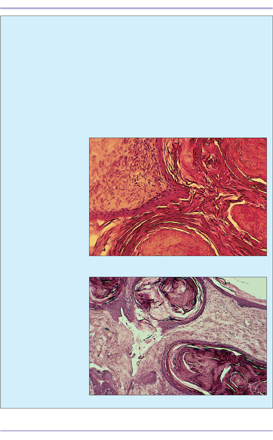
Squamous metaplasia of the nasal and pharyn-
geal mucosa (8.68 and 8.69) is a frequent clin-
ical condition in reptiles fed diets deficient in
vitamin A or `-carotene. Once this alteration
occurs, the lubricative mucoid glandular secre-
tion ceases and the affected animal becomes
more susceptible to respiratory and oropha-
ryngeal disorders.
Ulcerative stomatitis is one of the most com-
mon conditions found in the cranial alimentary
tract of captive snakes. This infectious inflam-
matory disease is caused by a variety of patho-
genic Gram-negative and some Gram-positive
bacteria. Depending upon the aetiologic agent,
the inflammatory response may be suppurative
or non-suppurative. In suppurative lesions, het-
erophil granulocytes predominate; in non-sup-
purative inflammations, heterophils may be
entirely absent.
Glossitis, pharyngitis, oesophagitis and gastri-
tis also occur in captive amphibians and reptiles.
8.68 Massive pharyngeal
hyperkeratosis in a desert
tortoise (Xerobates agassizi).
The pharyngeal glands are
replaced by pearl-like masses
of desquamated keratin. The
luminal epithelial surface is
thickened and covered by
dense keratin debris. A similar
alteration is seen in birds and
mammals suffering from
vitamin A deficiency.
H & E. ×12.5.
8.69 Cross-section of the
pharynx of a red-eared slider
turtle that was fed a diet
seriously deficient in `-carotene
or preformed vitamin A. The
pharyngeal glands display
squamous metaplasia and, as a
result, have lost their mucus-
secreting, goblet-cell-rich
glands, which have been
replaced by masses of
desquamated keratin debris
(1). The stratified squamous
epithelium lining the
pharyngeal lumen is thickened
and hyperplastic. H & E. ×12.5.
1
1
8.70
These inflammatory conditions are often caused
by items in the diet that injure the delicate
mucous membranes that cover the tongue or line
these cavities. In snakes, and to a lesser extent in
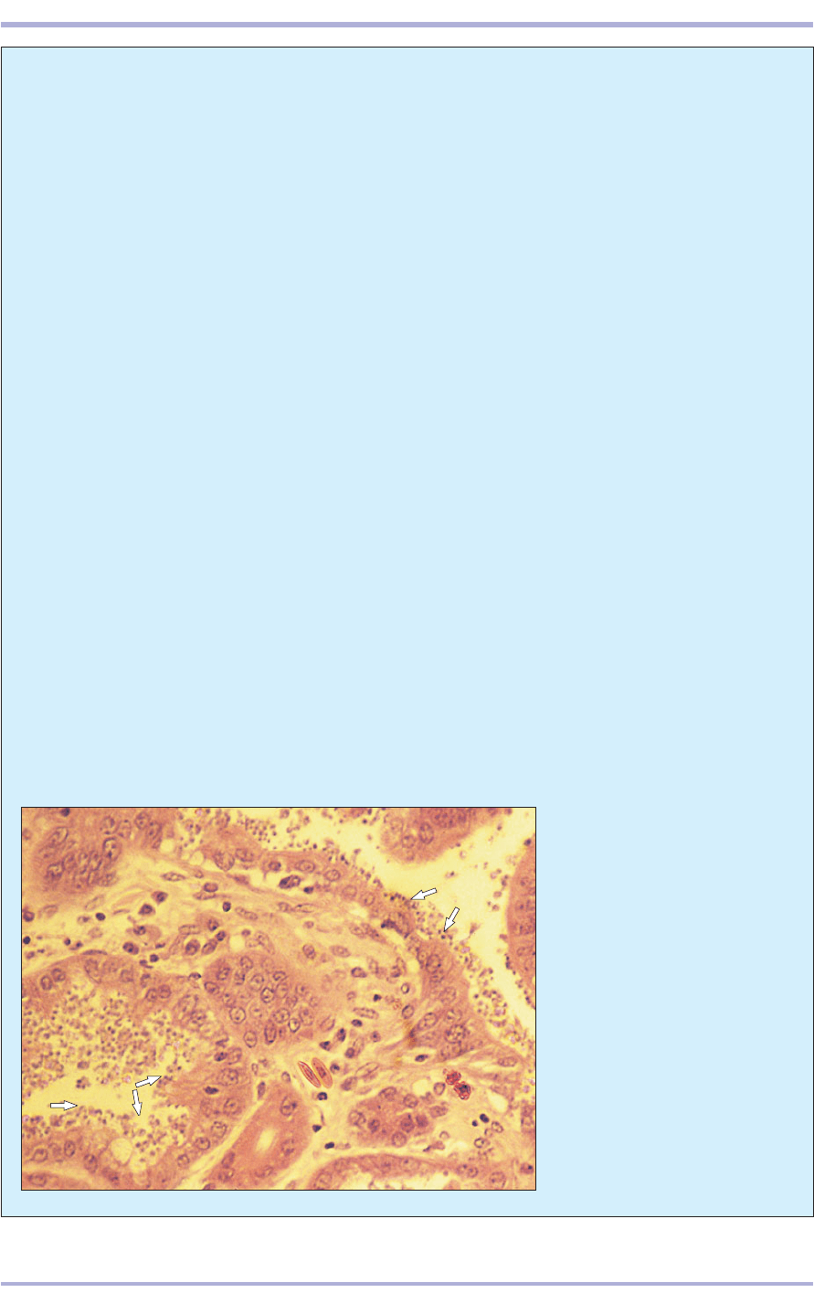
lizards, gastric cryptosporidiosis is a serious clin-
ical problem. Although typically termed ‘hyper-
trophic’ gastritis in the literature, the anatomical
and histological features of gastric cryp-
tosporidiosis in snakes are a marked hypertro-
phy of the muscular tunica comprising the wall
of the stomach, together with an atrophy of the
gastric mucosa. Gastric biopsy (or gastric lavage)
specimens of infected snakes reveal myriad num-
bers of protozoan organisms attached to the
brush border of the epithelial cells lining the gas-
tric lumen and gastric pits (8.70).
Usually, enteritis is accompanied by an over-
production of protective mucus by the goblet
cells. The inflammation may be suppurative, in
which heterophils are easily identified, or non-
suppurative, in which the predominant leuco-
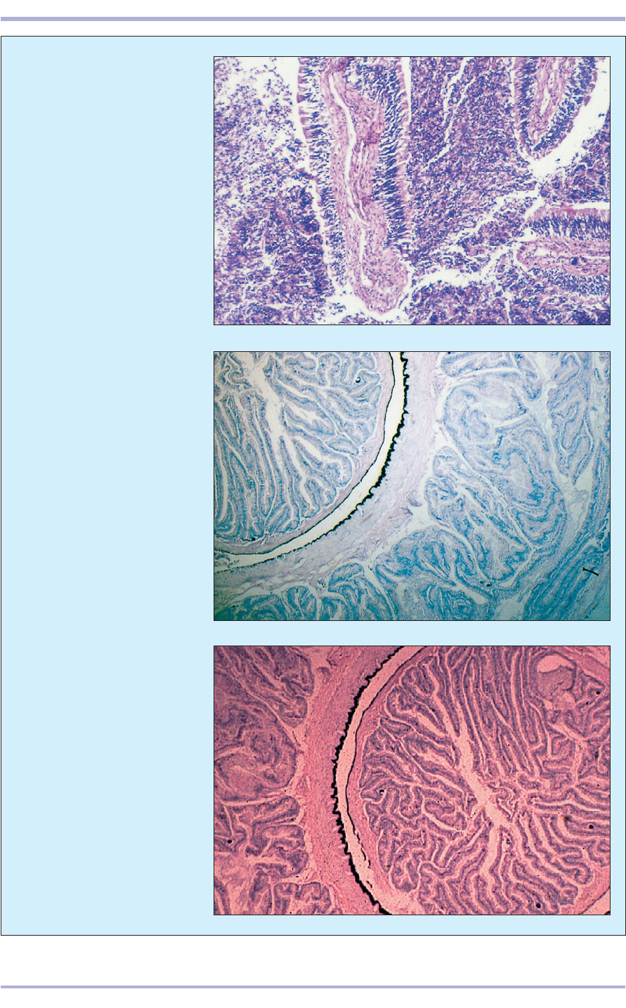
cytes are mononuclear (8.71). The aetiologic
agent may or may not be immediately apparent.
Intussusception (the telescoping of one seg-
ment of intestine into another, or into the stom-
ach) occurs relatively frequently in some reptiles,
particularly in iguanas and Old World
chameleons (8.72 and 8.73). The reasons for this
high incidence are unknown, but endoparasitism
and dietary problems, especially hypocalcaemia,
are suspected as predisposing factors.
Benign and malignant neoplasia of the stom-
ach and small intestine are relatively common in
captive reptiles, particularly snakes and lizards.
This may be a consequence of living considerably
longer while in captivity than under natural (wild)
conditions. Adenomata, carcinomata, leiomyoma
and leiomyosarcoma have been recorded in many
snakes and in fewer lizards.
Preneoplastic leukoplakia and invasive squa-
mous cell carcinoma have been described in che-
lonians. These proliferative lesions are similar to
those observed in mammals.
Adult green iguanas (which are folivorous
herbivores) have a simple stomach and a short
small intestine that transports the partially
processed leafy ingesta into the sacculated and
much expanded colon. Villous projections (8.67),
covered with pseudostratified, non-ciliated
columnar epithelium overlying a thin lamina pro-
pria and a core of smooth muscle and blood ves-
sels, extend into the colonic lumen and create a
larger surface area for the processing of cellulose
and absorption of nutrients.
122
Comparative Veterinary Histology with Clinical Correlates
8.70 Gastric cryptosporidiosis in
an Australian tiger snake
(Notechis scutatus). A myriad
number of round organisms
(arrowed) are attached to the
brush border of the mucosal cells
lining the gastric lumen and
gastric pits. H & E. ×250.
8.71
8.73
8.72
123
Digestive System
8.72, 8.73 Duodenal-jejunal
intussusception in a Fisher’s
chameleon (Chamaeleo fisheri;
8.72) and an iguana (8.73).
A segment of duodenum has
telescoped into the jejunum
causing the two serosal layers
to lie adjacent to each other.
H & E. ×12.5.
8.71 Non-suppurative enteritis
in a desert tortoise. Most of the
leucocytes are lymphoplasmacytic.
H & E. ×62.5.
8.74
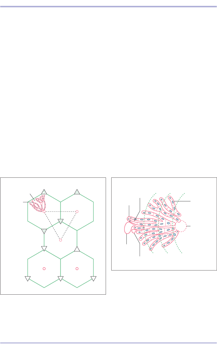
8.75 Liver acinus (functional unit). The liver parenchyma
is served by the terminal branch of the portal vein and
the hepatic artery. The acinus is divided into three
zones, which indicate the relative position of the cells in
realtion to the oxygen gradient. Hepatocytes in Zone 1
are closest to the fresh, oxygen-rich hepatic arterial
blood, and those in Zone 3 are further away. Equally,
cells in Zone 1 are first in line for toxins, etc., carried in
the portal blood, with Zone 3 the cells the least affected
by these.
8.74 Hepatic lobule. The classic lobule is the hexagon,
clearly seen in the figure by the outline (green) of
connective tissue). Portal areas (P) occur between the
lobules. A portal lobule is defined as the central
functional axis of the lobule (the black dotted triangle).
P
Sinusoid
Liver cord
cells;
hepatocytes
P
P
P
C
C
CC
C
P
P
P
P
Liver
The liver is the largest gland in the body. Blood
drains to it from the intestines in the hepatic por-
tal vein, and the products of digestion are metabo-
lized, harmful material detoxified, senescent
erythrocytes removed from the circulation and bile
secreted. The liver is surrounded by mesothelium.
The connective tissue capsule extends into the gland
and divides it into lobes and lobules. The structure
of the classic lobule is most clearly visualized in the
pig because of its plentiful array of connective tis-
sue dividing the liver into discrete hexagonal lob-
ules with a portal area at the corners of each
hexagon. This is not the case in the other domestic
animals, except under pathological conditions such
as cirrhosis. The portal areas (triads) occur between
three or more lobules and each contains one or
more branches of a hepatic artery, a hepatic portal
vein, a lymphatic vessel and a bile duct. The
parenchyma consists of polyhedral epithelial cells
of endodermal origin, the hepatocytes, arranged in
anastomosing rows separated by sinusoids con-
verging on the central vein. The sinusoids are lined
with fenestrated endothelial cells and macrophages,
124
8.75
Liver cord
Zone 3
Zone 2
Zone 1
Bile
canaliculis
Branch of the hepatic artery
Branch of
the hepatic
vein
Central
vein
part of the mononuclear phagocyte system. Blood
flows through the sinusoids to the central vein. This
in turn leaves the liver lobule to travel separately as
branches of the hepatic vein.
Bile is secreted by each hepatocyte into the bile
canaliculi, channels that are lined with the plasma
membranes of the hepatocytes, between adjoining
liver cells. It flows from there to a small bile duct in
the portal area. Where the bile duct is the central
functional axis of the lobule instead of the central
vein, the term ‘portal lobule’ is used. The liver aci-
nus is the smallest functional unit of the liver. It con-
sists of parenchyma served by a terminal branch of
the portal vein and the hepatic artery, and is drained
by two central veins and terminal branches of the
bile duct. It has functional and pathological signif-
icance. Bile ducts are lined with cuboidal epithelium
in the portal areas; the larger interlobular ducts are
lined with columnar epithelium (8.74–8.80).
Reptiles, amphibians and fish
The livers of fish, amphibians and reptiles are super-
ficially similar to those of mammals. However, there
are some differences. Many of the lower vertebrates
have abundant melanin pigment scattered through-
Comparative Veterinary Histology with Clinical Correlates
125
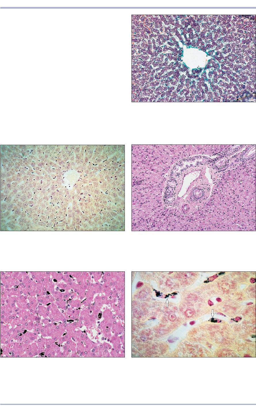
8.76 Liver (pig). The heaxagonal liver lobule is
delineated by the green strands of the interlobular
connective tissue. (1) Central vein. Masson’s trichrome.
×125.
8.76
8.77 Liver (ox). (1) Hepatocytes arranged in cords.
(2) Sinusoids lined by endothelial cells and macrophages
open into (3) the central vein. Safranin/haematoxylin.
×125.
8.77
8.78 Portal canal (triad) (sheep). (1) Branch of the bile
duct. (2) Hepatic artery. (3) Lymphatic vessel. (4) Hepatic
portal vein. (5) Liver cords. (6) Sinusoids. H & E. ×125.
8.78
8.80 Liver (dog). The hepatocytes lie in anastomosing
cords separated by sinusoids lined by macrophages
(arrowed). Safranin/haematoxylin after carbon injection.
×250.
8.80
8.79 Liver (dog). The macrophages have taken up the
injected carbon and appear as black areas between the
cords of hepatocytes. Carbon-injected with H & E
counterstain. ×125.
8.79
1
4
3
2
5
6
3
1
2
1
out the hepatocellular parenchyma (see Chapter 3).
Usually, this pigment is contained in melanophages
that are aggregated together in packet-like groups
of cells bearing fine dark-brown granules. In some
species the liver is arranged in narrow cords radi-
ating outward from a thin-walled central vein. It
has one or more portal triads consisting of an arte-
riolar branch of the hepatic artery and one or more
small bile ducts (as in mammals). In other species
the central veins are scattered randomly through-
out the liver and more than one portal triad or tri-
ads with multiple arterioles and bile canaliculi or
ducts are present.
An admixture of hepatocellular and pancreatic
tissues, thus forming a hepatopancreas, is present
in many fish and in some amphibians and reptiles.
Digestive System
8.81
8.838.82
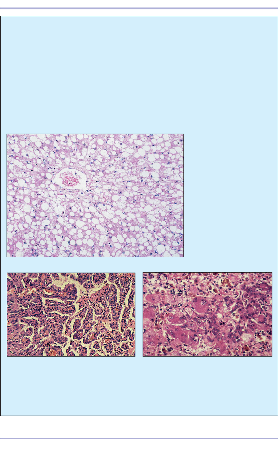
8.82 Cholangiocarcinoma in an Asiatic leopard
(Panthera pardus). The cuboidal to columnar
neoplastic cells form frond-like ducts and tubules with
thin walls composed of two cell layers separated by a
scant fibrovascular connective tissue stroma. H & E.
×125.
8.81 Hepatic lipidosis (horse).This
micrograph of horse liver shows a
central vein surrounded by
radiating cords of hepatocytes
that contain large, smoothly
round vacuoles which occupy most
of the cell and displace the
nucleus to the periphery. These
are fat vacuoles. The horse was
hyperlipidaemic with hepatic
lipidosis. H & E. ×250.
126
Comparative Veterinary Histology with Clinical Correlates
8.83 Giant cell hepatitis in a 5-year-old neutered
female Siamese cat. Note that many of the hepatocytes
are much larger than normal and frequently have
multiple nuclei. A mixed mononuclear and
polymorphonuclear leucocytic response is present.
H & E. Haemosiderin is present. ×125.
Clinical correlates
With its pivotal role in processing material carried
from the intestine via the portal system, the liver
is exposed to toxic factors and potentially harm-
ful micro-organisms passing from the gut.
Metabolic or nutritional disease (8.81), infectious
disease (see 8.84) and neoplasia (8.82), both local
and metastatic, can also affect the liver.
Inflammation of the liver is termed hepatitis
(8.83). Hepatic lipidosis, an excess fat storage in
the liver, is seen as a clinical problem in obese ani-
mals under physiological stress: often pregnant
pony mares, dairy cattle after parturition and ewes
carrying twins in late pregnancy (pregnancy tox-
aemia). Mobilization of large amounts of triglyc-
erides causes fatty acids to be presented to the liver
in excess of its capacity to handle them. This prob-
lem may be quite rapidly fatal and cases of sud-
den death due to liver rupture are not uncommon.
The liver has a great capacity for regeneration
of hepatocyte mass, but fibrosis is also a charac-
8.84
8.868.85
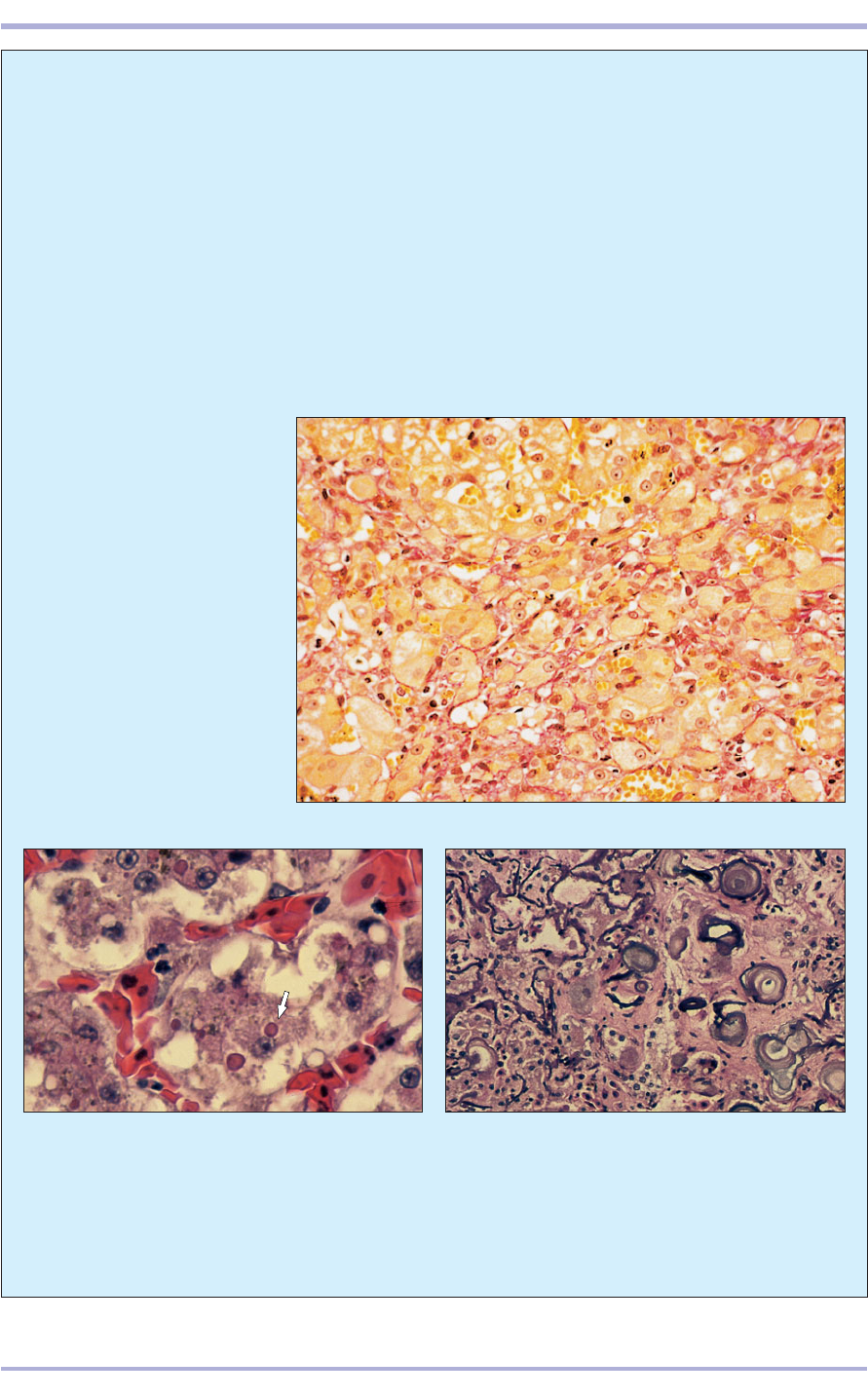
8.86 Nutrition related, massive hepatic mineralization
secondary to hypervitaminosis D
3
in an African leopard
tortoise (Geochelone pardalis). Most of the
hepatocellular parenchyma has undergone gross
alteration and is replaced by bone. As a consequence,
few normal hepatocytes remain. Several osteocytes
surrounded by concentric lamellae of compact bone
are present. H & E. ×125.
8.85 Viral hepatitis in a Colombian boa constrictor
(Boa C. constrictor). Most of the hepatocytes contain
eosinophilic, intracytoplasmic viral inclusion bodies
(arrowed), most of which are surrounded by narrow
clear ‘haloes’. H & E. ×250.
8.84 Micronodular cirrhosis in
a 1-year-old Cocker Spaniel. The
yellow-stained hepatocytes vary in
size and shape and the developing
fibrous tissue is stained red. With
this technique, red blood cells
stain yellow. Sirius Red. ×250.
127
Digestive System
teristic reaction of the liver to chronic injury. Any
hepatic injury severe enough to result in hepatic
necrosis results in some fibrogenesis, but pro-
gressive fibrosis can develop when the insult per-
sists or when the initial damage is severe and
provokes an extensive reaction. A canine liver
with micronodular cirrhosis (8.84) shows pro-
gressive fibrosis in which the normal architecture
of the liver is lost and cells are divided into small
groups surrounded by fibrous tissue.
Amphibians and reptiles
As with domestic animals, numerous chemical
and viral agents induce severe liver disease (8.85).
The hepatic parenchyma is sensitive to changes
in calcium and other minerals in the blood and,
under conditions of hypervitaminosis D
3
, may
undergo severe mineralization and even ossifica-
tion (8.86).
128
Gall bladder
The gall bladder (absent in the horse, dolphins, rhi-
nos and hippos) is a reservoir for bile and is
attached to the visceral surface or between the lobes
of the liver. The mucous membrane is folded in the
flaccid state, and the epithelial lining consists of tall
columnar cells with a striated border (8.87). Goblet
cells and mucus- and serous-secreting glands may
be present in ruminants. The muscularis externa is
a circular layer of smooth muscle and the other
serosa is continuous with the peritoneum.
The gall bladder of lizards and chelonians is
embedded in or surrounded by the liver, as it is in
mammals and birds (8.88). The gall bladder of
snakes is located at a variable distance from the liver
and is contiguous with the spleen and pancreas. A
long bile duct transports bile from the intrahepatic
bile duct(s) to the gall bladder for storage and even-
tual release into the duodenum.
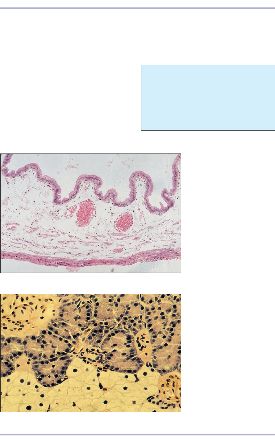
8.87 Gall bladder (cow). (1) Tall
columnar epithelium lining the
lumen. (2) Mucosal folds.
(3) Muscularis. (4) Serosa. H & E.
×12.5.
8.87
4
3
2
2
1
8.88 The liver and pancreas of some
fish, amphibians and reptiles are
fused or admixed with one another
and form a hepatopancreas.
Illustrated is a section of such a mixed
organ in an axolotl, a neotenic form
of the aquatic salamander
(Ambystoma maculatum). The
hepatocellular tissue is at the bottom
of this section; the exocrine
pancreatic portion is in the upper
right; an islet is in the upper left.
H & E. ×250.
8.88
Clinical correlates
The gall bladder can be a site of inflammation
(cholangitis), calculi (choleliths), neoplasia
and foreign bodies such as parasites (Fasciola
hepatica).
Comparative Veterinary Histology with Clinical Correlates
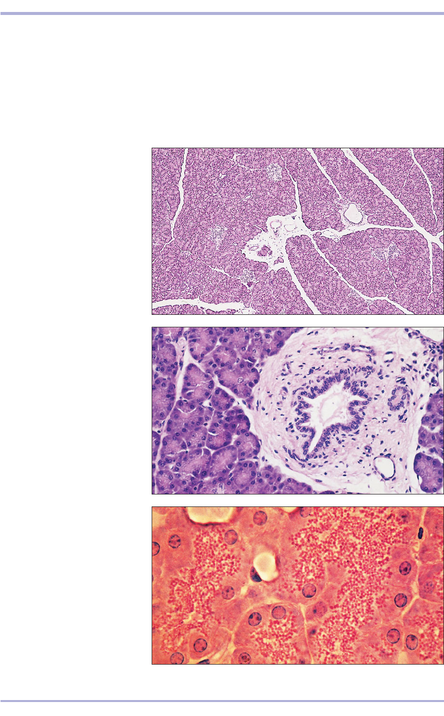
Pancreas
A fine connective tissue capsule extends into the
gland and divides it into lobules. The parenchyma
is composed of exocrine and endocrine tissue; both
are derived from the endoderm of the foregut. The
exocrine portion of the pancreas is a compound
tubuloacinar gland that secretes enzymes into the
duodenum. The acinar cells are tall columnar with
a basal nucleus in basophilic cytoplasm. Where the
secretory granules are stored the luminal cytoplasm
is eosinophilic. Projections of duct cells are com-
monly seen in the acinus; these are the centroacinar
cells that are typical of the pancreas. Smaller ducts
are lined with cuboidal epithelium and larger ducts
with columnar epithelium (8.89–8.91).
129
Digestive System
8.90 Pancreas (dog). (1) Serous
acinus with a centroacinar cell
(arrowed). (2) Interlobular connective
tissue. (3) Interlobular duct. H & E.
×125.
8.90
8.91 Pancreas (dog). (1) Nucleus lies
in the basal basophilic cytoplasm of
the serous cell. (2) Eosinophilic
granules (the secretion) lie in the
luminal cytoplasm. H & E. ×250.
8.91
2
1
3
2
1
1
8.89 Pancreas (dog). (1) Serous acini
of the exocrine pancreas.
(2) Interlobular connective tissue.
(3) Interlobular duct. (4) Pancreatic
islet, the endocrine pancreas. H & E.
×12.5.
8.89
2
3
4
1
8.94
8.93
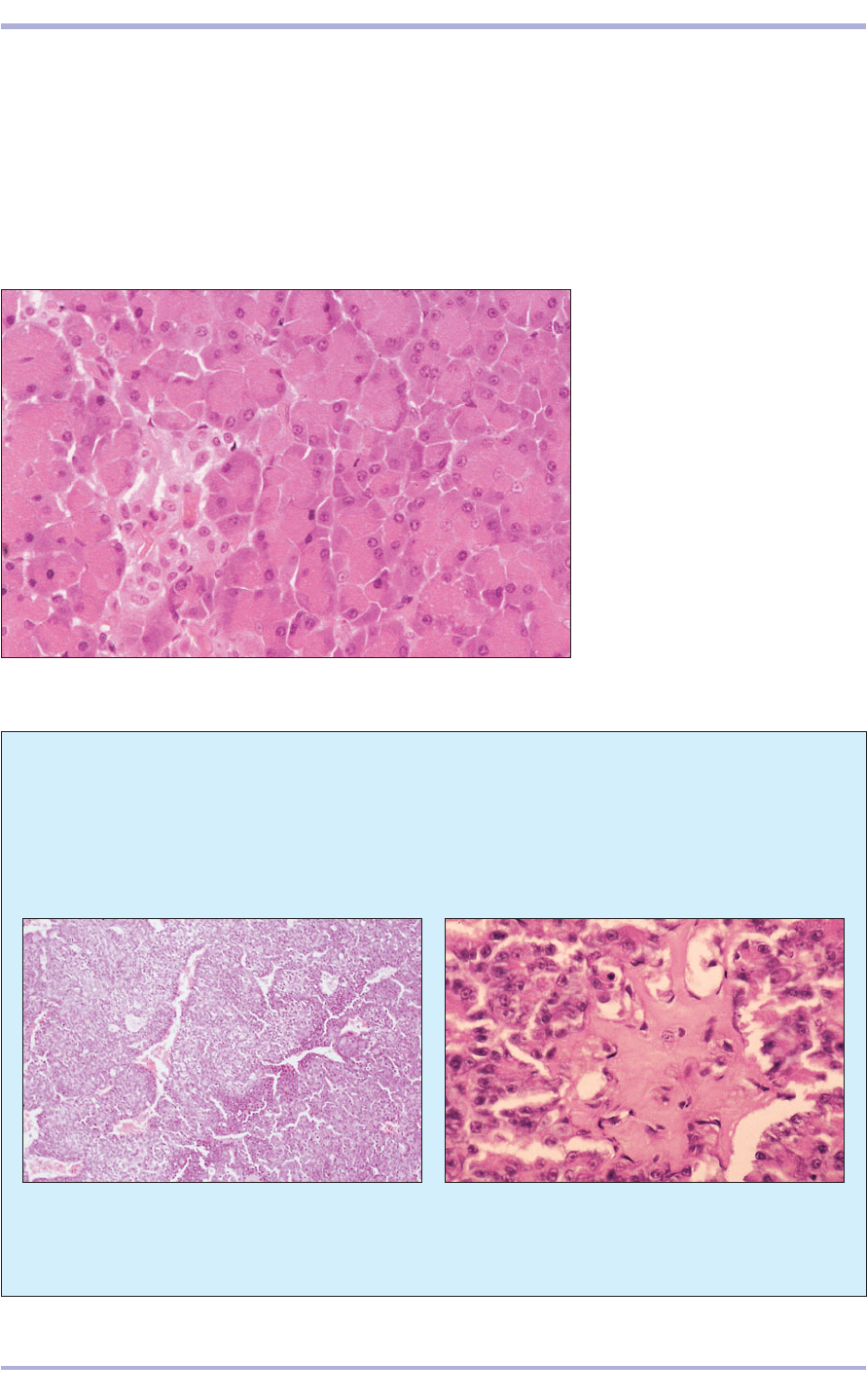
The endocrine pancreas is responsible for the
control of blood sugar concentrations, and isolated
groups of pale staining islet cells (pancreatic cells or
the islets of Langerhans) are found scattered among
the secretory units (8.92). These have two main cell
types: A, or alpha, cells secreting glucagon (a
polypeptide hormone secreted in response to hypo-
glycaemia or to stimulation by growth hormone);
and B, or beta, cells secreting insulin (a peptide hor-
mone released into the blood in response to a rise
in concentration of blood glucose or amino acids).
Rare D, or delta, cells secrete somatostatin and F
cells secrete pancreatic polypeptide. These cells
belong to the APUD cell group (see enteroendocrine
cells of the stomach).
130
Comparative Veterinary Histology with Clinical Correlates
Clinical correlates
Essentially all of the various pancreatic disorders
that occur in humans also occur in domestic mam-
mals and in the so-called ‘lower’ vertebrates
8.92 Pancreatic islet. Pancreas (cat).
(1) Pale staining endocrine cells form
cords associated with capillaries.
(2) Exocrine acinus. H & E. ×125.
8.92
1
2
8.94 Pancreatic islet amyloidosis in a neutered female
ocelot (Felis padalis). Essentially, this cat’s islets are
hyalinized and replaced with amorphous, eosinophilic
amyloid. H & E. ×125.
(8.93). Polycystic deformities, diabetes mellitus,
pancreatic amyloidosis (8.94), acute and
chronic pancreatitis, intraductal calculosis, and
both benign and malignant neoplasms are rec-
ognized in diverse species.
8.93 Pancreatic carcinoma in a 12-year-old dog. Part
of a nodular mass with a mostly solid pattern of poorly
differentiated or cuboidal-to-low columnar cells is
shown. H & E. ×62.5.
