Aughey E., Frye F.L. Comparative veterinary histology with clinical correlates
Подождите немного. Документ загружается.

151
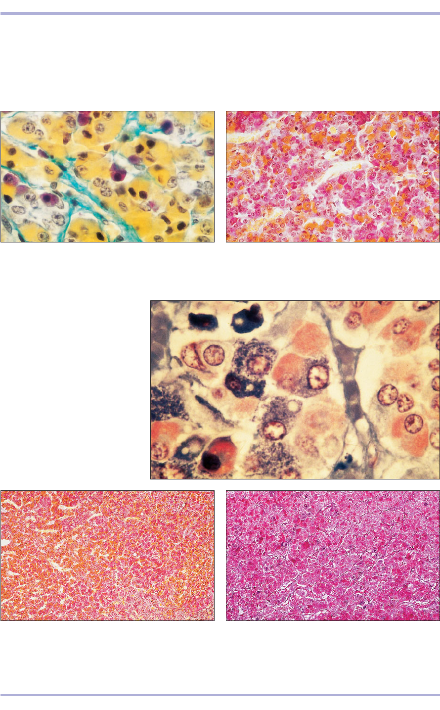
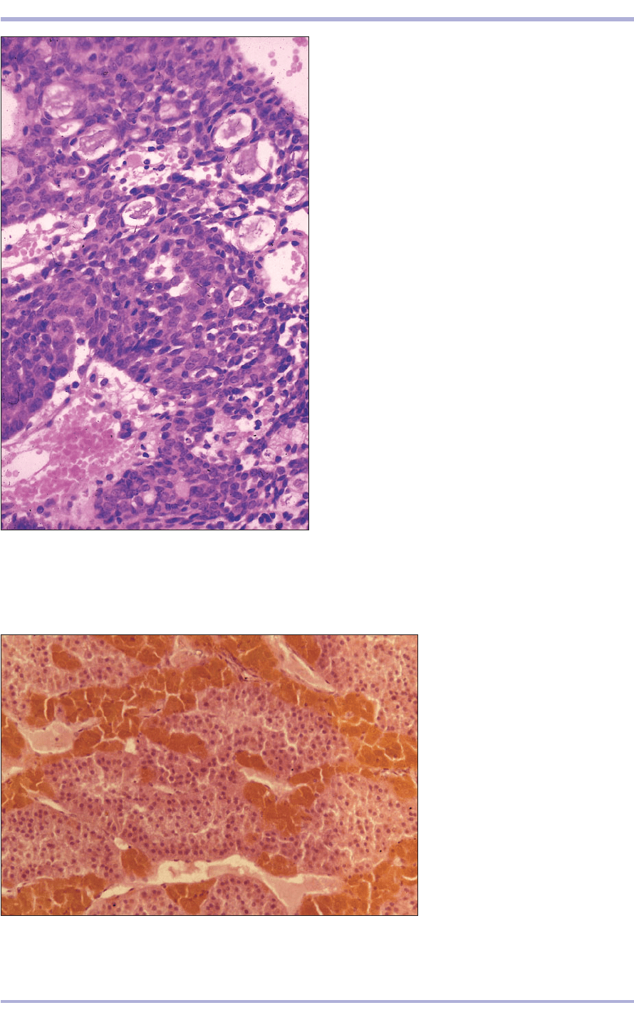
10.4 Pars distalis (cat). (1) Eosinophilic alpha cells.
(2) Basophilic beta cells. (3) Chromophobes. (4) Sinusoidal
blood vessels lie along the cords and clusters of secretory
cells. Orange G/acid fuchsin. ×250.
10.4
10.5 Pars distalis (horse). The orange staining A cells are
easily distinguished from the deep pink staining B cells.
Orange G/PAS. ×125.
10.5
10.6 Pars distalis (cow).
(1) Eosinophilic alpha cells.
(2) Basophilic beta cells.
(3) Chromophobe. (4) Sinusoid.
H & E. ×500.
10.6
10.7 Pars distalis (dog). A cells stain with orange G and
the B cells with acid fuchsin. Orange G/acid fuchsin. ×100.
10.7
10.8 Pars distalis (horse). A cells stain with orange G and
the B cells with acid fuchsin; the supporting connective
tissue is green. Orange G/acid fuchsin/light green. ×100.
10.8
1
1
2
4
3
3
4
3
2
chromophils have a strong affinity for dyes and are
divided into acidophils (alpha cells) and basophils
(beta cells) in H & E stained sections. The acidophils
can be further subdivided using special dyes such as
orange G (A cells, orangeophils) or azocarmine (B
cells, carminophils). Basophils are larger than acido-
phils, fewer in number and the cytoplasmic granules
stain strongly with PAS. The cells of the pars distalis
secrete hormones controlling the other endocrine
glands and also factors such as growth hormone.
Endocrine System
10.11
Neurohypophysis
The neurohypophysis is derived from a ventral
outpocketing of the diencephalon (the caudal part
of the forebrain) and is divisible into a pars ner-
vosa, median eminence and infundibular stalk.
The neurohypophysis is composed of numerous
unmyelinated nerve fibres with cell bodies that are
located in the supraoptic and paraventricular
nuclei of the hypothalamus. The axons converge
at the median eminence to form the hypothala-
mic<hypophyseal tract and pass through the
infundibular stalk to terminate on the endothelial
lining of the capillaries of the pars nervosa (10.12
and 10.13). The neurosecretions of these cells pass
down the axons and accumulate at the terminal
regions of the nerve fibres as neurosecretory
(Herring) bodies (10.14). The axons are supported
by pituicytes (neuroglial cells). Oxytocin (a stim-
ulant to the pregnant uterus and lactiferous ducts)
and vasopressin (antidiuretic) are the major hor-
mones produced by the nervosa.
Pars intermedia
The pars intermedia is well developed in domestic
animals and lies between the distalis and the ner-
vosa. In the horse these regions lie closely together.
In other domestic animals the pars intermedia and
152
Comparative Veterinary Histology with Clinical Correlates
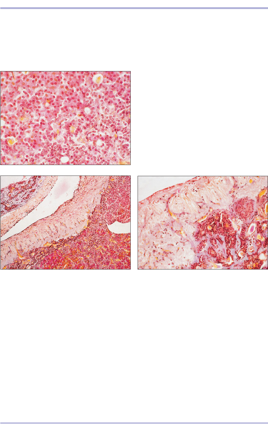
10.9 Pars intermedia (cat). The parenchymal cells are
arranged in (1) columns or in (2) follicles. (3) Blood
vessels. PAS/orange G/haematoxylin. ×250.
10.9
10.10 Pars intermedia/infundibular stalk (horse). (1) The
parenchymal cells are separated by blood vessels (orange-
staining erythrocytes). (2) The infundibular stalk is fibrous
in appearance with small blood vessels of the portal system.
(3) The third ventricle lined by ependyma. PAS/orange
G/haematoxylin. ×50.
10.10
10.11 Pars nervosa/infundibular stalk (horse). (1) Pars
tuberalis. (2) Infundibular stalk with portal vessels.
(3) Ependyma lining the third ventricle. PAS/orange
G/haematoxylin. ×125.
pars distalis are partially separated by the hypophy-
seal cavity. The parenchymal cells are basophilic
with a few acidophilic cells and are arranged in
columns or in follicles (10.9). The hormone secreted
by these cells stimulates melanocytes and controls
the degree of skin pigmentation.
Pars tuberalis
The pars tuberalis surrounds the infundibular stalk,
and together they form the hypophyseal stalk (10.10).
It is very vascular. The major blood vessels of the
hypothalamic<hypophyseal portal system lie in the
stalk, and allow the transfer of releasing factors
secreted in the brain to the target cells in the distalis.
The tuberalis cells are small and basophilic (10.11).
1
2
3
2
3
1
1
3
2
2
153
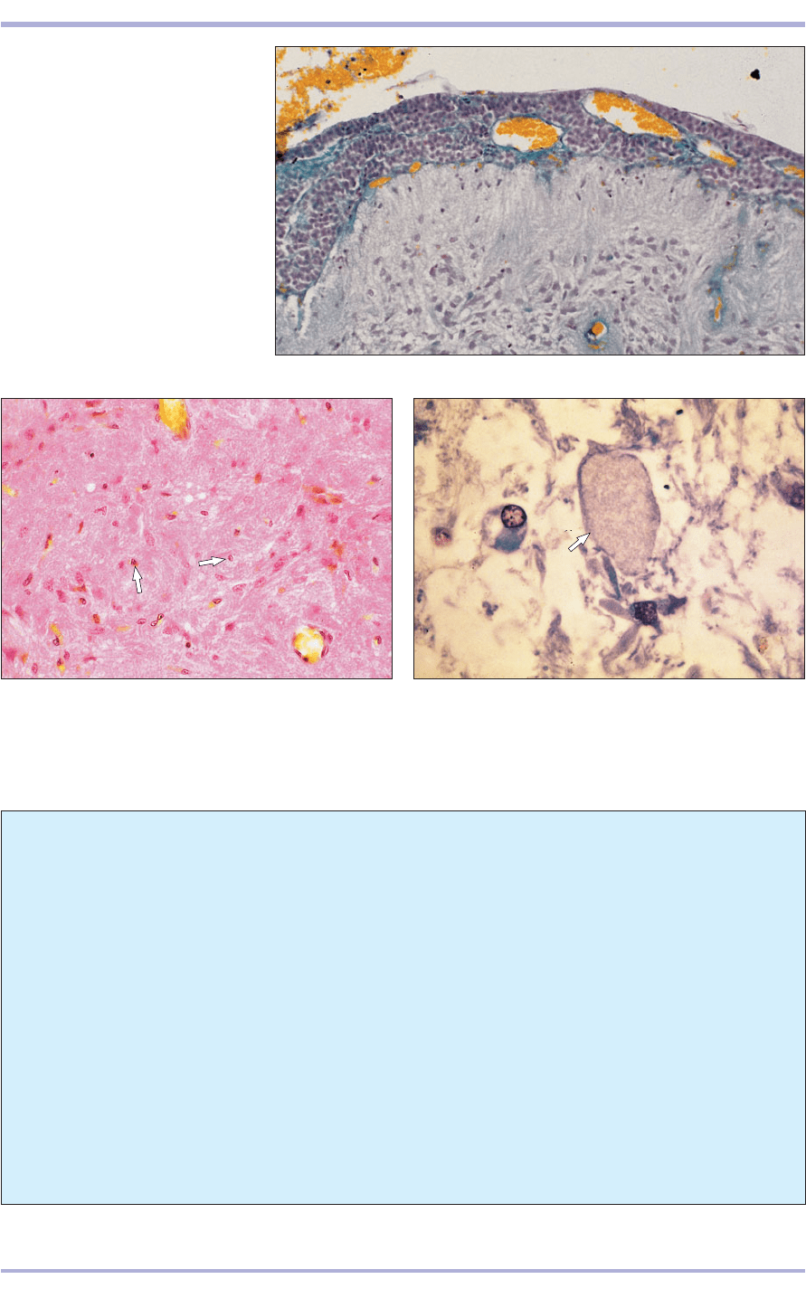
10.12 Pars nervosa (cat). (1) Pars
nervosa. (2) Pars intermedia.
(3) Infundibular recess. H & E. ×100.
10.12
10.13 Pars nervosa (cat). The pars nervosa has a
distinctive fibrous appearance; the nuclei of the pituicytes
are arrowed. H & E. ×125.
10.13
10.14 Pars nervosa (cat). The neurosecretion accumulates
in the terminal part of the axons of the hypothalamic<
hypophyseal tract to form neurosecretory bodies
(arrowed). H & E. ×250.
10.14
3
1
2
Clinical correlates
Spontaneous haemorrhage and subsequent
necrosis of the pituitary during late pregnancy
and parturition occur infrequently in humans
and rarely in domestic animals. Neoplasms
involving one or more lobes of the pituitary are
more common. Because of the location of the
pituitary gland and the limited space available
for an expanding lesion to occupy within the
skull, the clinical signs that attend pituitary
tumours are usually referable to pressure on
adjacent structures. If the optic chiasm is
affected, deterioration of vision extending to
blindness will ensue. More specifically, the signs
manifested by an animal with a hypophyseal neo-
plasm refer to hyper- or hyposecretion of one or
more pituitary tropic hormones.
Hormonally active tumours of the adenohy-
pophysis result in a clinical picture of corti-
costeroid excess known as Cushing’s disease,
while neoplasms of the neurohypophisis often
result in abnormalities of antidiuretic hormone
or oxytocin secretion. Neoplasms involving the
neurohypophisis usually result in either the
hypo- or hypersecretion of antidiuretic hor-
mone or oxytocin. Therefore, the first clinical
signs may be substantial changes in renal func-
tion, water loss or retention, and electrolyte
regulation.
Endocrine System
Thyroid gland
The thyroid gland is derived from an endodermal
outgrowth from the floor of the embryonic phar-
ynx. The thin capsule of dense irregular connective
tissue is continuous with the fine reticular fibres of
the vascular stroma. It is partially divided into lob-
ules by thin trabeculae. Each lobule consists of
numerous follicles lined with simple cuboidal
epithelium; the principal follicular cells secrete the
thyroid hormones (10.15 and 10.16). The secretion
is stored in the follicles as homogenous eosinophilic
colloid. The hormones cross back through the fol-
licular cells as required and enter the capillaries as
tri-iodothryonine and thyroxine (tetra-iodothyro-
nine). These regulate the metabolic activity of all of
the body cells and tissues. Large pale cells with an
eosinophilic cytoplasm, the parafollicular cells, lie
between the follicular cells (10.17). These cells pro-
duce thyrocalcitonin, an antagonist of parathyroid
hormone controlling blood–calcium concentrations.
The secretory activity of the thyroid is controlled
by thyrotrophin secreted by the pars distalis of the
pituitary gland.
154
Comparative Veterinary Histology with Clinical Correlates
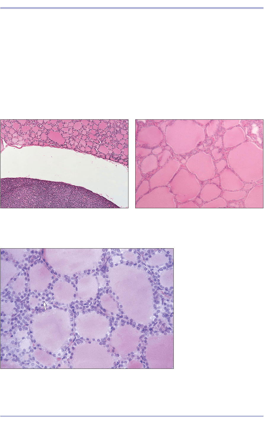
10.15 Thyroid/parathyroid gland (cat). (1) The thyroid
follicles are filled with colloid. (2) Densely basophilic cords
of parathyroid chief cells lie in the thyroid capsule. H & E.
×20.
10.15
10.16 Thyroid gland (dog). (1) The thyroid follicles show
considerable variation in size. (2) Vascular supporting
connective tissue. H & E. ×50.
10.16
10.17 Thyroid gland (sheep). The
thyroid follicle is lined by a simple
cuboidal epithelium. The clear
parafollicular cells are arrowed.
H & E. ×160.
10.17
1
1
1
2
1
2
10.18
Parathyroid gland
The parathyroids are derived from the endoderm of
the third and fourth pharyngeal pouches and are either
internal (embedded in the capsule of the thyroid) or
external (lying a variable distance away). A fine retic-
ular framework supports the cells and the blood ves-
sels. Cords of densely packed basophilic epithelial cells
range along the rich capillary bed (10.15 and 10.19).
155
Endocrine System
10.20 Parathyroid gland (cow). Clumps of oxyphil cells
(1) are present in the parenchyma associated with small
blood vessels. H & E. ×500.
10.20
10.19 Parathyroid gland (dog). Darkly staining basophilic
principal/chief cells are ranged along the capillary bed.
H & E. ×250.
10.19
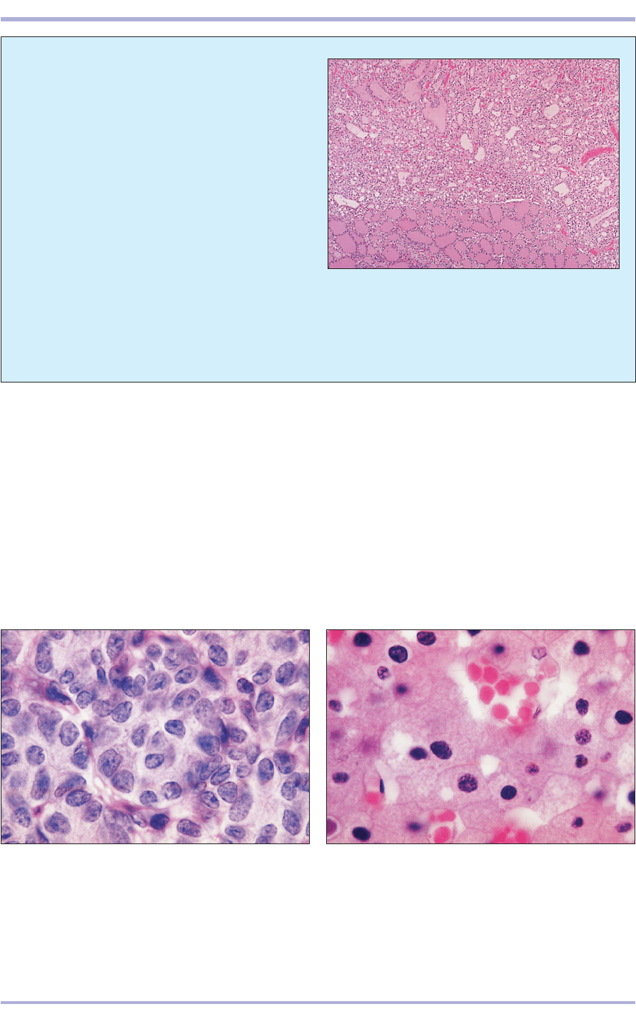
10.18 Thyroid adenoma (cat). Normal thyroid tissue,
at the base of the micrograph, is compressed by an
adenomatous growth that is more cellular and has
less colloid production. The cells are quite uniform
in appearance and form recognizable acinar patterns.
The mitotic rate is low. H & E. ×50.
They are of two types: chief cells and oxyphil cells.
Chief cells are the major source of parathyroid hor-
mone regulating calcium homeostasis. Oxyphil cells
are large cells with an acidophilic cytoplasm and a
pyknotic nucleus (10.20). They are found in the horse
and large ruminants but are rare in the other domes-
tic animals. Their function is unknown.
Clinical correlates
Feline hyperthyroidism (10.18) is one of the
most common endocrine diseases encountered
in veterinary practice. Typically, an elderly cat
presents with weight loss and polyphagia.
Vomiting and diarrhoea are also often reported.
Hyperthyroidism results from hypersecretion of
thyroid hormone by a hyperplastic or adenoma-
tous thyroid gland which is often palpably
enlarged (goitre).
1
1
Adrenal gland
The adrenal glands are paired and lie in the abdom-
inal cavity close to the craniomedial border of the
kidneys. The connective tissue capsule extends into
the gland as thin trabeculae of loose vascular retic-
ular connective tissue. The adrenal is divided into an
outer cortex derived from mesenchyme and an inner
medulla derived from neural crest cells (10.21).
The adrenal cortex produces three main groups of
hormones: glucocorticoids, which regulate carbohy-
drate metabolism; mineralocorticoids, which main-
tain electrolyte concentrations in the extracellular
fluid; and androgens, which possess the same mas-
culinizing effect as testosterone. It is divided into four
zones: the zona glomerulosa, the zona intermedia, the
zona fasciculata and the zona reticularis.
The zona glomerulosa (arcuata, multiformis) is
the outer layer immediately beneath the capsule. It
consists of curved cords or arcades of columnar cells
in the horses, carnivores and pigs (10.22 and
10.23), and as clusters of polyhedral cells in rumi-
nants. The cellular cytoplasm is acidophilic with a
small dark nucleus and secretes mineralocorticoids.
The zona intermedia (more common in the horse
and carnivores than in other domestic animals) lies
between the zona glomerulosa and the zona fascic-
ulata. The cells appear undifferentiated.
The zona fasciculata, the most extensive zone, is
formed of cuboidal or polyhedral cells arranged in
radial cords separated by a sinusoidal network of
blood vessels. The cytoplasm of these cells (also
called spongiocytes) may appear foamy after routine
processing and staining because of the loss of the
steroid glucocorticoid hormones (10.22–10.25).
The zona reticularis is the innermost zone next
to the medulla (10.26 and 10.27). The cells are
small, darkly staining anastomosing cords sur-
rounded by sinusoids. They secrete sex hormones.
The adrenal medulla produces adrenalin (epi-
nephrine). This hormone is a powerful vasopressor,
increasing cardiac output when the animal is dis-
tressed. It also regulates the sympathetic branch of
the autonomic nervous system and stimulates the
release of glucose from the liver. The medulla is com-
posed mostly of columnar or polyhedral APUD cells,
modified postganglionic sympathetic neurons that
take up and stain strongly with chromium salts and
have numerous brown granules in the cytoplasm.
(The chromaffin reaction demonstrates the presence
of adrenalin and noradrenalin.) In domestic mam-
mals an outer and inner zone of the medulla can
often be distinguished.
Other glands, such as the carotid and aortic bod-
ies, also demonstrate the chromaffin staining reac-
tion. Together with the adrenal medulla they are
known as the chromaffin system.
156
Comparative Veterinary Histology with Clinical Correlates
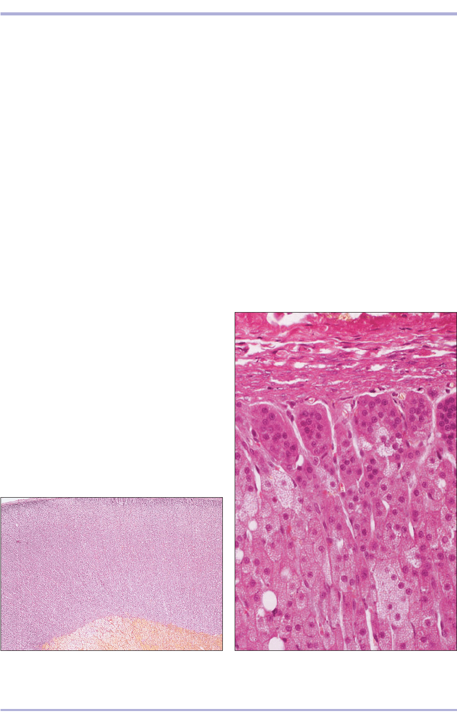
10.21 Adrenal gland (cat). The adrenal gland is divided
into an outer cortex and an inner medulla. H & E. ×5.
10.21
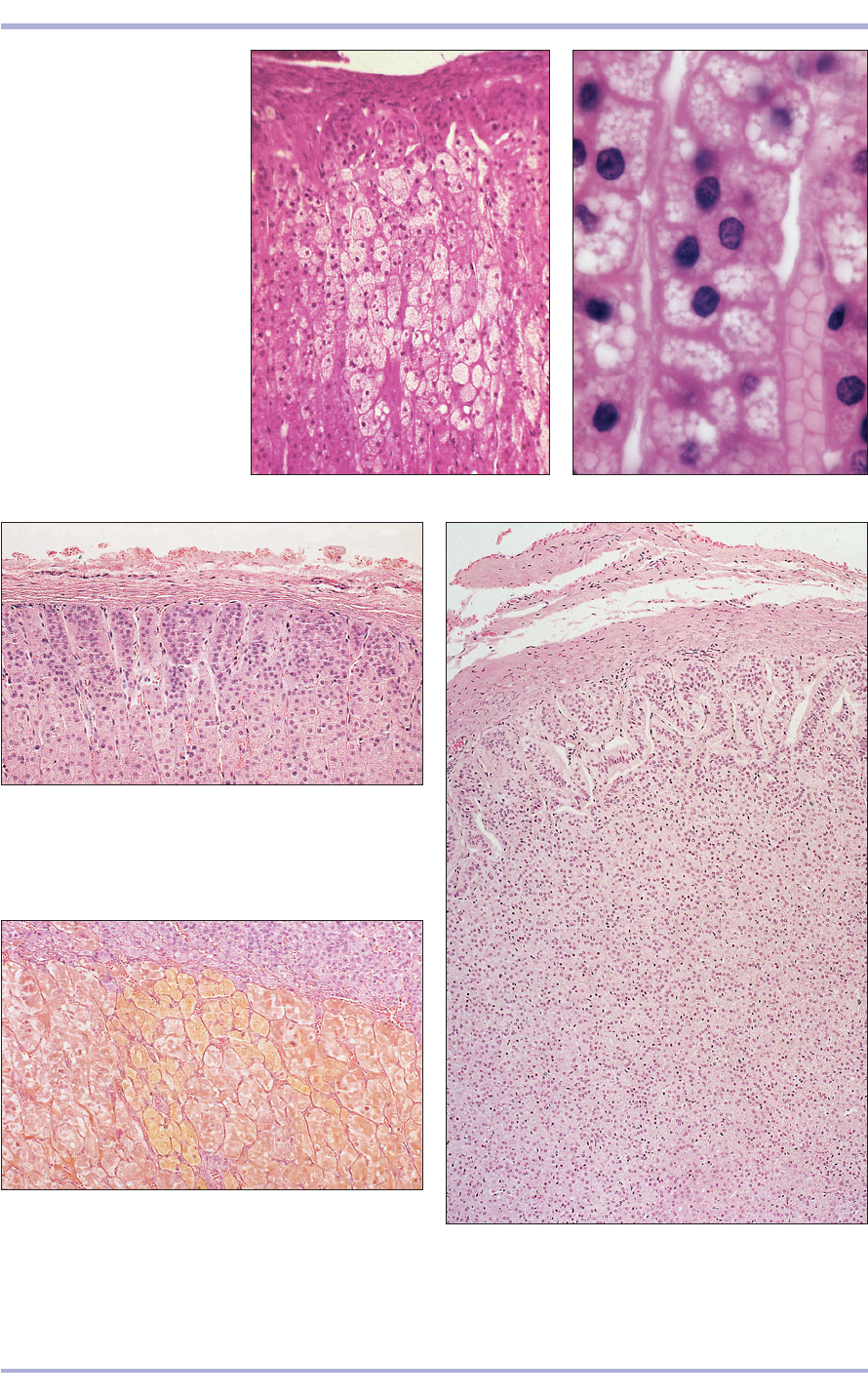
10.22 Adrenal cortex (cat). (1) Connective tissue capsule.
(2) Zona glomerulosa. (3) Zona fasciculata cells are
vacuolated spongiocytes. H & E. ×200.
10.22
3
1
2
157
10.23 Adrenal cortex (pig).
The vacuolated appearance
of the zona fasciculata cells
(spongiocytes) is evident.
H & E. ×250.
10.24 Adrenal gland (pig).
Spongiocytes line the sinusoids.
H & E. ×50.
10.23
10.24
10.25 Adrenal cortex (cat). (1) Connective tissue capsule.
(2) Zona glomerulosa cells are arranged in curved cords.
(3) Zona fasciculata cells are polyhedral arranged in long
radial cords. (4) Sinusoids. H & E. ×50.
10.25
10.26 Adrenal cortex/medulla (cat). (1) Zona reticularis
cells are arranged in anastomosing cords. (2) Adrenal
medulla sympathetic neuron cell bodies. The cytoplasm
is filled with golden brown granules, the chromaffin
reaction. Bouin’s fixation. ×100.
10.26
10.27 Adrenal gland (horse). (1) Zona glomerulosa.
(2) Zona fasciculata. (3) Zona reticularis. H & E. ×62.5.
10.27
1
2
3
1
2
2
1
2
3
4
Endocrine System

10.2910.28
Clinical correlates
Clinical manifestations of adrenal disease depend
on which cellular populations within the gland
are affected and to what extent. Function of the
adrenal cortex can be insufficient, leading to
hypoadrenocorticism (Addison’s disease). In dogs
the most common lesion of this type is idiopathic
bilateral adrenocortical atrophy in which all lay-
ers of the cortex are reduced in thickness and
there is reduced production of all classes of cor-
ticosteroids. Inflammation of the adrenal cortex
caused by a variety of microbial infections can
also lead to insufficiency. Clinically, a patient
often exhibits progressive loss of condition,
weakness and gastrointestinal signs but presen-
tation in a shock-like state of circulatory collapse
is also possible.
Hypersecretion of adrenal corticosteroids pro-
duces hypoadrenocorticism (Cushing’s syn-
drome), a clinical syndrome of steroid excess
characterized by skin and hair changes, polydip-
sia, polyphagia and weight gain with muscle
weakness. Causes include a functional adrenal
tumour or may be referable to a pituitary tumour
that overstimulates the adrenal glands. The typi-
cal skin biopsy changes in canine hyperadreno-
corticism are illustrated by 10.28. A primary
adrenal tumour from a Syrian hamster is shown
in 10.29, and is accompanied by a skin section
(10.30) from the same animal. In addition to the
expected findings of adnexal atrophy the skin
shows marked secondary hyperplastic changes,
possibly associated with self trauma.
Disorders of the adrenal medulla are less
commonly described but phaeochromocytomas
(10.31), tumours of the chromaffin cells, are the
most prevalent. These are usually recognized in
dogs or cattle. In dogs, around half of these
show malignant behaviour with metastasis to
regional lymph nodes and beyond. The major-
ity of phaeochromocytomas are not function-
ally active but an occasional tumour secretes
catecholamines.
158
Comparative Veterinary Histology with Clinical Correlates
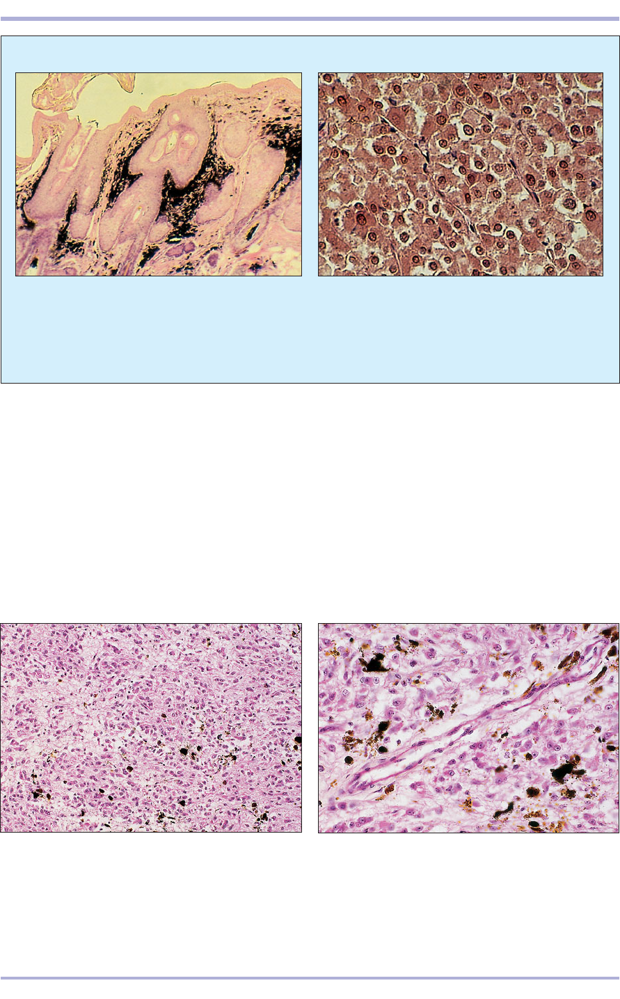
10.29 Adrenal cortical adenoma in a Syrian hamster
(Mesocricetus aurutus). The corticosteroid-secreting
cells are moderately pleomorphic, and a few mitotic
figures are seen. H & E. ×125.
10.28 Hyperadrenocorticism (dog). This skin section
is from an 8-year-old Whippet with hyperadreno-
corticism. The epidermis is thin, the hair follicles
inactive and their associated sebaceous glands
atrophic. There is dermal atrophy with reduction in the
number and thickness of the collagen bundles in the
deep dermis. No inflammatory reaction is present.
H & E. ×20.
10.3110.30
159
Endocrine System
10.32 Epiphysis cerebri (pineal gland; ox). The
pinealocytes are small epithelioid cells arranged in
(1) cords, (2) clusters or (3) follicles. Corpora arenaceae
(brain sand) are a feature of this gland. H & E. ×125.
10.32
10.30 A section of skin from the hamster illustrated in
10.29. The epidermis is hyperplastic, hyperkeratotic,
acanthotic and heavily pigmented. There is a
generalized atrophy of hairs and their precursors.
H & E. ×20.
10.33 Epiphysis cerebri (pineal gland; ox). (1) The pia
mater extends into the gland. (2) Corpoa arenaceae.
H & E. ×250.
10.33
1
2
1
2
3
Epiphysis cerebri
(pineal gland)
The pineal gland (epiphysis cerebri) is a dorsal
evagination of the diencephalon, attached by a
stalk to the dorsal wall of the third ventricle of the
cerebrum. It is covered by a capsule and trabecu-
lae of the pia mater and is divided into lobules by
connective tissue septa. The parenchyma is com-
posed primarily of pinealocytes, small epithelioid
cells with round nuclei and acidophilic cytoplasm,
supported by neuroglial cells. Corpora arenacea
are local calcified deposits present in the gland.
These are more numerous in older animals (10.32
and 10.33). This gland secretes melatonin and
serotonin, and responds to changes in daylight pat-
terns to influence the sexual rhythm of seasonal
breeders.
10.31 Phaeochromocytoma in a dog. The tumour cells
contain abundant red–brown granules and their nuclei
bear prominent nucleoli. Bouin’s fixation; H & E. ×200.
160
Avian endocrine system
The avian pituitary gland is similar to that of other
mammals except for the absence of the pars inter-
media. In the thyroid gland follicles are identical to
those of mammals, but the parafollicular cells are
located in a separate gland, the ultimobranchial
body (10.34), which is located close to the origin
of the carotid artery. There is no capsule. Vesicles
or cords of round basophilic cells lie in the con-
nective tissue surrounding the carotid artery. This
is the source of calcitonin in the bird and together
with parathyroid hormone it regulates calcium
metabolism.
In the parathyroid gland the parenchyma is com-
posed of irregular cords of principal/chief cells, sep-
arated by connective tissue and numerous sinusoids.
The adrenal gland has no clear division into cor-
tex and medulla. Instead, the parenchyma is com-
posed of intermingled cortical tissue and medullary
(chromaffin) tissue. The cortical cells are arranged
as anastomosing cords and are steroid-secreting.
The chromaffin cells are polygonal (10.35).
10.34 Ultimobranchial body (bird). The basophilic
cells are arranged in (1) vesicles or in (2) cords in the
connective tissue surrounding the carotid artery.
H & E. ×125.
10.34
10.35 Adrenal (inter-renal) gland
(bird). The chromaffin cells are filled
with golden brown granules and lie
in islands between the anastomosing
cords of steroid-secreting cells.
Bouin’s fixation. ×125.
10.35
1
1
2
Comparative Veterinary Histology with Clinical Correlates
