Aughey E., Frye F.L. Comparative veterinary histology with clinical correlates
Подождите немного. Документ загружается.

161
cells that are situated more centrally. Capillaries
and thin-walled venous sinuses are a variable fea-
ture, depending upon the taxon of animal being
studied.
The pars tuberalis (if it is present) consists of
small groups of cells forming follicle- or duct-like
structures on the ventral surface of the infundibu-
lar recess, in the region of the median eminence.
The pars nervosa forms a pit-like depression in
the infundibular recess in some reptiles. In others,
it lies more lateral or dorsolateral to the distal lobe.
It may or may not be divided into lobules by thin
connective tissue septa. The cells comprising the
pars nervosa tend to be finely granular and stain
paler than the pars distalis or pars intermedia
(10.36).
The staining characteristics of amphibian and
reptilian pituitary glands are generally comparable
with those observed in higher vertebrates.
Depending upon species and the laboratory stain-
ing protocols employed, acidophilic, basophilic,
amphophilic (the secretory granules staining both
erythrophilic and cyanophilic), chromophilic or ery-
throphilic glandular cells can be discerned. The pitu-
itary cells of some reptiles are strongly PAS-positive,
whereas they are not in reptiles of different fami-
lies. In addition, seasonal, age and sex differences
may further complicate the situation.
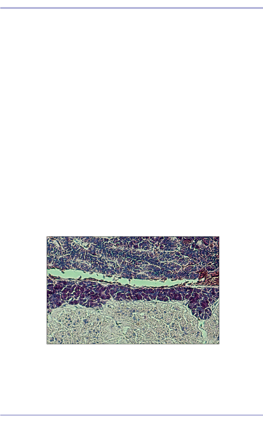
10.36 Whole mount sagittal section of the pituitary gland of a Children’s
python (Liasis childreni). The pars distalis is at the top; the pars intermedia is
represented as a variably narrow band of eosinophilic cells; the pars nervosa
is in the lower half. H & E. ×50.
10.36
Reptilian, amphibian and
fish endocrine system
Pituitary gland
The pituitary gland of amphibians and reptiles is
similar to that in mammals and birds, but there are
substantial differences between disparate families
of reptiles. It is beyond the scope of this text to
delineate each of them.
The adenohypophysis is composed of the inter-
mediate lobe (pars intermedia) and distal lobe (pars
distalis); the neurohypophysis is composed of the
median eminence and neural lobe (pars nervosa).
Neurosecretory perikarya of the supraoptic and
paraventricular nuclei form the hypothalamoneu-
rohypophyseal tract.
The intermediate lobe is closely juxtaposed to
the neural lobe and is joined to the distal lobe by
a narrow band of glandular tissue. The distal lobe
of most amphibians and reptiles tends to be flat-
tened and lies ventral to the pars nervosa and is
caudoventeral to the median eminence. It is com-
posed of cellular cords of cuboidal or prismatic
cells that are surrounded by a thin fibrovascular
connective tissue. This forms fine septa separating
adjacent cords of chromophilic cells that are situ-
ated peripherally from chromophobic (‘principal’)
Endocrine System
162
tissue (10.38). The thyroid often displays a marked
seasonal change in the shape and height of the fol-
licular epithelial cells and amount of colloid. They
are usually flattened and decidedly squamous or
may be tall columnar and pallisaded into a parallel
‘picket-fence’-like pattern and contain scanty colloid
during times of hypoiodine-induced hypothy-
roidism. Interfollicular ‘C’ cells are located in the
interstitial connective tissue that separates adjacent
follicles (10.39). Usually, the intrafollicular colloid
is amorphous and agranular, but it can also be dis-
tinctly granular.
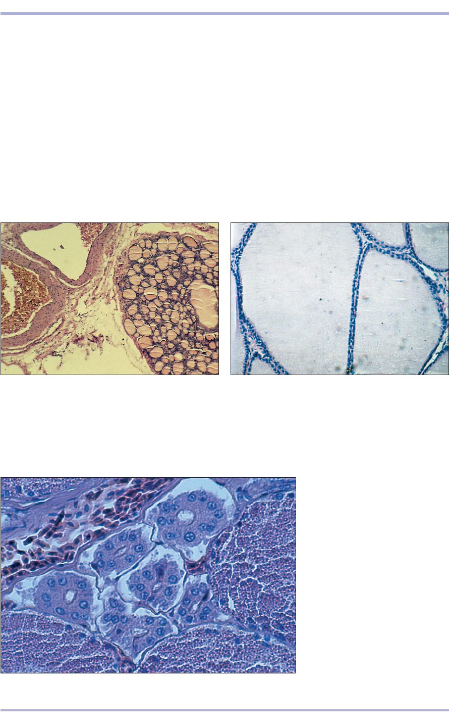
10.39 Interfollicular (‘C’) cells in
the thyroid of a hog-nosed snake
(Heterodon platyrhinos). These
endocrine cells are arranged into
small diameter tubuloacinar follicles
with tiny central lumens. The
thyroidal follicular colloid in this
section is unusually granular.
H & E. ×200.
10.39
10.38 Thyroid from a red-eared slider turtle (Trachemys
scripta elegans). The histological characteristics of the
reptilian gland change seasonally and may range from
follicles lined by cuboidal to a much flattened squamous
epithelium. H & E. ×125.
10.38
10.37 Whole mount section of the thyroid (1) of a Central
American rattlesnake (Crotalus durissus bicolor). The
thyroid lies immediately adjacent to the the heart base and
is readily identified by its characteristic pink, colloid filled
follicles. One aortic arch (2) and a jugular vein (3) are
immediately to the left of the thyroid. H & E. ×20.
10.37
Thyroid gland
The thyroid glands of most amphibians and reptiles
are located just cranial to the cardiac outflow tract,
often lying between or immediately adjacent to the
twin aortic arches, cranial vena cava and pulmonary
vasculature (10.37). In amphibians the thyroid is
present as paired lobes. In reptiles it is usually a sin-
gle organ, although bilobed glands with a thin isth-
mus joining the two lobes have been observed. The
colloid-filled follicles are highly variable in size and
are separated by a fine fibrovascular connective
1
2
3
Comparative Veterinary Histology with Clinical Correlates
10.41
Parathyroid gland
Fish lack a parathyroid gland. Rather, the ultimo-
branchial body is highly developed and serves as the
major regulator of calcium and phosphorus metab-
olism. The parathyroid glands of most amphibians
and reptiles are present as one or (usually) two pairs
of lobes that are located in the caudal cervical
region, often adjacent to the thymus or jugular
veins. The cells may be cuboidal to prismatic or
polyhedral and contain small intracytoplasmic gran-
ules that usually stain pale pink with H & E. They
are formed into small lobules by thin fibrovascu-
lar connective tissue septa that penetrate the gland
from the surrounding capsule (10.40). Under some
conditions, clear or foamy appearing spaces may be
scattered throughout the glandular tissue.
Adrenal glands
The adrenal tissues of fish, amphibians and many
reptiles are significantly different. The adrenal glands
(sometimes referred to as inter-renal or intrarenal
tissue) of most fish are embedded in the cranial kid-
neys, where they surround the larger blood vessels
and may be intermixed with haematopoietic tissue.
There are two kinds of adrenal cells: medullary or
chromaffin cells and sympathetic nerve-like para-
ganglion cells. Amphibians possess discrete paired
adrenals composed of three major cell types: chro-
maffin ‘medullary’ cells that are of neurectodermal
origin; ‘cortical’ cells; and Stilling cells that are of
mesodermal origin. Although it is known that the
cortical cells secrete adrenal corticosteroids, the
163
function of Stilling cells is not known. Some inves-
tigators consider the Stilling cell to be a distinct
glandular organ.
In reptiles the paired adrenals usually lie imme-
diately adjacent to the kidneys or, in some instances
where the kidneys are located within the pelvic
canal, they lie medial to the gonads. Two major cell
types are found: dark staining chromaffin or
medullary cells that secrete catacholamines; and pale
staining cortical steroidogenic cells. In mammals,
these cells are confined to the cortical and medullary
zones. In reptiles, the location of the cells tends to
be less circumscribed; the chromaffin cells are in
islands or nests scattered throughout the steroido-
genic cortical cells. The cytoplasm of cortical cells
often contains small clear lipid-like vacuoles (10.41).
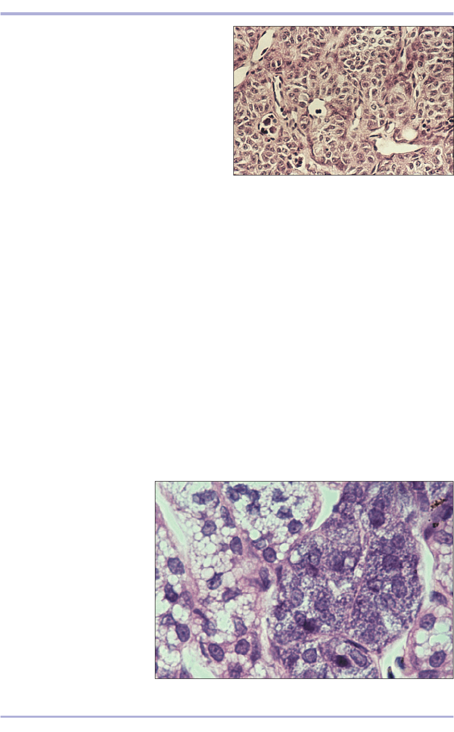
10.40 Parathryoid gland of a desert tortoise (Xerobates
agassizi). The secretory cells are formed into small lobules
bound by thin fibrovascular connective tissue septa.
H & E. ×125.
10.40
2
1
10.41 Adrenal gland of a desert
tortoise (Xerobates agassizi). The
kidney is in the lower right; the
adrenal is on the left. Rather than
being confined to a discrete cortex
and medulla, the deeply staining
chromaffin cells (1) and the pale
spongiocytes (2) are grouped into
nest-like aggregates that are
admixed throughout the gland.
H & E. ×200.
Endocrine System
164
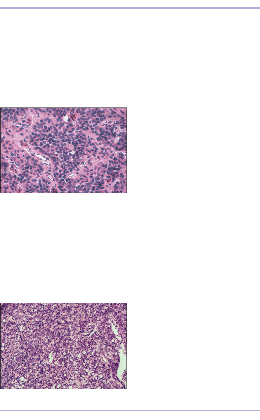
10.43 Ultimobranchial body of a boa constrictor (Boa c.
constrictor). Many of the secretory epithelial cells that
comprise this glandular organ contain large, clear, lipid
vacuoles. Typically, these glands are formed from solid
sheets of cells with little separation by connective tissue
septa. Small blood vessels penetrate the tissue. H & E. ×50.
10.43
Pineal gland
In teleost fish the pineal gland is hollow and con-
sists of columnar epithelial cells of three types: sen-
sory, sustentacular and ganglion-like. It resembles
a sensory organ; some primitive cyclostomes actu-
ally possess a pineal covered by a relatively clear
lens-like structure that admits light.
In amphibians the pineal gland is more solid than
in fish and arises as a median outgrowth on the dor-
sal surface of the brain. Like the gland in fish, it is
photosensitive. In response to darkness it secretes
melatonin, which causes peripheral melanophages
to aggregate, thus lightening the integument’s
colour.
In reptiles the pineal gland is solid and composed
of glandular secretory cuboidal to low columnar
epithelial cells that are arranged into lobules sepa-
rated by delicate fibrovascular septa that are exten-
sions from the surrounding capsule (10.42).
Preliminary investigations have revealed at least
some inter-relationships between the pineal and
other endocrine glands, especially the thyroid. The
pineal gland of lizards lies slightly rostral to the
parietal foramen. Whether sufficient light can enter
the skull through the parietal eye and the parietal
foramen (in those species that possess them) is con-
jectural, but appears to be possible.
10.42 Pineal gland of a Burmese python (Python molurus
bivittatus). The cuboidal epithelial cells have indistinct cell
membranes and large round nuclei. They are arranged
into lobule-like groups that are separated from each
other by delicate fibrovascular septa. H & E. ×125.
10.42
consists of tall columnar or pseudostratified colum-
nar epithelium formed into spherical vesicle-like
structures. They are paired in some amphibians, sin-
gle in some salamanders, and absent in some frogs
and in caecilians.
In reptiles the ultimobranchial bodies are paired
and function in calcium and phosphorus regulation.
They also have a relationship to thyroid function in
some reptilian species. They are believed to secrete
calcitonin in response to blood calcium concentra-
tions. Reptilian ultimobranchial bodies are composed
of cuboidal to low columnar glandular epithelial cells
with finely granular pink staining cytoplasm, often
containing clear lipid-like vacuoles (10.43).
Ultimobranchial body
In the lower vertebrates the ultimobranchial bodies
play an important role that is shared in the higher
vertebrates by the parathyroid and thyroid. In
teleost fish the ultimobranchial body is well devel-
oped and lies in the transverse septum between the
ventral surface of the oesophagus and liver, imme-
diately caudal to the heart. The glandular tissue
Comparative Veterinary Histology with Clinical Correlates
Clinical correlates
Thyroid gland
As is the case with the higher vertebrates,
hypothyroidism, goitre, thyroid inflammation and
neoplasia occur occasionally in the lower verte-
brates. Clinical hyperthyroidism is relatively com-
mon in domestic cats. Dietary hypothyroidism is
relatively common in semiaquatic turtles, espe-
cially those kept as pets by persons living in so-
called ‘goitre belts’ where humans suffer from
chronic hypothyroidism unless they receive sup-
plemental iodine. Herbivorous chelonians, espe-
cially those originating from oceanic volcanic
islands, appear to be particularly prone to dietary
hypothyroidism.
Parathyroid gland
Captive reptiles often display the clinical signs
of secondary hypoparathyroidism that are
induced by being fed either food containing
insufficient available calcium or, more com-
monly, food containing an improperly low cal-
cium : high phosphorus ratio. The parathyroid
glands from these animals become hyperplas-
tic and hypertrophied. With chronicity,
changes consistent with adenomatous hyper-
165
Endocrine System
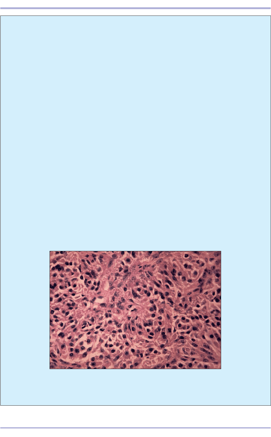
plasia are seen: the glandular cells become
pleomorphic, distorted and may lose their nor-
mal granularity. Sometimes, actual primary
hyperparathyroidism occurs. The cells then
assume frankly pleomorphic shapes, their
nuclear chromatin becomes more dense, occa-
sional bipolar cells and mitotic figures may be
observed, and the lobules become distorted or
break through their septal confines and coa-
lesce (10.44).
Adrenal gland
For discussion of this, see pages 158–9.
Pineal gland
One benign pinealoma has been reported in a
green iguana (Iguana iguana). It was found at
necropsy and was grossly and microscopically
pigmented. It was composed of branching papil-
lary fronds of streamer-like, very elongated
columnar epithelial cells borne on thin stalks of
fibrovascular connective tissue that projected into
the lumen of cystic spaces within the mass.
Destruction of the pineal gland might be
expected to disrupt the normal diurnal<nocturnal
behaviour pattern, and perhaps the colour, of an
animal subjected to this experimental protocol.
10.44 Parathyroid adenoma from a desert tortoise that was displaying
clinical manifestations of primary hyperparathyroidism, including severe
osteopenia. The tumour cells are pleomorphic, hyperchromatic and
arranged irregularly. H & E. ×160.
10.44

Ultimobranchial body
The paired ultimobranchial bodies of reptiles
undergo hypertrophy as a consequence of
chronic hypocalcaemia. They have been
reported to involute spontaneously with ageing
in some normal lizards. Functional hyperpla-
sia (or adenomata) of the ultimobranchial bod-
ies can cause excessive mobilization of calcium
from skeletal stores. Hypersecretion of calci-
tonin, with subsequent hypercalcaemia, may
result in osteopenia and, occasionally, renal
tubular calcium urolithiasis.
166
Comparative Veterinary Histology with Clinical Correlates
The male reproductive system consists of the paired
testes suspended in the scrotal sacs, a continuous
duct system for the storage and transport of the
male gamete, the spermatozoon, and an ejaculatory
organ, the penis. The accessory sex glands secrete
into the urethra and provide a suitable fluid
medium to transport the spermatozoa during ejac-
ulation and in the female reproductive tract.
Testis
The testis is a compound tubular gland with an
exocrine and an endocrine function. The exocrine
function is the production of spermatozoa by the
lining epithelium of the seminiferous tubules. The
endocrine function is the production of the male
sex hormone, testosterone, by the specific inter-
stitial (Leydig) cells in the intertubular connective
tissue. The testis is covered by mesothelium con-
tinuous with the visceral layer of the tunica vagi-
nalis. A thick dense connective tissue capsule, the
tunica albuginea, encloses the testis. A variable
amount of smooth muscle may be present. The
tunica vasculosa is the inner vascular layer of the
tunica albuginea.
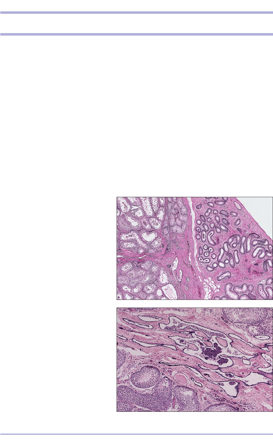
The capsule is reflected into the median plane of
the testis to form a partition, the mediastinum, and
gives off loose vascular connective tissue, the septula
testis, to divide the testis into lobules to support the
seminiferous tubules (11.1 and 11.2). The coiled
seminiferous tubules are lined with a multilayered
seminiferous epithelium of spermatogenic cells and
sustentacular (Sertoli) cells. They rest on a basement
membrane and are surrounded by a lamellated con-
nective tissue with myoid elements. The specific
interstitial (Leydig) cells are found in the loose vas-
cular connective tissue separating the tubule.
167
11. MALE REPRODUCTIVE SYSTEM
11.1 Testis (cat). (1) Efferent ducts invested by
connective tissue. (2) Tunica albuginea. (3) Rete
testis. (4) Mediastinum testis. (5) Seminiferous
tubules. H & E. ×20.
11.1
11.2 Testis (cat). (1) Rete testis lying in the
mediastinum. (2) Seminiferous tubules leading
into (3) tubuli recti lined by sustentacular cells.
H & E. ×50.
11.2
1
3
2
3
5
1
2
4
Production of
spermatozoa
In the prepubertal male there are two cell types: the
sustentacular cell and the spermatogonium, the
immature male germ cell. The sustentacular cells are
tall columnar, extending from the basement mem-
brane to the lumen of the tubule, with a pale vesic-
ular basal nucleus and a prominent nucleolus. As the
name suggests, the sustentacular cells support the
later stages in the development of spermatozoa.
Spermatogonia lie next to the basement membrane
and are small round cells with a dark staining
nucleus. At puberty they move away from the mem-
brane and undergo mitotic divisions. When these
cease the cells go through a period of growth, the
deoxyribonucleic acid is replicated and the cells
become tetraploid primary spermatocytes. The
nucleus is granular and the coiled chromosomes give
a dense staining reaction. The primary spermatocyte
divides meiotically to form two secondary sperma-
tocytes, which each divide immediately to form two
haploid spermatids. These are small cells with a
spherical nucleus and lie close to the lumen of the
tubule. The spermatids move into recesses in the sus-
tentacular cells and metamorphose into spermato-
zoa, shedding the excess cytoplasm into the lumen of
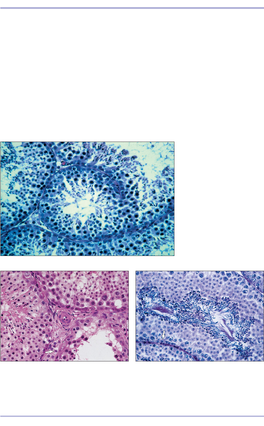
the tubule (11.3–11.5).
168
Comparative Veterinary Histology with Clinical Correlates
11.3 Testis (bull). The seminiferous
tubules are lined by a multilayered
epithelium. (1) Spermatogonia.
(2) Primary spermatocytes.
(3) Spermatids. (4) Developing
spermatozoa. (5) Interstitial tissue
separates the tubules. H & E. ×160.
11.3
11.4 Testis (bull). Vascular interstitial tissue lies between
the seminiferous tubules; specific interstitial cells are
vacuolated (arrowed). H & E. ×200.
11.4
11.5 Testis (dog). All the stages of spermatogenesis are
seen in the lining epithelium. (1) Spermatogonia.
(2) Primary spermatocytes. (3) Spermatids. (4) Developing
spermatozoa. The sustentacular cells are arrowed.
H & E. ×200.
11.5
1
2
3
4
1
1
3
5
4
2
11.7
Structure of spermatozoa
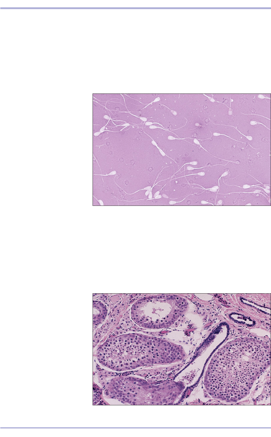
The mature spermatozoon consists of a head and
a tail (11.6). The head contains the condensed hap-
loid nucleus. The anterior two-thirds is covered by
the acrosomal cap, a derivative of the Golgi appa-
ratus containing the enzyme hyaluronidase, which
is required for penetration of the ovum at fertil-
ization. The tail (a flagellum) has the characteris-
169
11.6 Spermatozoa (bull). The
spermatozoa consist of a head and a
tail (flagellum). Unstained
spermatozoa were live and stained
spermatozoa were dead at the time
of staining. Nigrosin–eosin. ×625.
11.6
2
3
1
Tubuli recti rete testis,
efferent ducts
The seminiferous tubules, at their terminal seg-
ments, are joined by a transitional zone, lined by
sustentacular cells, to straight tubules (tubuli recti),
which are continuous with a network of anasto-
mosing channels that form the rete testis (11.7). The
11.7 Testis (cat). (1) Seminiferous
tubules. (2) Straight tubules. (3) Rete
testis. H.& E. ×50.
rete testis has a simple squamous or cuboidal epithe-
lium. It is surrounded by the loose connective tissue
of the mediastinum and is drained by between seven
and 20 efferent ducts (see 11.1). The ducts are lined
with simple columnar or pseudostratified epithe-
lium, and some of the cells are ciliated. The lumen
of each duct is wide and the lamina propria is sparse
connective tissue (with some smooth muscle in the
stallion).
tic structure of flagellae and cilia, with two central
microtubules and more peripheral doublets mak-
ing up the axial filament complex. The proximal
part is surrounded by an end-to-end helix of mito-
chondria to provide the energy during movement.
Sperm are non-motile and immature at this stage.
The peritubular myoid elements push the mixture
of sperm and debris down the seminiferous tubule
towards the mediastinum.
Male Reproductive System
Epididymis and ductus
deferens
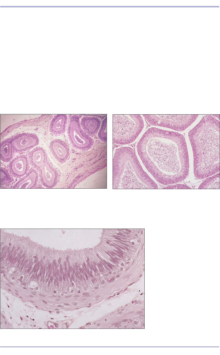
The epididymis, where sperm mature and become
motile (11.8–11.10), is a single, long coiled duct
lined with pseudostratified epithelium. Long cyto-
plasmic processes, the stereocilia, project into the
lumen. The epithelium rests on a connective tissue
lamina propria and there is a variable amount of
circular smooth muscle. The latter increases in vol-
ume as the tail of the epididymis approaches the
ductus deferens.
The ductus deferens has a very thick muscular
wall and a small lumen, and it is lined with pseu-
dostratified columnar epithelium resting on a deli-
170
11.8 Epididymis (bull). The single coiled ductus
epididymis is lined by a pseudostratified columnar
epithelium resting on a vascular lamina propria
continuous with the supporting connective tissue of the
head of the epididymis. Spermatozoa are present in the
lumen of the tubule. H & E. ×50.
11.8
11.9 Epidiymis (bull). Pseudostratified columnar
epithelium lines the tubule; the lumen is filled with
spermatozoa. H & E. ×100.
11.9
11.10 Epidiymis (bull). (1) Lumen of
the tubule. (2) Luminal cytoplasm
extends into the lumen as
stereocilia. (3) Nuclear layer of the
epithelial cells. (4) Vascular lamina
propria. (5) Muscularis. (6) Vascular
connective tissue. H & E. ×250.
11.10
cate lamina propria containing collagen and elastic
fibres. The mucosa is arranged in longitudinal folds,
and the muscularis is composed of smooth muscle
fibres, presenting a variety of arrangements. In the
domestic animals it is often difficult to determine spe-
cific layers of muscle. Circularly arranged fibres pre-
dominate, and a fibrous adventitial coat is present
(11.11 and 11.12). The ductus deferens terminates
at the colliculus seminalis of the proximal urethra.
Near to this junction it dilates to form an ampulla in
the stallion, bull, ram and dog. In the boar and tom-
cat, simple tubular glands are present in the lamina
propria. The lamina propria and submucosa are tall
columnar and lined with glandular secretory units.
The secretion may calcify in the lumen to form cor-
1
2
4
5
6
3
Comparative Veterinary Histology with Clinical Correlates
