Aughey E., Frye F.L. Comparative veterinary histology with clinical correlates
Подождите немного. Документ загружается.

181
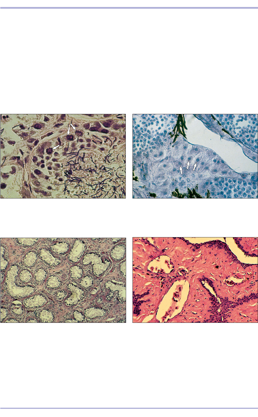
11.38 Section of a seminiferous tubule of a mature
green iguana (Iguana iguana). Three spermatogonia are
shown in mitosis (arrows). Primary spermatocytes (1) and
many tailed spermatozoa (2) lie within the centre of the
tubule. H & E. ×200.
11.38
11.39 A section of the testis of a panther chameleon
(Chamaeleo pardalis). A nest of large interstitial cells
containing finely granular cytoplasm can be seen in the
lower third of this image (arrows). H & E. ×200.
11.39
Reptiles utilize internal fertilization. Male chelo-
nians possess a single erectile penis that is often heav-
ily pigmented and is covered by lightly keratinized
and mucous epithelium. Male snakes and lizards pos-
sess paired erectile intromittant organs, called
hemipenes, that contain fibrovascular tissue that,
when involuted, invaginates. When erect the outer
surface is covered with keratinized epithelium.
Additional flamboyant spines, flounces and other sex-
ual adornments are species specific. The hemipenes
receive lubrication from mucous glands that are
located within the lumen of the hemipenial sheath. In
some reptiles, modified sebaceous glands are associ-
ated with the hemipenes and their sheaths (11.41).
Male crocodilians possess a relatively small erec-
tile penis that superficially resembles that of higher
vertebrates; it even has a glans-like swelling at its
distal end. The surface is covered with lightly ker-
atinized epithelium without horny projections.
Male tuataras lack an erectile intromittant
organ. Internal fertilization is accomplished by
cloacal apposition during which semen is trans-
ferred to the female and enters the proctodeum por-
tion of the cloaca.
1
1
2
11.40 Inactive testis of a green sea turtle (Chelonia
mydas). The seminiferous tubles are lined only by
sustentacular cells and a few spermatogonia. H & E. ×100.
11.40
11.41 Inverted (detumescent) hemipenis of a Carolina
anole lizard (Anolis carolinensis). The organ is composed
of spongy fibrovascular tissues containing many arterial
and venous channels (1), and stratified lightly cornified
squamous epithelium (2). Lobules of glandular tissue,
similar to sebaceous glands, lie at the base of each
hemipenis and furnish lubrication and scent-rich
secretions to the erectile intromittant organ.
H & E. ×50.
11.41
1
2
2
Male Reproductive System
182
Comparative Veterinary Histology with Clinical Correlates
11.4411.43
Clinical correlates
True hermaphroditism occurs occasionally in
reptiles but is usually only discovered at
necropsy. The condition can involve one gonad
of each sex, one or more ovotestes or separate
masses of ovarian and testicular tissue.
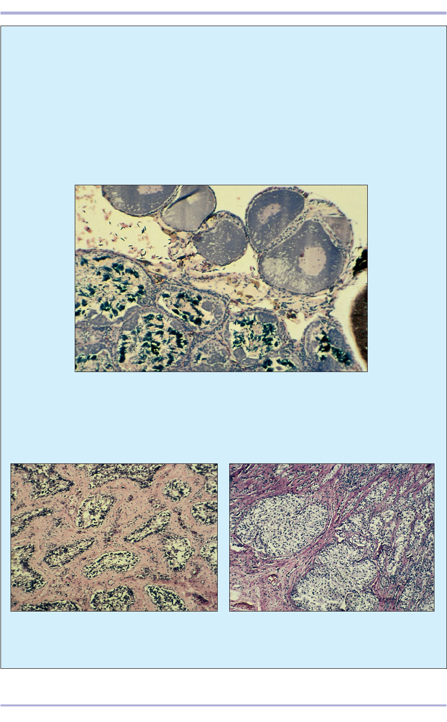
A Bidder’s organ is normally present in male
bufonid toads. This interesting structure is a cap-
like mass of ovarian tissue attached to the cra-
nial pole of the testis (11.42). It is unclear why
androgenic and ovarian hormone secretion do
not each suppress their opposites.
As in domestic animals, the reptilian or
amphibian testis can be the site of inflammatory
disorders, atrophy and fibrosis (11.43), and neo-
plastic disease (11.44). Malignant tumours are
more prevalent than benign variants.
11.42 Bidder’s organ in a western toad (Bufo boreas). Although similar to
hermaphroditic gonadal tissue, Bidder’s organ is a normal constituent found
in some bufonid amphibians and consists of a cap-like assemblage of
pigmented ovarian tissue, including yolked follicle-like structures attached
to the cranial pole of each testis. The presence of ovarian tissue does not
suppress spermatogenesis. H & E. ×50.
11.43 Testicular fibrosis in a blood python (Python
curtus). The seminiferous tubules are atrophic and
widely separated from each other by wide bands of
mature fibrocollagenous connective tissue. H & E. ×20.
11.44 Sustentacular (Sertoli) cell tumour from a
snake. Note large nests of pale-staining sustentacular
cells. H & E. ×100.
11.42
12.1
183
The female reproductive system consists of the
ovaries, uterine tubes (oviducts or Fallopian tubes),
uterus, vagina and vestibule. The external genitalia
are the vulva, labia, clitoris and external urethral
orifice.
Ovaries
Cortex and medulla
The ovaries have an exocrine function, the produc-
tion of the egg or female gamete, and an endocrine
function, the production of female sex hormones.
Each ovary is divided into an outer parenchymatous
zone cortex and an inner vascular zone medulla
12. FEMALE REPRODUCTIVE SYSTEM
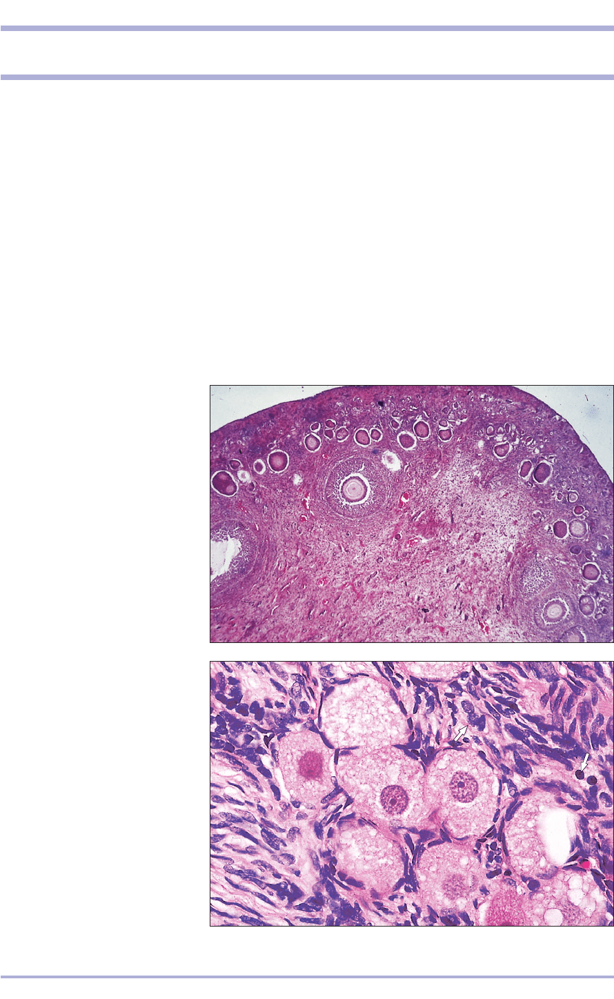
12.1 Ovary (cat). (1) Parenchymatous
zone with primary follicles (2) and
secondary follicles (3). (4) Vascular
zone. H & E. ×7.5.
(12.1), except in the mare where there is no zonal
division. The cortex of each ovary is covered by a
simple squamous or cuboidal epithelium that is con-
tinuous with the mesothelium of the visceral peri-
toneum (the germinal epithelium), except at the hilus
where blood vessels and nerves enter and exit from
the gland. Beneath the epithelium is a layer of dense
connective tissue, the tunica albuginea. Deep to that
is the ovarian or cortical stroma, containing ovarian
follicles in various stages of development. The ovar-
ian stroma consists of spindle-shaped cells arranged
in whorls surrounding the ovarian follicles. In car-
nivores, particularly the cat, numerous specific inter-
stitial cells are present in the stroma. They are small,
round, epithelioid cells with a round nucleus and
they stain poorly (12.2).
12.2 Ovary. Parenchymatous zone
(cat). (1) Primary follicles; the large
oocyte is surrounded by flattened
nurse cells. (2) Cellular ovarian
stroma. The specific interstitial cells
are arrowed. H & E. ×250.
12.2
1
1
2
3
4
2
1
4
The medulla is highly vascularized and consists
of fibroelastic connective tissue and some smooth
muscle (12.3). Channels, lined with (densely stain-
ing) cuboidal epithelium and called the rete ovarii,
are conspicuous components of the medulla in car-
nivores and ruminants (12.4). They are derived from
the mesonephric tubules during embryogenesis.
Development of the follicles
Primordial follicles are the least developed and most
numerous follicles of the ovary, lying just below the
tunica albuginea. Each consists of a primary oocyte
surrounded by a layer of simple squamous follicle
(nurse) cells (see 12.2). The primary oocytes origi-
nate in the late embryonic/early postnatal ovary
from oogonia and are surrounded by a flattened
layer of nurse cells. Further development is arrested
until puberty, when a regular cycle of events results
in the passage of one or more follicles into the
lumen of the uterine tube during reproductive
cycles. A number of follicles begin to grow, and the
primary oocyte accumulates yolk and enlarges from
60 +m to between 100 and 120 +m in diameter.
Concomitantly, an acidophilic, translucent mem-
brane, the zona pellucida, forms around the oocyte
(12.5). The nurse cells become cuboidal, then
columnar, then stratified and begin to accumulate
fluid in the intercellular spaces. The follicle now
comes under the influence of follicle-stimulating
hormone from the pituitary gland and continues to
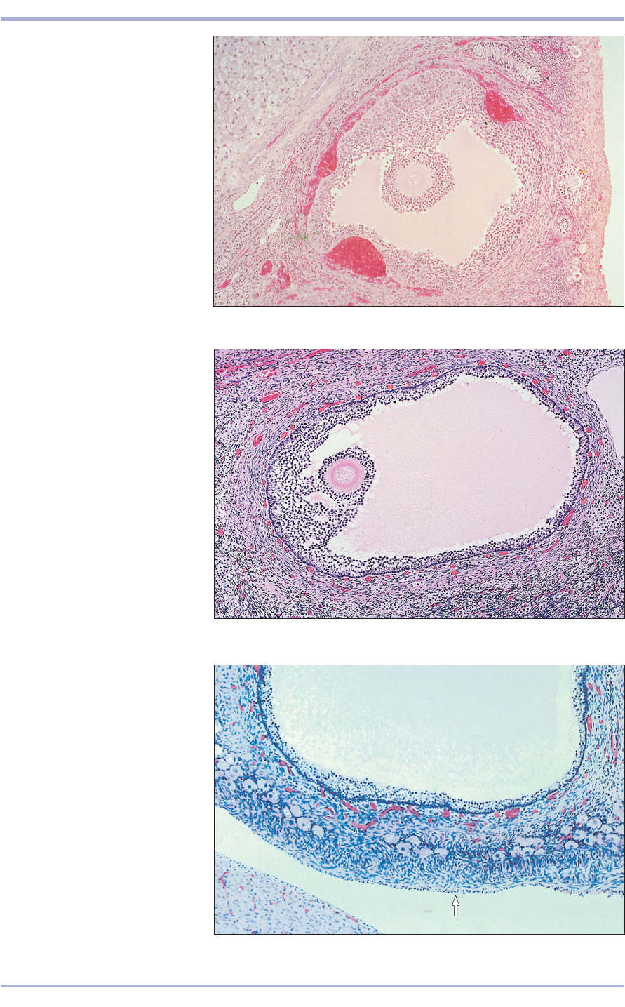
grow to become a secondary follicle, with a C-
shaped, fluid filled space, or antrum (12.6). Its cells
are now called the granulosa layer. The follicle
secretes oestradiol, the female sex hormone that
prepares the endometrium to receive the fertilized
egg. The ovarian stroma condenses around the
developing follicle to form an inner layer, the theca
interna, which is cellular and vascular, and an outer
layer, the theca externa, which is composed of
fibrous connective tissue.
The follicle continues to increase in size, mov-
ing to the surface of the ovary to become a vesicu-
lar [tertiary (mature) Graafian] follicle. The oocyte
is surrounded by a multilayer of granulosa cells, the
cumulus oophorus (12.7 and 12.8). Ordinarily, a
mature vesicular follicle contains a single oocyte.
The follicles of certain animals (carnivores, sows
and ewes) may, however, contain up to six oocytes.
Maximum size is reached just before ovulation.
184
Comparative Veterinary Histology with Clinical Correlates
12.3 Ovary. Vascular zone (bitch). (1) The ovarian stroma
is very vascular. (2) Smooth muscle cells. H & E. ×125.
12.3
12.4 Ovary. Vascular zone (sheep). Tubules of the reti
ovarii are arrowed. H & E. ×125.
12.4
12.5 Ovary. Parenchymatous zone (sheep). (1) The oocyte
is surrounded by the granulosa layer. (2) Ovarian stroma.
H & E. ×125.
12.5
2
1
2
1
185
12.6 Ovary. Secondary follicle
(bitch). (1) The oocyte is surrounded
by the amorphous zona pellucida.
(2) The granulosa cells are stratified.
(3) Fluid accumulates to begin
antrum formation. (4) Ovarian
stroma concentrates around the
follicle to form the theca. H & E.
×62.5.
12.6
12.7 Ovary. Tertiary follicle (cow).
(1) The oocyte surrounded by the
zona pellucida is embedded in a
mound of granulosa cells, the
cumulus oophorous. (2) Theca or
capsule of ovarian stroma. H & E.
×62.5.
12.7
12.8 Ovary. Parenchymatous zone
(cat). The cuboidal epithelium is
arrowed. (1) Tunica albuginea is a
thin layer of connective tissue.
(2) Primary follicles. (3) Vescicular
(tertiary) follicle with a large fluid
filled antrum (4) lined by granulosa
cells (5) and encapsulated by the
vascular theca interna (6) and the
fibrous theca externa (7). Gomori’s
trichrome. ×62.5.
12.8
3
4
5
7
6
2
1
1
2
1
2
4
3
Female Reproductive System
Ovulation
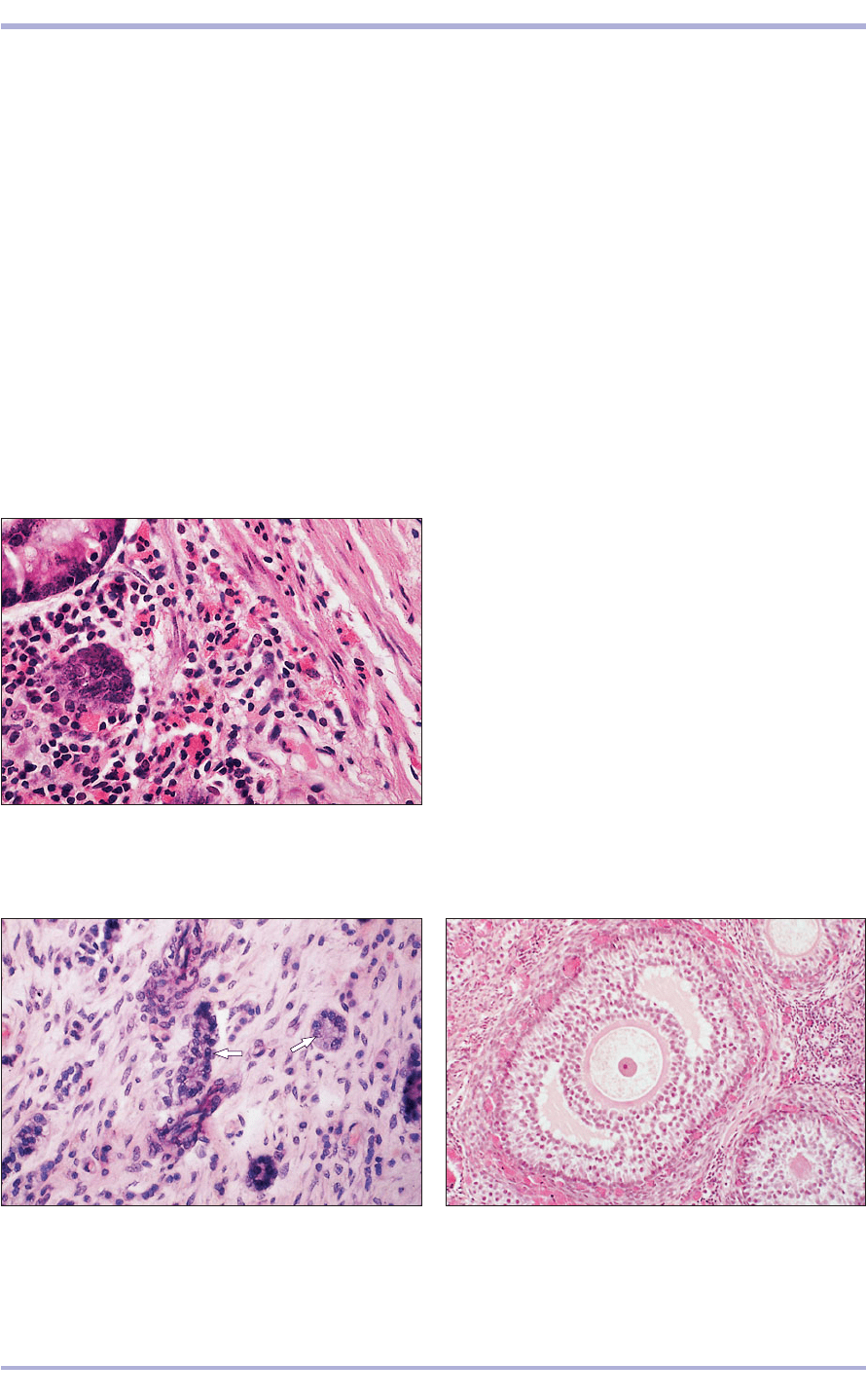
At ovulation the primary oocyte divides into sec-
ondary oocytes, one cell retaining most of the cyto-
plasm; completion of meiosis occurs at fertilization.
The follicle breaks open, releasing the oocyte, which
passes into the uterine tubes. The follicle collapses
and the granulosa cells, together with those of the
theca interna, hypertrophy, expanding into the cav-
ity to form the granulosa lutein and theca lutein cells
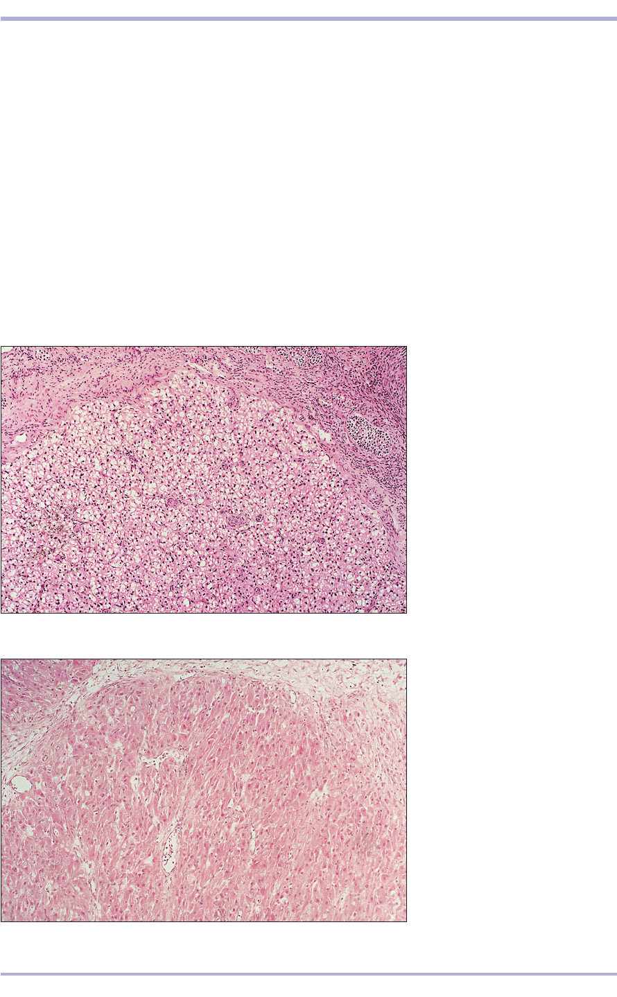
of the corpus luteum (12.9 and 12.10). They are
arranged in long cords separated by vascular con-
nective tissue. The luteal cells secrete progesterone
which, with the oestradiol, prepares the uterus for
possible conception. With regression of the corpus
luteum, stromal elements move in and replace the
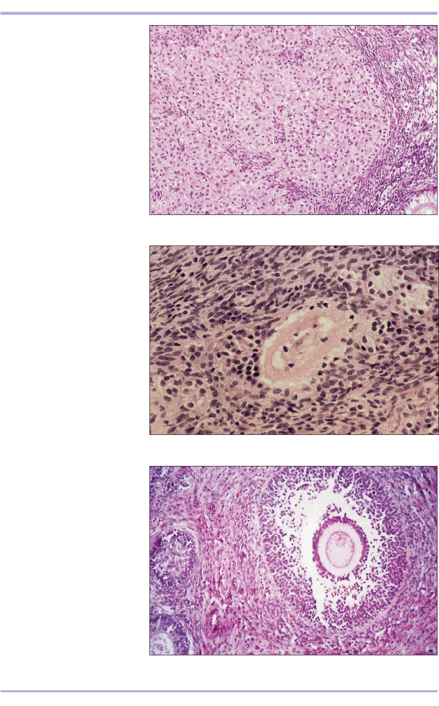
dead cells with collagen to form a scar: the corpus
albicans (12.11 and 12.12). Although many pri-
mordial follicles begin the process outlined above,
few become mature. The majority undergo a degen-
erative regression. They are called anovular or atretic
follicles and can be recognized by the irregular out-
line of the follicle and the separation of the granu-
losa cells (12.13). Cells of the theca interna
hypertrophy and the zona pellucida becomes
swollen. Eventually, the entire follicle is resorbed.
186
Comparative Veterinary Histology with Clinical Correlates
12.9 Ovary. Parenchymatous zone
(sheep). (1) The pale vacuolated cells
are part of the corpus luteum.
(2) Ovarian stroma. H & E. ×62.5.
12.9
12.10 Ovary. Parenchymatous zone
(sow). (1) Cords of large densely
stained luteal cells are separated by
(2) blood vessels. H & E. ×125.
12.10
1
1
2
1
2
187
12.11 Ovary. Parenchymatous zone
(sheep). (1) Ovarian stroma. (2) Dense
white connective tissue. (3) Active
fibroblasts replacing degenerating
luteal cells. H & E. ×125.
12.11
12.12 Sheep. Parenchymatous zone
(sheep). The central pale staining
area is dense white connective tissue
(scar tissue) and represents a later
stage in the development of a corpus
albicans. H & E. ×250.
12.12
12.13 Ovary. Parenchymatous zone
(bitch). Atretic follicle. (1) The
granulosa cells are separating; spaces
are present between them. (2) The
oocyte is no longer in contact with
the lining granulosa cells. H & E.
×125.
12.13
1
2
3
2
1
Female Reproductive System
3
Uterine tubes (oviducts)
The uterine tubes are long flexuous musculo-
membranous tubes. They consist of an expanded
cranial end, the infundibulum, which is close to the
ovary; a middle segment, the ampulla; and a cau-
dal narrow part or isthmus that opens into the ipsi-
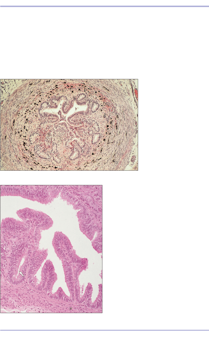
lateral horn of the uterus. The epithelium is simple
or pseudostratified columnar with secretory and
188
12.14 Transverse section (TS) uterine
tube (sheep). (1) The lumen is lined
by a pseudostratified epithelium
thrown into deep folds. (2) The
lamina propria has deposits of dark
brown pigment. (3) The muscularis
is for the most part circular smooth
muscle fibres. (4) The serosa is
loose vascular connective tissue
with prominent blood vessels.
H & E. ×62.5.
12.14
12.15 TS uterine tube (cow). The lining epithelium is
pseudostratified columnar with ciliated and non-ciliated
cells.The deep mucosal folds are cut in transverse section
giving the appearance of mucosal glands (arrowed).
H & E. ×125.
12.15
ciliated cells. The lamina propria has longitudinal
folds giving it a glandular appearance. In some
breeds of sheep (blackface and crosses), pigment
cells are present. The muscularis is mainly a layer
of circular smooth muscle, thickening at the junc-
tion with the uterine horn, with an outer layer of
longitudinal muscle. The serosa is loose vascular
connective tissue with prominent blood vessels
(12.14 and 12.15).
2
2
3
4
1
Comparative Veterinary Histology with Clinical Correlates
189
Uterus
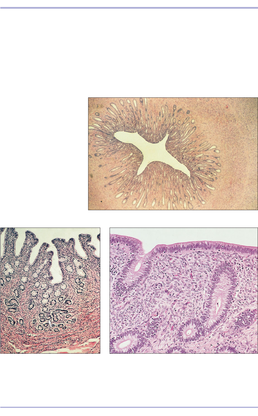
The uterus is bicornuate, with right and left horns
(cornua), a body (corpus) and a neck (cervix)
(12.16–12.23, all illustrations in transverse sec-
tion). The uterine wall in the cornua and corpus
has three layers: an inner endometrium (mucosa),
a middle myoemetrium (muscularis) and an outer
perimetrium (serosa).
Endometrium
The epithelial lining of the endometrium is simple
cuboidal or columnar in the mare and carnivores,
and pseudostratified in the sow and ruminants. In
ruminants, ciliated cells may be present. The lam-
ina propria is a deep layer of vascular connective
tissue, with simple tubular endometrial glands that
open into the lumen of the uterus. These glands are
12.16 TS uterus (cat).
(1) Endometrium with simple straight
tubular glands. (2) Myometrium with
circular and longitudinal smooth
muscle. H & E. ×7.5.
12.16
12.18 TS uterus (bitch). (1) The lining epithelium is simple columnar.
(2) The endometrial glands are lined by tall columnar epithelial cells
with a basal nucleus. (3) The lamina propria is very cellular with
many small blood vessels. H & E. ×125.
12.18
12.17 TS uterus (mare). Only the
endometrium is present. The lining
epithelium is simple columnar; the
endometrial glands are slightly coiled
at the base (arrowed). H & E. ×62.5.
12.17
2
1
1
2
Female Reproductive System
3
190
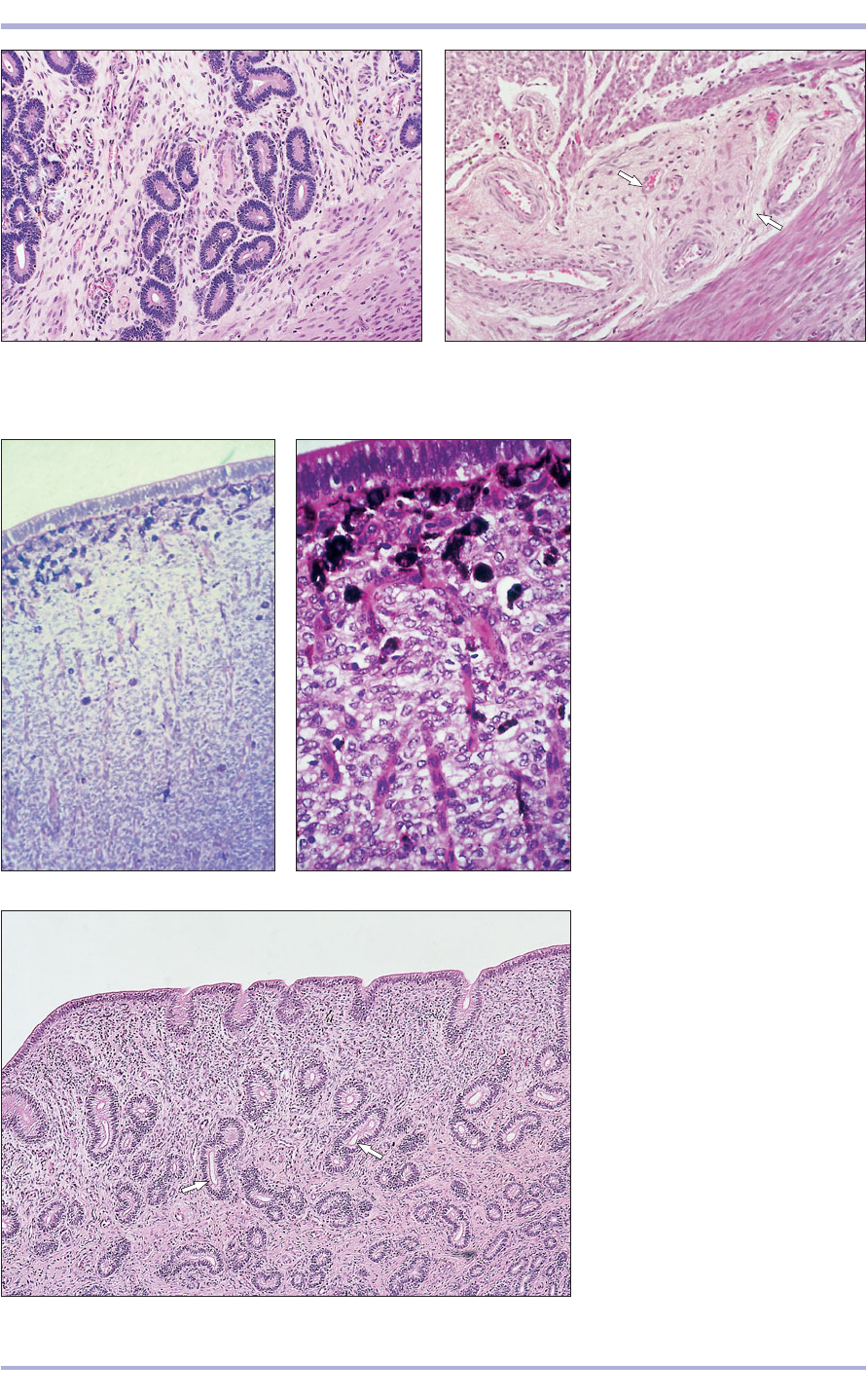
12.19 TS uterus (cow). (1) The endometrial glands are
markedly coiled. (2) Thick layer of myometrium. H & E.
×100.
12.19
12.21 TS uterus (sheep). Only part of
a caruncle is present; the connective
tissue is very cellular and well
vascularized. H & E. ×125.
12.22 TS uterus. Caruncle (sheep).
Local deposits of pigment (melanin)
are present in the connective tissue
of some breeds of sheep. H & E. ×250.
12.21
12.20 TS uterus (cow). The myometrium is split by the
stratum vasculare (arrowed.). H & E. ×100.
12.20
12.22
12.23 TS uterus. Intercaruncular area
(sheep). The endometrial glands
(arrowed) are confined to the
intercaruncular area. H & E. ×100.
12.23
1
2
Comparative Veterinary Histology with Clinical Correlates
