Aughey E., Frye F.L. Comparative veterinary histology with clinical correlates
Подождите немного. Документ загружается.

12.84
12.83
211
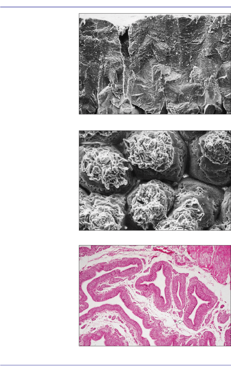
12.82 TS eggshell with the
membranes removed. (1) Gaseous
pore. (2) Cuticle. (3) Multiple layers
of calcite containing organic matrix.
(4) Mammillary layer. Scanning
electron micrograph. ×160.
12.82
1
2
1
2
3
4
12.83 Mammillary layer of the
eggshell. This is the organic
component. The individual structural
units are the mammillae arranged as
(1) mammillary caps and
(2) mammillary cones. Scanning
electron micrograph. ×320.
12.84 Vagina (bird). A
pseudostratified columnar epithelium
lines the vagina; small tubular glands
(sperm-host glands) are found in the
lamina propria (arrowed). H & E.
×62.5.
Female Reproductive System

12.86
Reptilian, amphibian and
fish female reproductive
system
Paired ovaries are typical in fish, amphibians and rep-
tiles, and the histology of oogenesis is similar to that
in mammals (12.85 and 12.86). The ovaries are com-
posed of germinative, stromal, vascular and nervous
tissue. The ova begin their development as oogonia
that are mitotically derived from successive genera-
tions of oocytes. Diploid primary oocytes then
undergo meiotic division to become primary oocytes,
and primary polar bodies that are discarded. Another
reduction division yields haploid ova, and secondary
polar bodies that also are discarded.
The ovum is encircled by a cell membrane, a vari-
ably narrow zona pellucida and a layer of follicle
cells. During vitellogenesis, the yolk is added. This
process occurs after a variable period of time after
ovulation. Although the ova of most amphibians are
uninuclear, some species are known to produce mult-
inucleated ova. However, before fertilization all of
the nuclei, except one, become inactivated. Reptile
ova typically have a uninuclear ovum. However, bin-
ucleated ova are produced occasionally and, after
being fertilized, may yield twin embryos.
After ovulation, the corpora lutea, then the cor-
pora albicans and finally the corpora atretans
replace the ovulated follicles.
The appearance of reptilian oviducts varies
depending on the species and whether the female
is egg laying (oviparous) or live bearing (ovivivip-
arous or viviparous). However, they are readily rec-
ognizable. The histological features of the tubular
oviduct changes with each segment as it courses dis-
tally from the infundibulum. It is lined by ciliated,
often mucus-secreting, glandular columnar epithe-
lium for at least part of its length. The thin walls of
the oviducts contain alveolar or tubuloalveolar
glands that secrete albumin and shell substrate onto
the yolked egg as it descends. The cuboidal to
columnar epithelial cells comprising these glands
tend to be characterized by their distinctive
eosinophilic cytoplasmic granularity (12.87 and
12.88). The caudal oviduct of many reptiles also
contains straight or branched crypt-like depressions
that are surrounded by cuboidal epithelium with
eosinophilic granular cytoplasm (12.89). In many
species these glandular crypts serve as spermathe-
cae in which spermatozoa are nourished and stored
for prolonged periods of time. The oviducts of
viviparous and oviviviparous reptiles are thick-
walled, muscular and vascular, and contain glands
with secretions that help nourish the developing
embryo(s). They often exhibit marked plaiting,
which facilitates their distension during gravidity or
pregnancy. These modified oviducts are called the
‘uterus’ by some authorities.
The embryos of some viviparous reptiles develop
a primitive vascular placenta. In the lizard (Xantusia
vigilis) it is disc-shaped; in others it is more diffuse.
Although it was long thought that embryonic devel-
opment did not occur in shelled eggs until they were
deposited and exposed to atmospheric oxygen, it
has been demonstrated that significant embryonic
development can occur before egg deposition in
some species.
212
Comparative Veterinary Histology with Clinical Correlates
12.85 Ovary and proximal oviduct (fimbrium) of a desert
tortoise (Xerobates agassizi). H & E. ×25.
12.85
12.86 Corpus luteum (arrowed) of a leopard gecko
(Eublepharis macularius). (1) Granulosa lutein cells.
(2) Primordial follicles. (3) Oocyte cytoplasm. (4) Tunica
albuginea. (5) Theca externa. (6) Stroma. H & E. ×62.5.
1
3
5
2
4
6
2
213
Female Reproductive System
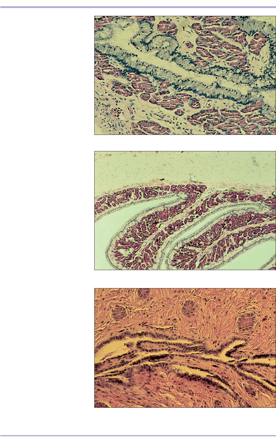
12.88
12.87 and 12.88 Shell gland portion
of the oviduct of a Pacific pond turtle
(Clemmys marmorata). The luminal
lining epithelium is pseudostratified
columnar and contains numerous
goblet cells. Subjacent to the mucosa
are lobules of shell-secreting glands
with characteristic eosinophilic
granule-containing cells. H & E. ×127.
12.87
12.89 Caudal oviduct from a sexually
mature female green iguana (Iguana
iguana). This portion of the oviduct is
characterized by numerous shallow
crypts that connect with small lobules
of glandular epithelial cells. These
crypts are thought to be sites of
sperm storage and nourishment that
sustain spermatozoa for prolonged
periods that may exceed 4–6 years in
some reptiles. H & E. ×62.5.
12.89
12.90
12.92
12.91
Clinical correlates
Ovarian and oviductal (or ‘uterine’) inflammation
and infections are less common in fish, amphib-
ians and reptiles than in mammals. Rupture of
yolked eggs with subsequent leakage of yolk into
the coelomic cavity is, however, a relatively fre-
quent reproductive disorder in oviparous reptiles.
Once lipid has gained entry into the vascular sys-
tem, it is disseminated widely in the form of yolk-
lipid emboli throughout the body (12.90 and
12.91).
Ovarian neoplasms have been observed in fish
and amphibians, but are less prevalent than in
mammals, birds and reptiles. As with mammals,
relatively common reptilian ovarian neoplasms are
granulosa cell tumours (see 12.68), luteal tumours
and thecal cell tumours (see 12.69). Dys-
germinoma (see 12.70) and teratoma (12.92)
occur less often. The teratoma is distinguished
by usually having tissue from all three germ lay-
ers present: hair (or scales), bone, cartilage,
teeth, muscle (of all three types), glandular tis-
sue, nerve and brain-like structures, and so on.
214
Comparative Veterinary Histology with Clinical Correlates
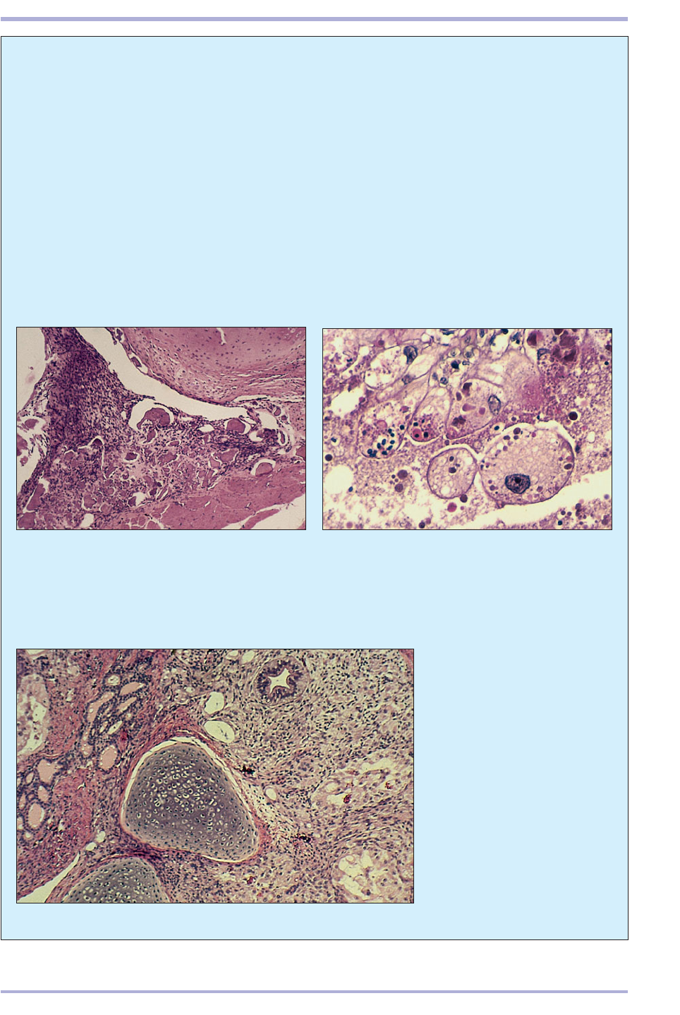
12.92 Ovarian teratoma from a
green iguana (Iguana iguana).
This section illustrates (1) two
masses of cartilage, (2) a duct-like
structure, (3) an aggregate of
thyroid-like follicles filled with
pink staining protein resembling
colloid, and (4) nervous tissue.
H & E. ×62.5.
12.91 Yolk serocoelomitis in a green iguana (Iguana
iguana). The smooth muscle tunic of the viscus organ
is covered with yolk, and an inflammatory exudate
is comprised of macrophages with engulfed lipid.
H & E. ×125.
12.90 Egg-yolk serocoelomitis from an iguana. The
intense exudative reaction to yolk characterized by
numerous histiocytic macrophages with engulfed yolk
lipid. H & E. ×62.5.
1
1
4
2
3
3
4
Two properties of cytoplasm are typified by nervous
tissue: irritability – the ability to react; and conduc-
tivity – the ability to transmit the elicited response.
The nervous system is subdivided into the cen-
tral nervous system (CNS), which comprises the
brain and spinal cord, and the peripheral nervous
system (PNS), which covers all other nervous tissue
and acts to interconnect all the tissues of the body
with the CNS. The nervous system is derived from
a specialized region of surface ectoderm along the
dorsal midline of the flat embryonic disc. The ecto-
dermal cells thicken to become neural ectoderm.
During flexion, the neural folds form and the lips
fuse to become the neural tube. This is incorporated
into the developing embryo to form the brain and
spinal cord. A separate population of neural ecto-
dermal cells remains separate from the
neural tube. This is the neural crest and
forms the PNS.
The basic cellular unit of nervous tis-
sue is the neuron. This comprises a large
cell body (from 0.4 to 12 +m in diame-
ter) with a single nucleus and a promi-
nent nucleolus. Basophilic granules
(Nissl’s granules) are present in the cyto-
plasm and represent rough endoplasmic reticulum
(13.1). The function of the neuron is to receive and
transmit impulses. The dendrites are branching
processes at the receiving end, and the axon is the
single, long process for onward transmission of the
impulse. Information passed along a chain of neu-
rons is transmitted from axon to dendrite at a spe-
cialized junction: the synapse.
Multipolar neurons are by far the most common
type. The single axon arises from one pole at a gran-
ule-free area of the neuron body: the axon hillock.
One or two dendrites arise from the opposite pole
and branch extensively. Examples of these neurons
are found in the CNS, in the cerebral and cerebel-
lar cortex, and in the spinal cord (13.2). Bipolar
neurons have one axon and one main dentrite, with
215
13. NERVOUS SYSTEM
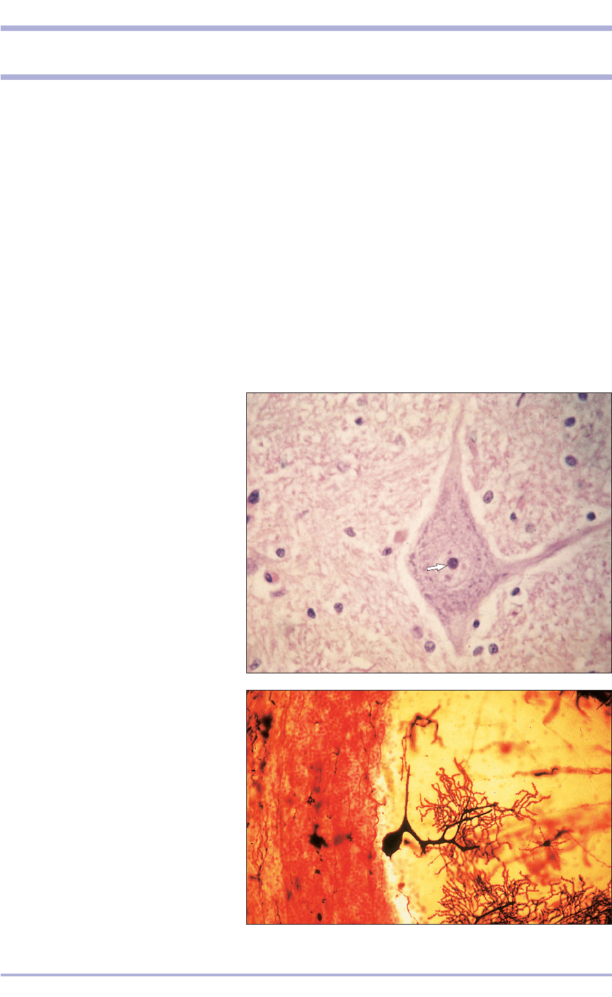
13.1 Motor neuron. Trigeminal nerve
(dog). (1) Nucleus and nucleolus (arrow).
(2) Basophilic granules (Nissl’s granules).
(3) Axon hillock. (4) Neuroglial cells.
H & E. ×200.
13.1
1
2
3
4
4
13.2 Multipolar neuron. Cerebellar cortex
(dog). (1) Cell body. (2) Branching processes.
Golgi’s silver. ×125.
13.2
2
1
216
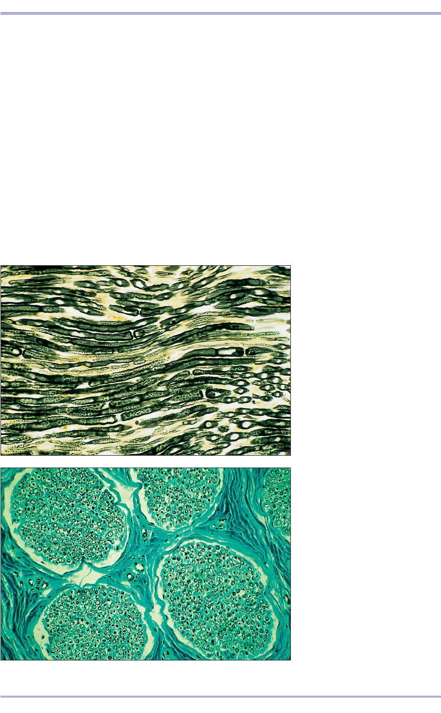
13.3 Longitudinal section (LS)
peripheral nerve (cat). (1) Axon.
(2) Neurilemmal sheath. (3) Node.
Osmic acid. ×200.
13.3
13.4 Transverse section (TS)
peripheral nerve (dog). (1) Bundles
of axons cut in transverse section,
fascicles. (2) Epineurium.
(3) Perineurium. (4) Endoneurium.
Masson’s trichrome. ×25.
13.4
little branching, and are found in the retina.
Pseudounipolar neurons are so-called because a sin-
gle process arises from the cell body and divides into
two. These cells are found in the dorsal root gan-
glia and in the sensory ganglia of the cranial nerves.
The axon with its covering myelin sheath, the
neurilemma, constitutes the nerve fibre. The neurilem-
mal sheath is composed of individual cells that form
a continuous investment from the origin of the axon
almost to the peripheral termination (13.3). These
cells produce a lamellar system of membranes with
a high fat content. In the PNS the neurilemmal sheath
is produced by neurolemmocytes, and in the CNS by
neuroglial cells: oligodendrocytes. The degree of the
investment varies; heavily invested fibres are known
as myelinated and minimally invested fibres as non-
myelinated. The axon has no sheath at the peripheral
nerve endings; these are naked fibres. The individual
sheaths divide the nerve fibre into segments. Where
adjoining cells meet, a node (of Ranvier) is formed so
that each internodal segment represents an investing
neurilemmal cell. The high fat content of the sheath
means that fat stains are best used to demonstrate the
myelinated fibres (13.3).
Peripheral nervous
system
The PNS is derived from the neural crest and is
composed of nerves and ganglia. Nerves are com-
posed of nerve fibres and ganglia are localized
groups of nerve cells. The individual fibres are
gathered into bundles or fascicles by a connective
tissue sheath, the epineurium, and form an
anatomical nerve. The epineurium extends into the
1
4
2
1
3
3
3
1
2
Comparative Veterinary Histology with Clinical Correlates
1
217
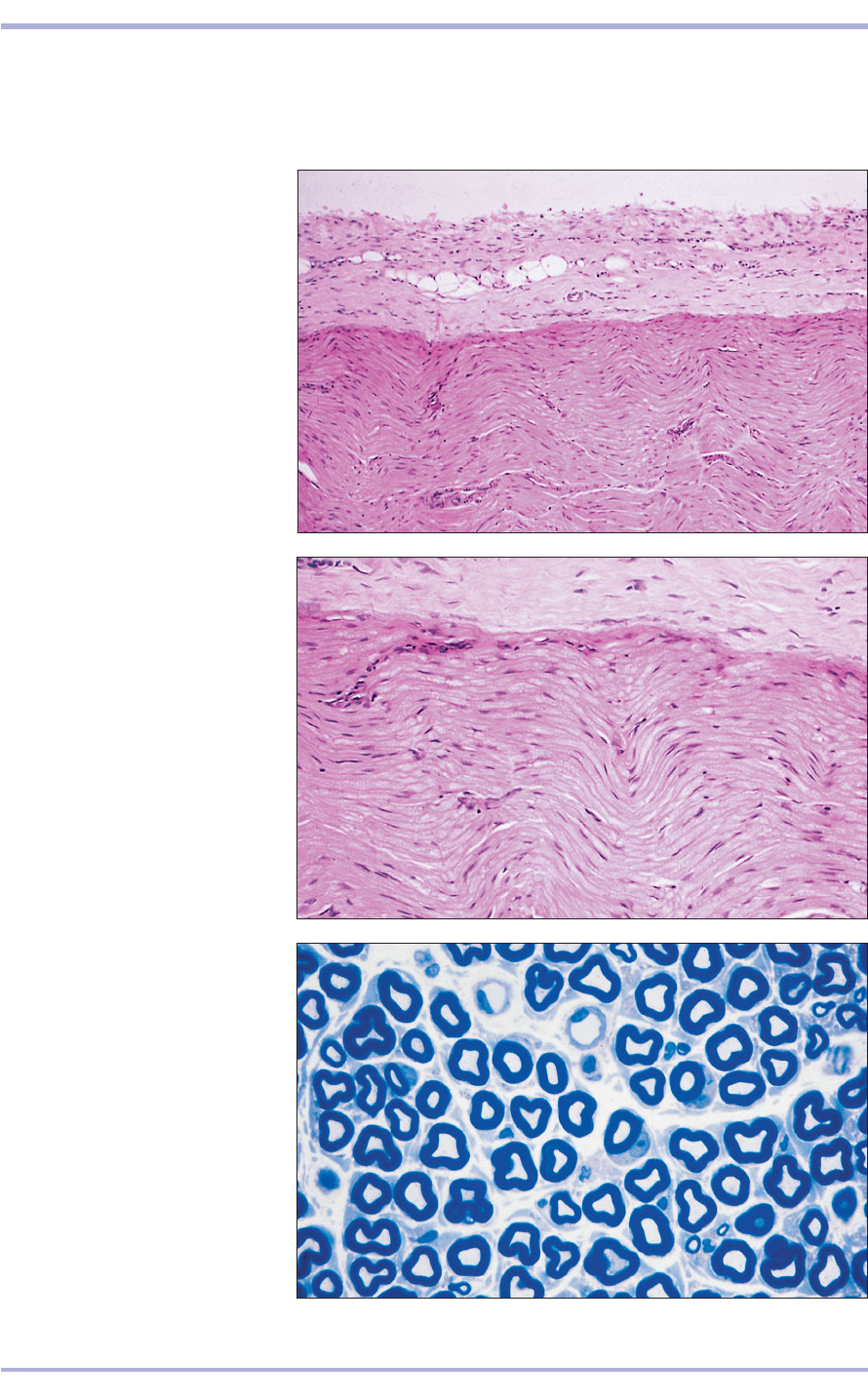
13.5 LS peripheral nerve (dog).
(1) Epineurium with fat cells.
(2) Nuclei of the neurilemmal cells.
(3) Axons cut longitudinally; note
the wavy appearance. H & E. ×62.5.
13.5
1
2
3
13.7 TS laryngeal nerve (horse).
(1) Perineurium with blood vessels.
(2) Endoneurium. (3) Axons. Toluidine
blue. ×62.5.
13.7
13.6 LS peripheral nerve (dog).
(1) Epineurium. (2) Axons.
(3) Neurilemmal cell nuclei.
H & E. ×125.
13.6
1
1
2
3
1
2
3
nerve and subdivides the fascicles with a vascular
connective tissue investment: the perineurium.
This in turn surrounds each nerve fibre with fine
vascular connective tissue: the endoneurium
(13.4–13.7). In the dorsal root ganglion the neu-
rons are peripherally arranged, with the nerve
Nervous System
218
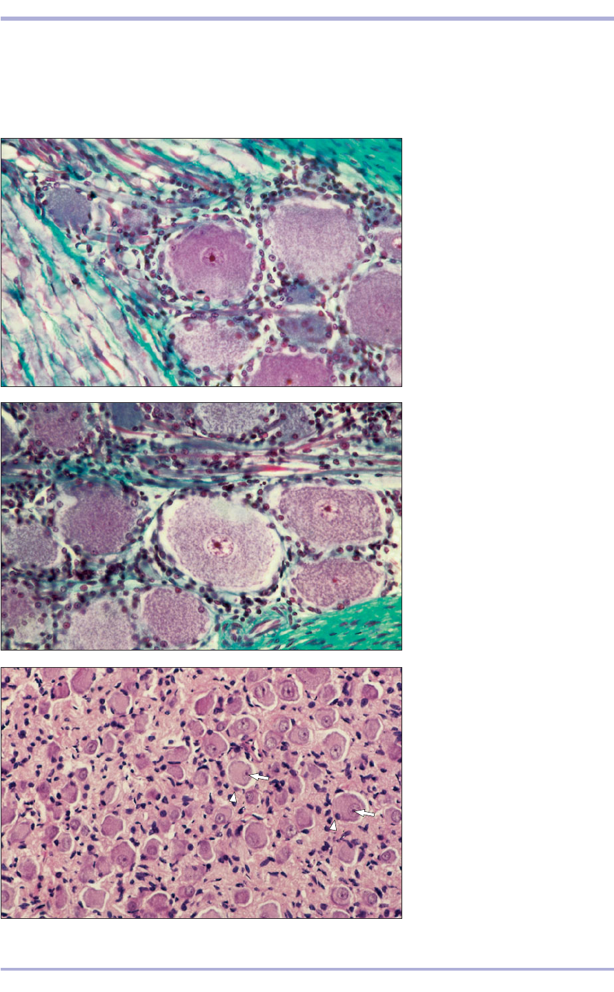
13.8 Dorsal root ganglion (cat).
(1) Neuron bodies. (2) Nerve fibres
and supporting neuroglial cells.
Masson’s trichrome. ×160.
13.8
13.9 Dorsal root ganglion (cat).
(1) Neuron body. (2) Satellite cells;
these are supporting neuroglial cells.
Masson’s trichrome. ×160.
13.9
13.10 Sympathetic ganglion (dog).
(1) Neuron body with eccentric
nucleus (arrow) and satellite cells
(arrowhead). H & E. ×125.
13.10
1
1
1
2
1
2
fibres in the centre of the ganglion (13.8). The
large neuron bodies are surrounded and supported
by satellite cells (13.9). In the sympathetic ganglion
the neurons are evenly distributed, the cell bodies
are smaller and the nucleus is eccentrically placed
in the cell (13.10). Parasympathetic ganglia are
small groups of neurons within the terminal tissue
(13.11).
Comparative Veterinary Histology with Clinical Correlates
219
13.11 Parasympathetic ganglion.
Stomach (pig). (1) Neuron body.
(2) Nerve fibres and supporting
neuroglial cells. H & E. ×250.
13.11
1
2
Nervous System
Central nervous system
The brain and spinal cord are enveloped in connec-
tive tissue membranes, the meninges. The outer
dense membrane is the dura mater; the middle fine
cobweb-like membrane is the arachnoid; and the
inner, very vascular, areolar membrane is the pia
mater. The arachnoid and pia mater are continuous
and called the leptomeninges (13.12). The central
fluid filled canal and the ventricles of the brain are
lined with a columnar epithelium: the ependyma.
The tela choroidea lies in the thin roof plate of the
third and fourth ventricles. Its surface is covered
with modified ependymal cells that secrete
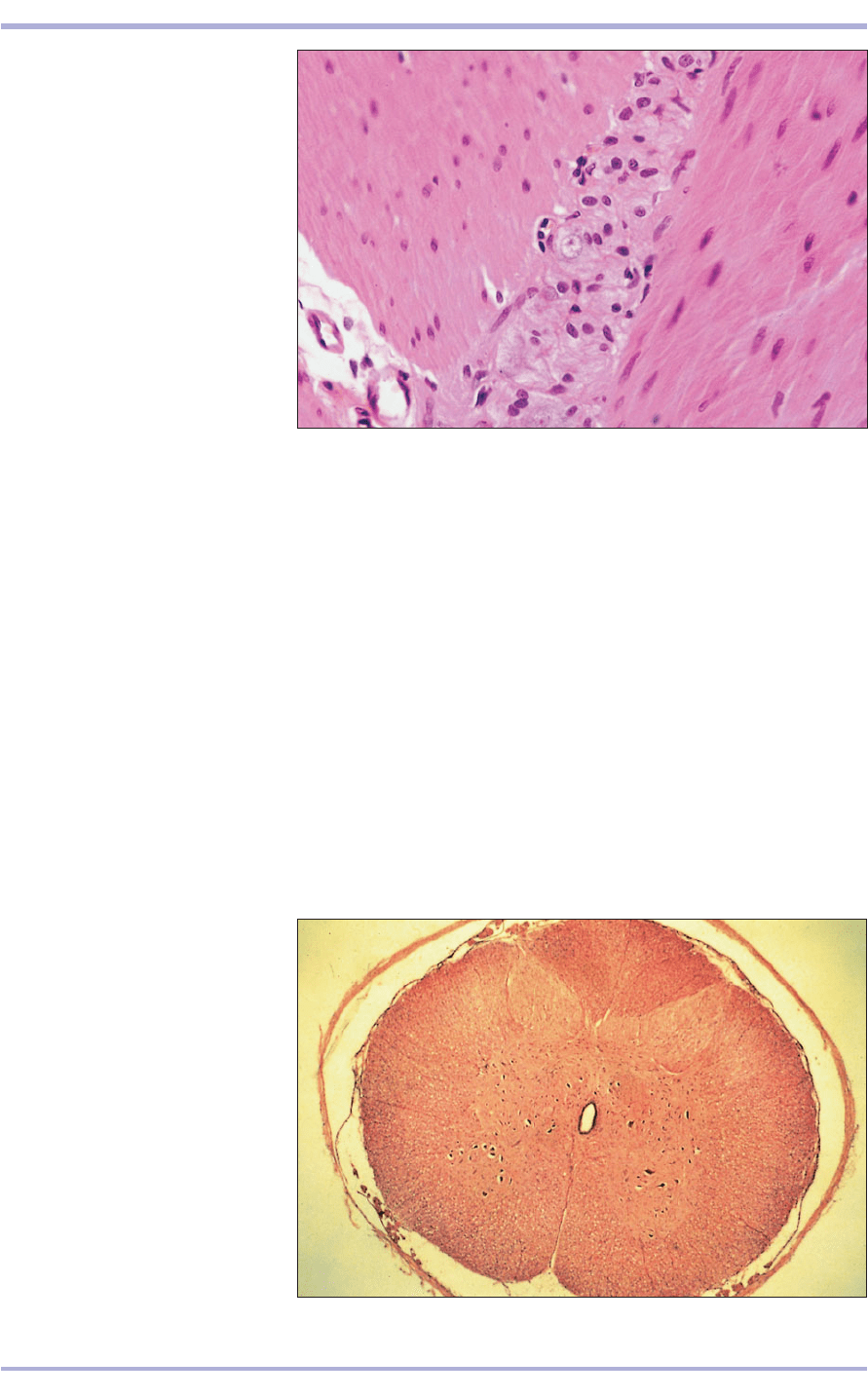
13.12 TS spinal cord (cat). (1) Dura
mater. (2) Arachnoid. (3) Pia mater.
(4) Spinal canal lined by ependyma.
H & E. ×7.5.
13.12
1
2
3
4
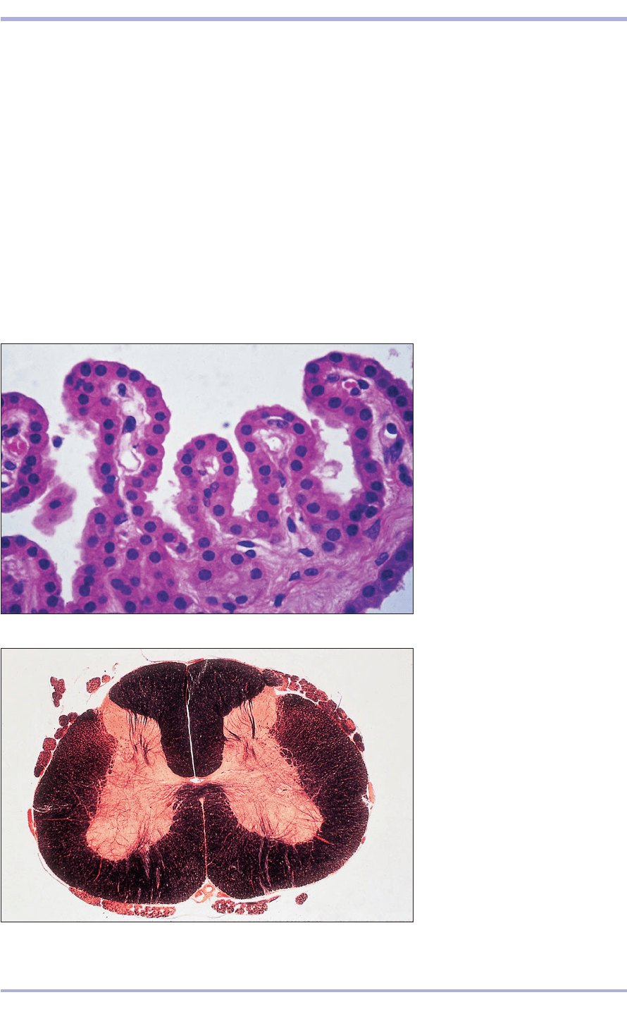
cerebrospinal fluid (13.13). The pia mater coats the
entire surface of the CNS, extends into the fissures
and sulci and is separated from the ependyma only
by the basal lamina.
In the CNS, collections of functionally related
neurons are called nuclei and collections of nerve
fibres are called tracts. The fibres have an investing
sheath cell: the oligodendrocyte. The presence of fat
gives a white glistening appearance and the fibres
are called white matter. By contrast, the cell bodies
appear grey and form the grey matter (13.14).
Neuroglial cells are the supporting cells of the
CNS, taking the place of the connective tissue of
other systems. Oligodendrocytes invest the axon,
provide the myelin sheath and act as supporting
satellite cells. Astrocytes are stellate cells with long
processes. In fibrous astrocytes the processes are
thin with minimal cytoplasm, whereas in proto-
plasmic astrocytes they are thick and fleshy
(13.15–13.18). The cell processes extend to the
blood vessels in the pia mater, acting as anchors
and transferring nutrients. The blood vessels are
220
Comparative Veterinary Histology with Clinical Correlates
13.14 TS spinal cord (cat). (1) Black
areas are the fat stained white
matter. (2) Pink stained areas are the
cellular grey matter. Osmic acid. ×7.5.
13.14
13.13 TS choroid plexus in the
fourth ventricle (cat). (1) Ependyma.
(2) Capillaries. H & E. ×160.
13.13
2
2
1
1
2
2
1
