Aughey E., Frye F.L. Comparative veterinary histology with clinical correlates
Подождите немного. Документ загружается.

231
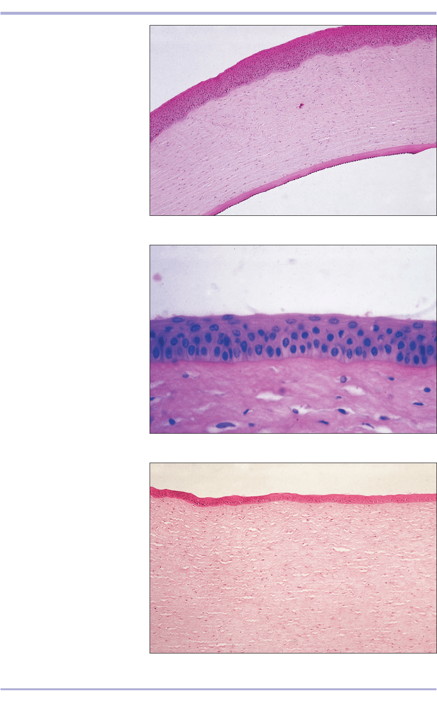
14.12 Cornea (horse). (1) Stratified
squamous epithelium of the anterior
surface. (2) Substantia propria.
(3) Simple cuboidal endothelium of
the posterior surface resting on a
thick basement membrane
(Descemet’s). H & E. ×62.5.
14.12
14.13 Cornea. Anterior surface
(dog). (1) Stratified squamous
epithelium of the anterior surface.
(2) Basement membrane (Bowman’s).
(3) Substantia propria. H & E. ×125.
14.13
14.14 Cornea. Posterior surface
(dog). (1) Substantia propria.
(2) Basement membrane
(Descemet’s). (3) Simple squamous
endothelium. H & E. ×62.5.
14.14
1
2
3
3
1
2
2
1
3
Special Senses
14.17
inner surface is attached to the middle tunic, the
choroid, by a layer of delicately pigmented connec-
tive tissue, the lamina fusca (14.15). The optic nerve
passes through the sclera at the lamina cribrosa
(14.16).
The choroid is vascular and has numerous
melanocytes. The ciliary body is an anterior con-
tinuation of the choroid that extends to the base
of the iris. The inner surface is a continuation of a
non-light-sensitive retina: the pars ciliaris retinae.
The loose connective tissue of the stroma contains
the ciliary muscle, bundles of smooth muscle fibres.
An inner non-pigmented choriocapillary layer con-
tains a capillary network that supplies the retina
(14.17<14.20). The choroid of some species has an
iridescent reflecting tissue layer: the tapetum
lucidum (dog, 14.15; cat, 14.19). This gives their
232
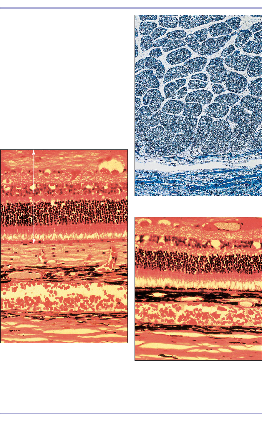
14.15 Eye. Tapetum fundus (dog). (1) Sclera composed of
dense white fibrous tissue. (2) Lamina fusca. (3) Chorioid
with capillaries. (4) Tapetum cellulosum. (5) The
photosensitive retinal layers are (6) pigment layer,
(7) rods and cones, (8) nuclei of the rods and cones,
(9) outer synaptic layer, (10) bipolar nerve cell nuclei,
(11) inner synaptic layer, (12) optic nerve cells, (13) optic
nerve fibres. H & E. ×160.
14.15
14.16 Optic disc (horse). (1) Scleral connective tissue.
(2) Optic nerve bundles. H.& E. ×25.
14.16
14.17 Eye. Non-tapetum fundus (dog). The retinal layers
are (1) pigment layer, (2) rods and cones, (3) nuclei of the
rods and cones, (4) outer synaptic layer, (5) bipolar nerve
cell nuclei, (6) inner synaptic layer, (7) optic nerve cells,
(8) optic nerve fibres. H & E. ×160.
2
1
3
4
5
6
7
8
1
2
2
3
4
2
1
13
12
11
10
9
8
7
6
5
Comparative Veterinary Histology with Clinical Correlates
233
Special Senses
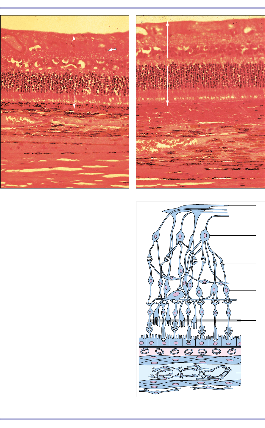
14.18 Eye. Non-tapetum fundus (cat). (1) Sclera
composed of dense white fibrous tissue. (2) Lamina fusca.
(3) Choroid with capillaries. (4) The photosensitive retinal
layers are (5) pigment layer, (6) rods and cones, (7) nuclei
of the rods and cones, (8) outer synaptic layer, (9) bipolar
nerve cell nuclei, (10) inner synaptic layer, (11) optic nerve
fibres. Optic nerve cells are arrowed. H & E. ×160.
14.19 Eye. Tapetum fundus (cat). (1) Sclera.
(2) Choriocapillaris. (3) Non-pigmented tapetum
cellulosum. (4) The photosensitive retinal layers are
(5) pigment layer, (6) rods and cones, (7) nuclei of the
rods and cones, (8) outer synaptic layer, (9) bipolar nerve
cell nuclei, (10) inner synaptic layer, (11) optic nerve
fibres. H & E. ×160.
14.20 Eye. Layers of the retina. (1) Vessel layer of the
choroid. (2) Connective tissue/tapetum lucidum.
(3) Choriocapillaris. (4) Pigment epithelium.
(5) Photoreceptors, layers of rods and cones. (6) External
limiting membrane. (7) Outer nuclear layer. (8) Outer
plexiform layer. (9) Inner nuclear layer. (10) Inner
plexiform layer. (11) Ganglion cell layer. (12) Nerve
fibre layer. (13) Internal limiting membrane.
14.18
14.19
14.20
1
3
4
11
4
11
10
9
8
7
6
5
3
2
1
10
9
8
7
6
5
2
13
12
11
10
9
8
7
6
5
4
3
2
1
eyes the property of shining in the dark, and allows
incident light two opportunities to stimulate the
retinal receptors. It is located between the chorio-
capillary and vascular layers of the choroid in the
dorsal portion of the eye. In ungulates and the horse
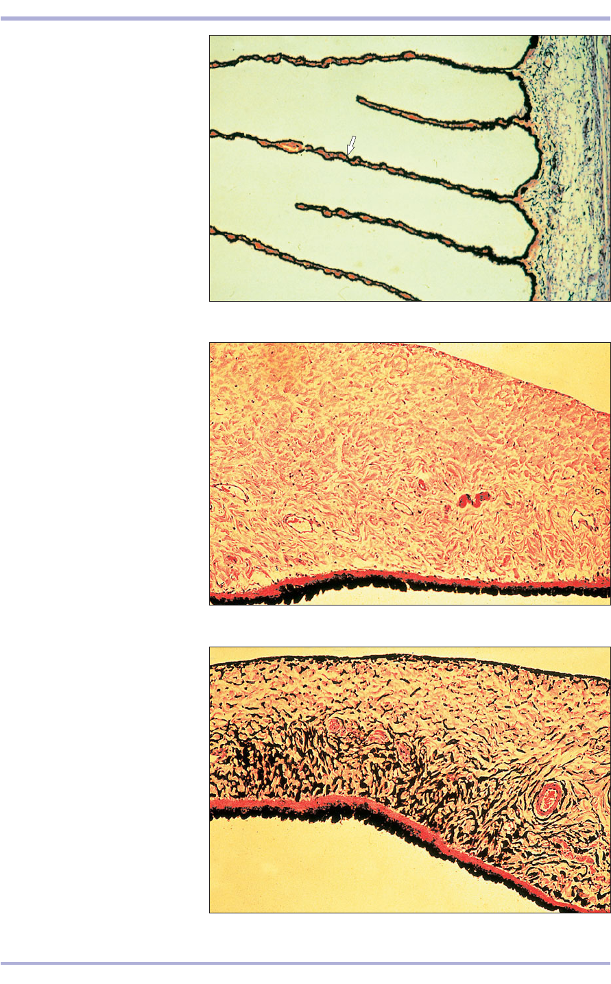
a fibrous area of the choroid forms the tapetum
fibrosum (14.21). In carnivores, a cellular layer
forms the tapetum cellulosum (14.19).
The ciliary processes form a circle of radial folds
surrounding the lens. This epithelial basal layer con-
sists of pigmented columnar cells and the surface
layer of non-pigmented columnar cells (14.22).
The iris, the most anterior part of the uveal layer,
is a muscular diaphragm rostral to the lens and
pierced centrally by an opening: the pupil. The base
of the iris is attached to the ciliary body. The con-
nective tissue stroma supports many blood vessels
and contains pigment cells. The epithelium is con-
fined to the posterior surface, and is the most ante-
rior part of the non-light-sensitive retina: the pars
iridica retinae. Both layers of epithelial cells are pig-
mented. The anterior surface is non-epithelial, and
is covered with fibrocytes and melanocytes (14.23
and 14.24).
The retina is the innermost layer of the wall of the
eye. The photosensitive portion lines the inner surface
of the eye posteriorly from the ora ciliaris retinae, the
point of transition between the photosensitive and
non-photosensitive areas of the retina. The latter con-
sists of two layers of non-light-sensitive epithelium
beginning at the ora serrata forming the pars iridica
retinae and the pars ciliaris retinae. From the choroid
to the cavity of vitreous humour the 10 layers (14.15)
of the photosensitive area are as follows:
• pigment epithelium (the inner layer of the embry-
onic cup);
• layer of rods and cones (modified dendrites that
act as photoreceptors);
• outer limiting membrane, formed by neuroglial
processes;
• outer nuclear layer (nuclei in the cell bodies of
the rods and cones);
• outer synaptic (plexiform) layer (connecting the
rod and cone neurons with the dendrites of the
bipolar neurons);
• inner nuclear layer of bipolar neurons relaying
the impulses received at layers 2 to 8.
• inner synaptic (plexiform) layer (linking the
axons of the bipolar neurons and the dendrites
of the optic nerve cells);
• ganglion cell layer (optic nerve cells);
• nerve fibre layer (the axonal processes of the
optic nerve cells converge at the optic disc and
become myelinated to form the optic nerve; this
sieve-like part of the sclera is the lamina
cribrosa);
• inner limiting membrane, the expanded extrem-
ity of the neuroglial cells in layer 3.
Layers 2 to 10 are derived from the outer layer of
the embryonic optic cup.
234
Comparative Veterinary Histology with Clinical Correlates
14.21 Eye. Tapetum fibrosum (horse). (1) Pigment layer of the retina.
(2) Compact layer of fibrous connective tissue, the tapetum fibrosum.
(3) Choroid with blood vessels and some pigment cells. H & E. ×100.
14.21
2
3
1
235
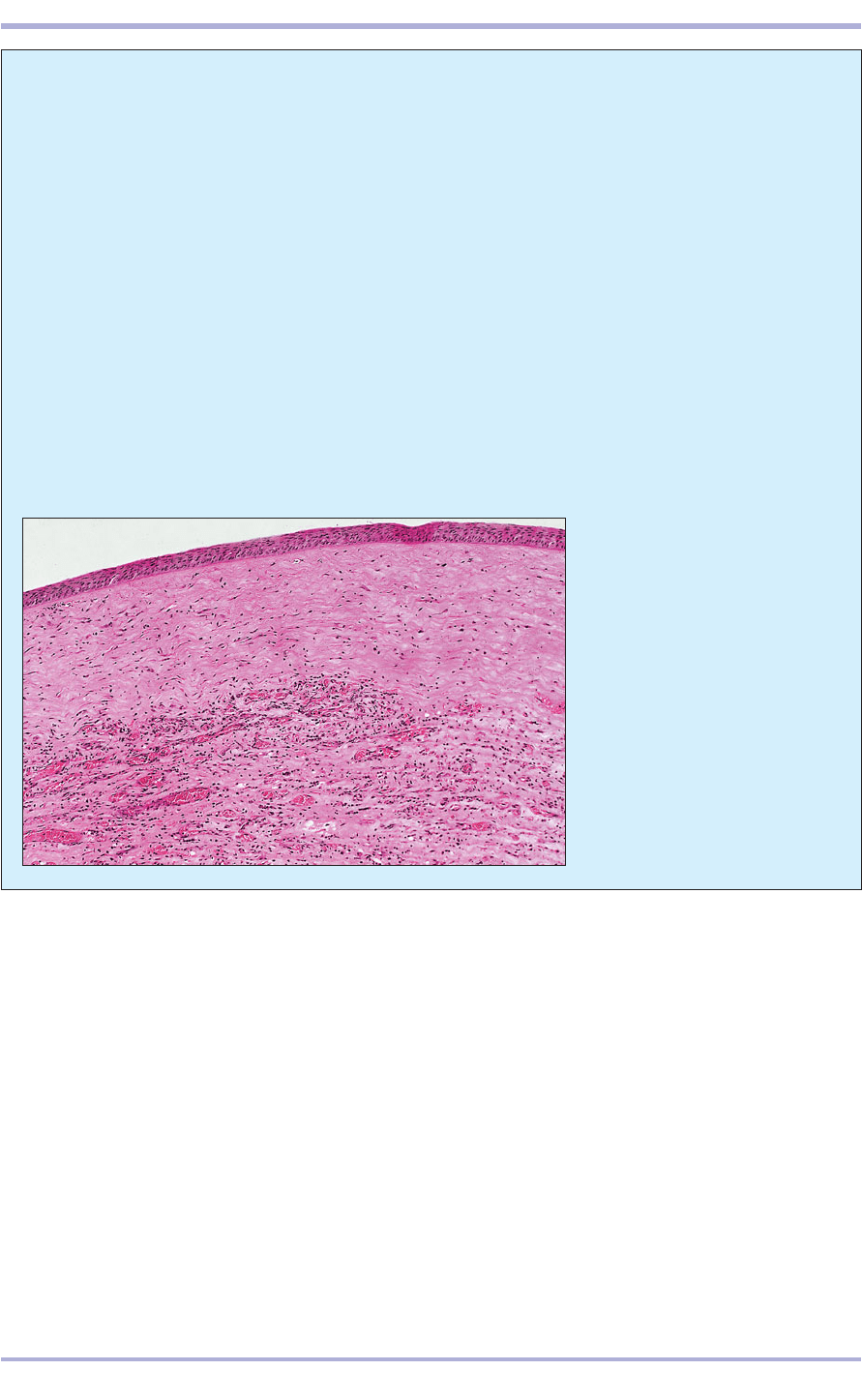
14.22 Ciliary processes (dog). (1) The
long ciliary processes extend from the
ciliary body, a localized expansion of
the vascular coat. The epithelium is
arrowed. (2) Ciliary muscle. H & E.
×62.5.
14.22
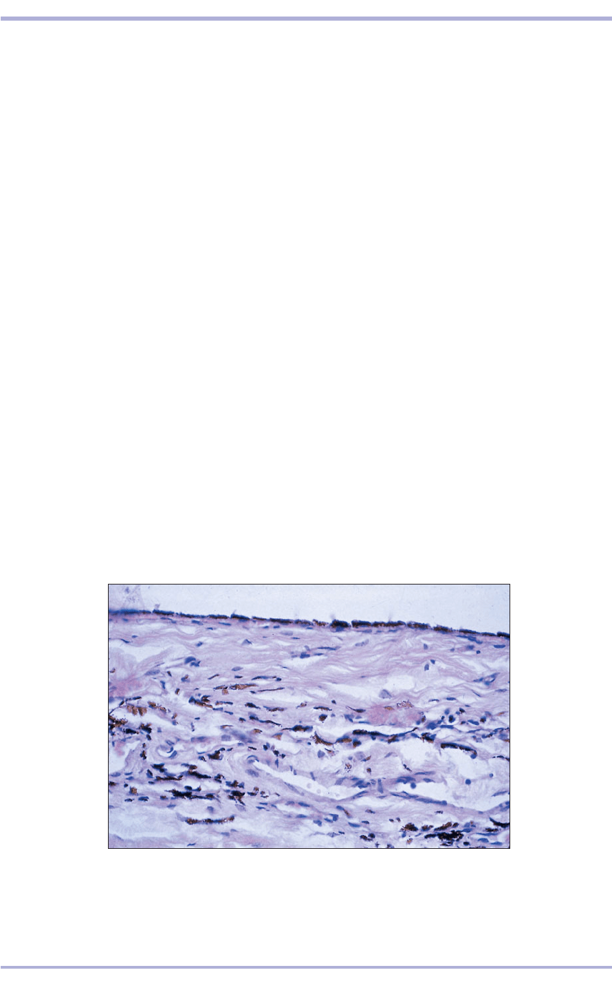
14.23 Iris (dog). (1) The anterior
surface is covered by flattened
fibrocytes. (2) The core of the iris is
vascular connective tissue. (3) The
posterior surface epithelium is two
layers of cells and part of the retina.
Pigment is present. H & E. ×62.5.
14.23
14.24 Iris (dog). Compare this with
14.23 and note the pigment; this
eye will be much darker. H & E. ×62.5.
14.24
1
2
3
2
1
Special Senses
14.25
Avian eye
The avian sclera has a ring of overlapping scleral ossi-
cles enclosed in dense connective tissue (14.26 and
14.27). The avian cornea is similar to the mammalian
but has a more prominent basement membrane. The
choroid is thick and well-vascularized with numer-
ous pigment cells; there is no tapetum. The avian
retina has the same number of layers as the mam-
malian, but is avascular. The cones contain oil
droplets, thought to enhance colour vision. The
pecten oculi is a thin, folded, heavily pigmented
membrane projecting into the vitreous humour from
the optic disc (14.28). The lens is composed of a body
Clinical correlates
Numerous inherited ocular abnormalities are rec-
ognized in purebred dogs. In some cases control
or monitoring schemes exist. Such defects may
affect structures of the globe or associated tis-
sues, such as the eyelids. Some dog breeds have
exaggerated palpebral fissure shapes which can
predispose to entropion and may require surgi-
cal correction. Dermoids, foci of cutaneous-type
differentiation on the cornea or conjunctiva that
may produce hair and other adnexal structures,
are recognized in all species.
Inflammatory lesions of the eyes are named
according to the structures affected and may
result from infectious agents, chemical irritation
or trauma. Ocular lesions may be part of a gen-
eralized disease syndrome, as in keratitis asso-
ciated with malignant catarrhal fever (MCF) in
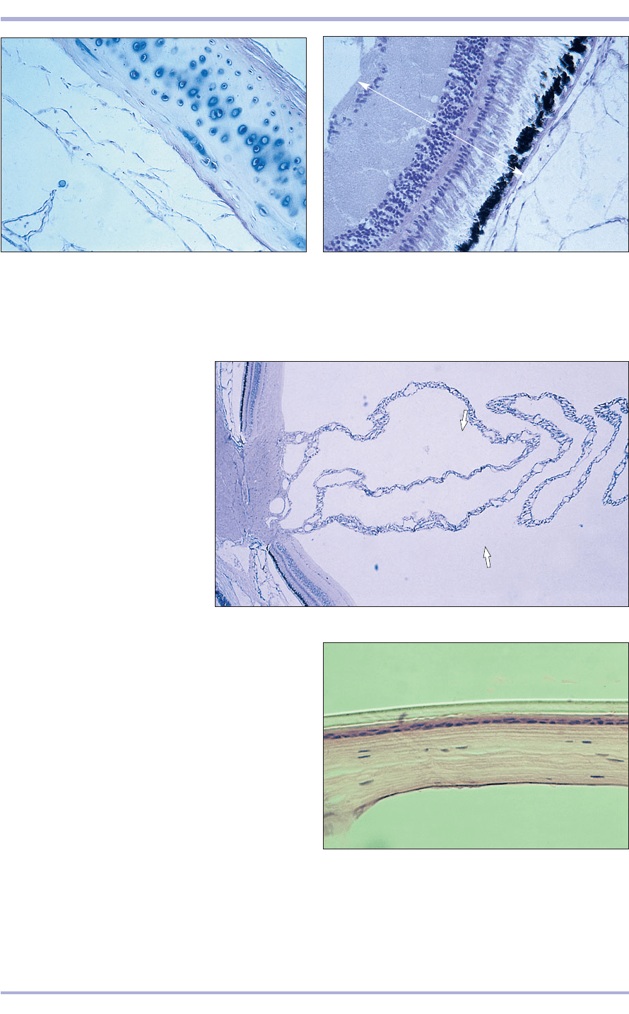
cattle (14.25). Migrating parasitic larvae can
reach the eye with potentially damaging conse-
quences, and occasional incidents of human vis-
ceral larva migrans caused by infections with
intermediate stages of ascarid parasites
(Toxocara canis) occur. Thelazia species of spi-
uroid worms inhabit the conjuctival sacs and
lacrimal glands of horses, cattle and in North
America dogs and cats also.
Important primary neoplasms of the ocular
structures include squamous cell carcinoma,
tarsal gland tumours and melanomas. The eye
may also be affected in cases of multicentric lym-
phosarcoma in several species and may be
affected by metastatic tumour spread.
236
Comparative Veterinary Histology with Clinical Correlates
14.25 Keratitis (cow). This
section, illustrating keratitis, or
inflammation of the cornea, is
from a cow with MCF. The effects
of this highly fatal, sporadic
herpes virus infection of
ruminants are multisystemic and
characterized by vasculitis. The
normally avascular cornea shows
neovascularization – the
production of a capillary network,
fibroplasia, oedema and
infiltration by mononuclear
inflammatory cells around the
vessels. H & E. ×62.5.
and an annular pad forming a ring around the equa-
tor of the lens. The lens fibres of the annular pad are
arranged radially, but in the lens body the fibres run
parallel to the optical axis.
Deep (Harderian) gland
This gland lies on the ventral caudomedial aspect
of the eyeball. The gland cells are vacuolated. Large
numbers of plasma cells are present seeded from the
cloacal bursa. These cells discharge antibodies
mixed with the gland secretion into the conjuncti-
val sac, thus providing local immunity. Lacrimal
glands are also present.
237
14.26 Avian eye. (1) Sclera with hyaline cartilage.
(2) Choroid. H.& E. ×62.5.
14.26
14.27 Avian eye. (1) Choroid. (2) Retinal layers are
(3) pigment layer, (4) rods and cones, (5) nuclei of the
rods and cones, (6) outer synaptic layer, (7) bipolar nerve
cell nuclei, (8) inner synaptic layer, (9) optic nerve cells,
(10) optic nerve fibres. H.& E. ×125.
14.27
14.28 Avian eye. The pecten
(arrowed) is a heavily folded,vascular
pigmented membrane projecting
into the vitrous humour from the
posteroventral surface of the eye.
(1) Optic nerve. (2) Retina.
(3) Choroid. H.& E. ×12.5.
14.28
1
3
2
1
2
3
5
9
10
6
7
4
8
1
2
Reptilian, amphibian
and fish eyes
Fish, amphibians, reptiles and birds are relatively
close phylogenetically to each other. Generally, the
histological features of their eyes are similar, but
there are some notable exceptions.
The pupillary shapes in fish, amphibian and
some reptilian eyes vary considerably between fam-
ilies within a given class.
Some species possess nictitating membranes;
others do not. Snakes and some lizards lack move-
able eyelids, and their corneas are covered with a
tertiary spectacle, an optically clear keratinized
epidermal tunic that is derived from the integu-
ment. This structure is shed and renewed with
each moult of the epidermis (14.29). Like many
14.29 Cornea and tertiary spectacle of a regal (ball)
python (Python regius). The thin keratinized spectacle
(1) is contiguous with the periorbital integument, and it
is shed and renewed with each moult. (2) Anterior
epithelium. (3) Descemet’s membrane. (4) Cornea
stroma. H & E. ×125.
14.29
3
1
2
4
Special Senses
14.32
birds, numerous reptiles have eyes supported in
part by scleral ossicles. The crystalline lens in the
eyes of fish, amphibians and reptiles is histologi-
cally similar to that in mammals. The posterior
segment of the eye, particularly the visual epithe-
lium, varies markedly within each class. The
238
14.30 Whole mount section of the eye of a small yucca
night lizard (Xantusia vigilis), a species that lacks
moveable eyelids. H & E. ×7.5.
14.30
14.31 Visual epithelium of a panther chameleon
(Chamaeleon pardalis). (1) Conus papillaris. (2) Retina.
(3) Optic nerve. H & E. ×20.
14.31
layers of the retina and choroid are similar to
those in mammals (14.30 and 14.31). In many rep-
tiles, particularly diurnal species, a vascular and
heavily pigmented conus papillaris projects ante-
riorly into the globe from the head of the optic
nerve (14.31).
2
3
1
Clinical correlates
Essentially, all of the clinically significant oph-
thalmic disorders affecting mammalian eyes can
affect the eyes of lower vertebrates. However,
some conditions are unique to some lower ver-
tebrates because their eyes are characterized by
structures that are lacking in the mammalian eye;
for example, inflammation of the tertiary spec-
tacle and the subspectacular space could only
occur in an animal whose eyes are lidless and
covered with an epidermally derived spectacle
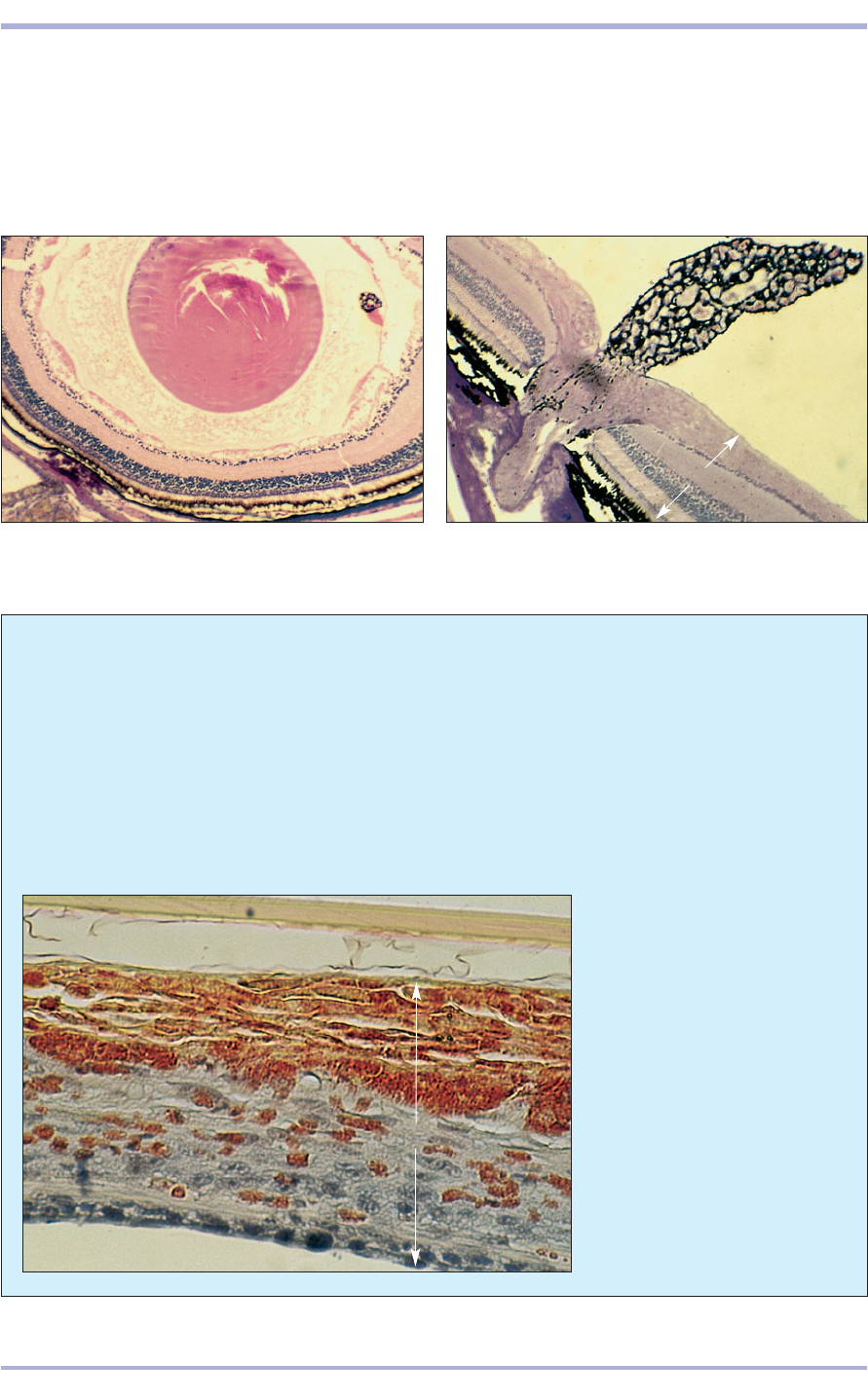
(14.32). Some species are more prone to certain
ophthalmic disorders induced by their incorrect
captive diets. Examples of such lesions are the
lipid and calcified corneal opacities of some
frogs. Cholesterol deposits occasionally develop
in the corneas of some reptiles.
14.32 Suppurative keratitis
in a mountain kingsnake
(Lampropeltis zonata). The
corneal stroma (1) contains
numerous heterophilic
granulocytic leucocytes.
(2) Tertiary spectacle.
(3) Cornea. Congo Red. ×160.
2
1
3
Comparative Veterinary Histology with Clinical Correlates
14.34
239
14.33 External ear canal (dog). (1) Stratified squamous
keratinized epithelium lines the canal. (2) Hair follicles.
(3) Sebaceous glands. (4) Sweat glands. (5) Elastic
cartilage. Masson’s trichrome. ×20.
14.33
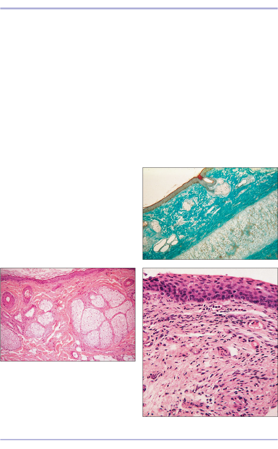
14.34 Ear canal (dog). (1) Stratified squamous
keratinized epithelium. (2) Hair follicles. (3) Special
sebaceous glands secreting cerumen. (4) Sweat glands.
H & E. ×50.
14.35 Tympanic membrane (goat). (1) External surface
covered by stratified squamous epithelium. (2) Dense
connective tissue. H & E. ×125.
14.35
Ear
The ear has three divisions: external, middle and
internal.
External ear
The external ear comprises the auricle (pinna), the
auditory canal and the tympanic membrane. The
auricle consists of a central plate of elastic cartilage
covered by skin rich in sebaceous glands. There are
also some sweat glands and a variable amount of
hair (14.33). The auditory canal is a rigid tunnel
lined with thin skin; the upper portion is supported
by elastic cartilage, the remainder by bone. It con-
tains large coiled sweat glands, some fine hair and
a few sebaceous glands that secrete cerumen (ear
wax; 14.34).
1
2
1
2
2
3
4
1
2
3
4
5
Middle ear
The auditory canal ends at the tympanic membrane,
which separates the external ear from the middle
ear. The external surface of the membrane is cov-
ered by skin and the internal surface by a simple
squamous or cuboidal epithelium. The centre con-
sists of dense collagen bundles (14.35).
Three auditory ossicles, the malleus, the incus and
the stapes, form a chain of small bones from the tym-
panic membrane to the oval window in the petrous
part of the temporal bone. Sound is transmitted by
the ossicles to the fluids of the internal ear and gen-
erate movement of the delicate basilar membrane of
the cochlea. The expanded rostral part of the tym-
panic cavity forms the auditory (Eustacian) tube and
connects the middle ear and the pharynx [see also
Chapter 7, Gutteral pouch (7.6)].
Special Senses
14.37
Internal ear
The internal ear consists of the osseous (bony)
labyrinth and the membranous labyrinth. The
osseous labyrinth is a space within the temporal
bone and consists of the vestibule, semicircular
canals and cochlea. The membranous labyrinth is
inside the osseous labyrinth and consists of the utri-
cle and saccule (within the vestibule), the semicir-
cular ducts (within the semicircular canals) and the
cochlear duct (within the cochlea). It contains
endolymph and is separated from the walls of the
osseous labyrinth by perilymph. It adheres to the
wall of the osseous labyrinth by means of fine con-
nective tissue strands derived from the connective
tissue lamina propria of the lining endothelium.
This endothelium is replaced at certain points by
neuroepithelial cells (14.36). In the semicircular
canals, local expansions of the ampullae house sen-
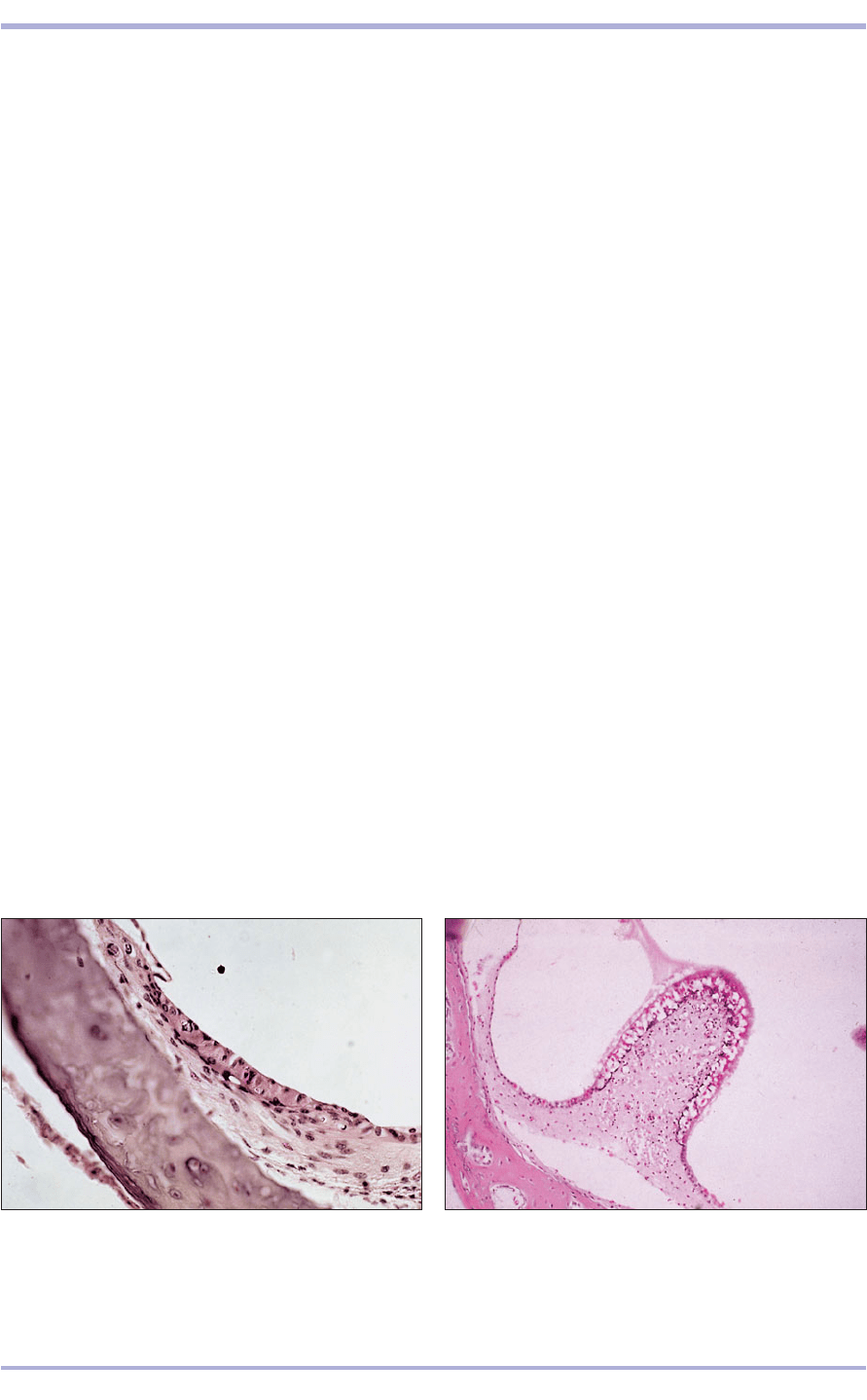
sory structures: the cristae ampullaris. The neu-
roepithelial sensory hair cells and supporting
(sustentacular) cells of each crista are covered by a
gelatinous cupola (14.37). When the latter is
deflected during rotational movements of the head,
the sensory cells are stimulated and impulses sent
to the brain.
Both the utricle and saccule are lined in part by
maculae, patch-like collections of sensory hair cells
and supporting cells. Maculae are lined with
mesothelium and covered with a gelatinous otolithic
membrane, in which are embedded calcium car-
bonate crystals: the otoliths. As the membrane shifts
in response to gravity acting upon the otoliths, sen-
sory cells of the maculae are stimulated. They
enable the animal to determine the position of its
head in space and to assess linear acceleration and
deceleration (14.38). The sensory cells are sur-
rounded by terminals of the vestibular nerve.
The osseous cochlea surrounding the cochlear
canal in a spiral around a central pillar of bone,
the modiolus, contains in turn the spiral lamina,
a thin shelf of bone that travels up the modiolus.
The canal is divided into three compartments: the
dorsal scala vestibuli and ventral scala tympani,
which contain perilymph and are lined with squa-
mous epithelium, and the cochlear duct between
them (14.39). The floor of the duct is formed from
the fibrous basilar membrane and the roof from
the vestibular (Reissner’s) membrane, and consists
of two layers of simple squamous epithelium. The
acoustically sensitive spiral organ (of Corti) rests
on the basilar membrane and is composed of
uroepithelial hair cells and sustentacular cells. The
lower surface of the membrane, facing the ventral
scala tympani, is lined with simple squamous
epithelium. The bases of the cells are widely sep-
arated and the apices enclose to form a triangular
space, the inner tunnel, containing a gelatinous
substance and the cochlear nerve fibres. Overlying
the spiral organ and extending from the spiral lim-
bus (an elevation of protective tissue above the
spiral lamina) is the tectorial membrane, resting
on the cilia of the hair cells. The cilia are displaced
when the basilar membrane vibrates in response
to sound waves passing through the fluid-filled
scalas. Nerve terminals form a web around the
bases of the hair cells and transmit stimuli to the
spiral ganglion of bipolar neurons. The axons
form the cochlear division of the eighth cranial
nerve (14.40).
240
Comparative Veterinary Histology with Clinical Correlates
14.36 Inner ear (cat). (1) Specialized neuroepithelial cells
in the vestibule continuous with (2) endothelial cells
lining the labyrinth. H & E. ×125.
14.36
14.37 Inner ear. Crista (cat). (1) Bone of the osseous
labyrinth. (2) Neuroepithelial (hair) cells and supporting
cells of the crista continuous with (3) endothelium lining
the membraneous labyrinth. (4) Cupula. H & E. ×125.
1
2
3
4
1
2
