Aughey E., Frye F.L. Comparative veterinary histology with clinical correlates
Подождите немного. Документ загружается.

221
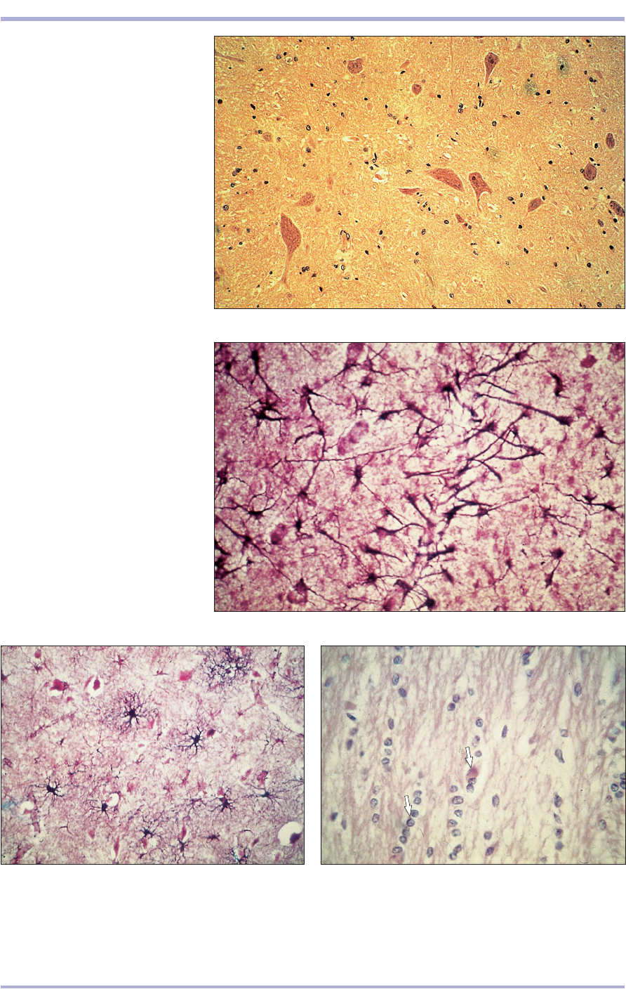
13.15 Spinal cord (dog). (1) Neuron.
(2) Nuclei of protoplasmic astrocytes.
H & E. ×125.
13.15
13.16 Spinal cord (dog). (1) Neuron.
(2) Nuclei of protoplasmic astrocytes.
H & E. ×125.
13.16
2
1
2
1
13.17 Spinal cord (dog). (1) Neuron. (2) Nuclei of
protoplasmic astrocytes. (3) Fibrous astrocytes. H & E.
×125.
13.17
13.18 Corpus callosum (dog). Oligodendrocyte nuclei in
orderly columns (arrowed) providing the neurilemmal
sheath in the central nervous system. H & E. ×125.
13.18
1
1
2
3
2
Nervous System
13.21
The cerebral and cerebellar cortices are highly
specialized folded areas of outer grey matter with
layers of neurons of various sizes and supporting
cells, as well as a central core of white matter
(13.20–13.25).
222
Comparative Veterinary Histology with Clinical Correlates
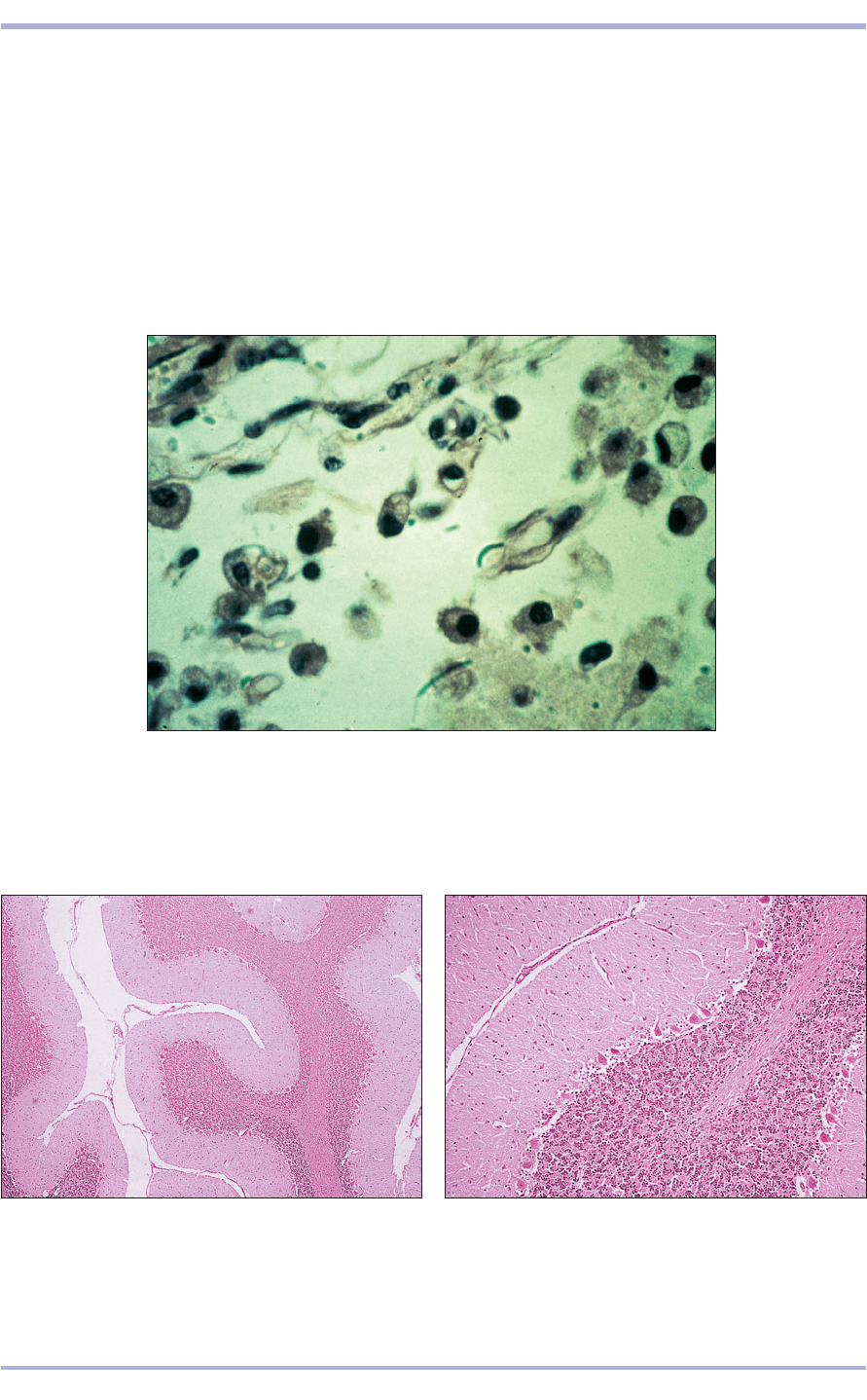
13.19 Microglial cells in the central nervous system (cat). H & E. ×256.
13.19
13.20 Cerebellum (cat). (1) Outer cellular layer of grey
matter and (2) inner fibrous layer of white matter. H & E.
×25.
13.20
13.21 Cerebellum (cat). (1) Inner granular layer of small
neurons. (2) Middle Purkinje layer of large neurons.
(3) Outer molecular layer with few neurons and many
branching processes. H & E. ×100.
1
2
3
2
1
1
lined by a continuous endothelium. There is a well-
developed basal lamina that, with the protoplas-
mic astrocyte processes, forms the formidable
blood<brain barrier. Microglia are the phagocytic,
scavenging cells of the CNS (13.19).
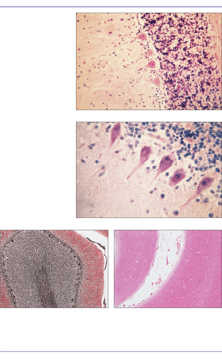
13.24 Cerebellum (cat). Each folium has (1) a central core
of white matter, the nerve fibres and supporting cells,
and (2) a surface cellular layer of grey matter. Cajal’s
silver. ×62.5.
13.24
223
13.22 Cerebellum (sheep).
(1) Granular layer. (2) Purkinje cell
layer. (3) Molecular layer. H & E. ×125.
13.22
1
2
1
2
3
13.23 Cerebellum (dog). Large
Purkinje cells are arrowed. H & E.
×250.
13.23
13.25 Cerebral cortex (cat). (1) Meninges. (2) White
matter. (3) Grey matter. H & E. ×100.
13.25
1
3
2
Nervous System
224
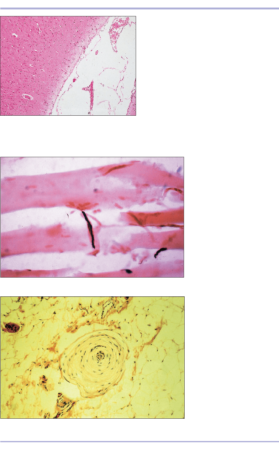
13.26 Cerebral cortex (cat). Cellular grey matter.
H & E. ×25.
13.26
13.27 Motor end plate in striated
muscle (dog). (1) Striated muscle
fibre. (2) Terminal nerve fibre. Cajal’s
silver. ×250.
13.27
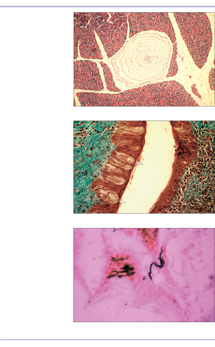
13.28 Pacinian corpuscle. Bladder
(dog). (1) Pacinian corpuscle.
(2) Loose areolar connective tissue.
H & E. ×20.
13.28
Nerve endings and receptors are specialized ter-
minal parts of axons or dendrites. They include
motor end plates, pressure receptors (such as
Pacinian corpuscles) and taste buds (13.26–13.31).
Nerve tissue is so characteristic that it is readily
identified, even in invertebrates.
1
2
1
2
Comparative Veterinary Histology with Clinical Correlates
225
13.29 Lamellar (Pacinian) corpuscle
in a serous gland (dog).
H & E. ×20.
13.29
13.30 Circumvallate papilla. Tongue
(cow). Taste buds are arrowed.
Masson’s trichrome. ×160.
13.30
13.31 Cat skin to illustrate nerve
endings stained black with Cajal’s
silver. ×250.
13.31
Nervous System
13.34
13.33
13.32
226
Comparative Veterinary Histology with Clinical Correlates
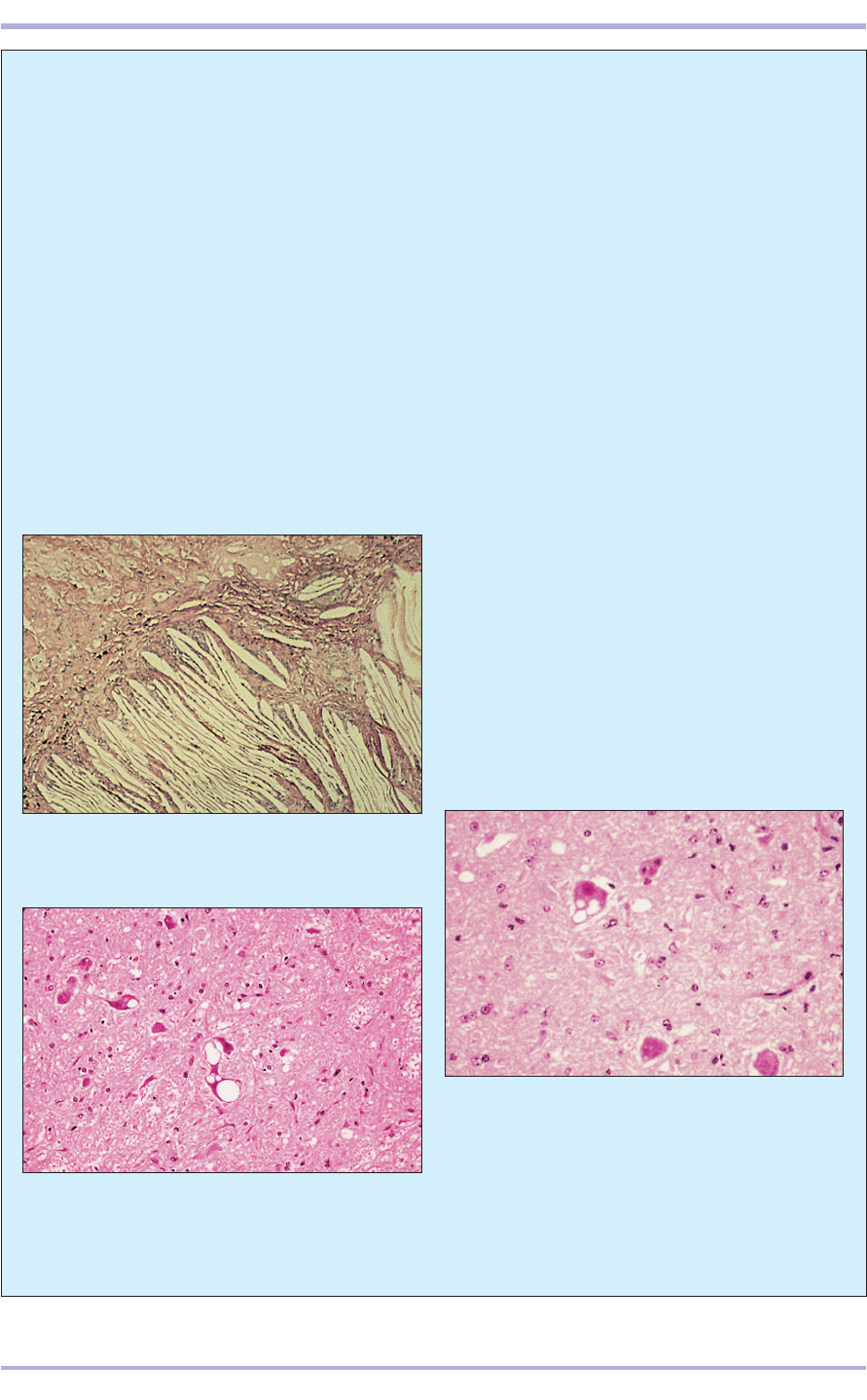
13.34 Bovine spongiform encephalopathy (BSE, cow).
This section is taken from the midbrain of a cow with
BSE. The histological changes are similar to those found
in scrapie, with vacuolation of neuronal cell bodies in
target areas of the brain, which include in particular the
medullary nuclei. Some vacuolation of the neuropil is
present in this section. The severity is variable between
cases. Special precautions, including treating the tissues
with formic acid to inactivate the BSE agent, are
employed when processing these tissues. H & E. ×125.
from almost every type of cell within the nervous
system have been recognized. The functional dis-
tinction between benign and malignant neo-
plasms within the CNS is not as significant as in
other systems, as any space-occupying lesion,
even the most biologically benign, has serious
consequences. Even non-neoplastic lesions, such
as equine cholesteatoma (13.32) in which a mass
of cholesterol crystals surrounded by inflamma-
tory cells located in the choroid plexuses can pro-
duce space-occupying effects, in particular
hydrocephalus caused by obstruction of ventric-
ular drainage.
Transmissible spongiform encephalopathies
(TSE) are an unusual group of infectious central
nervous disorders, although the nature of the
infective agent, which differs from conventional
agents like bacteria and viruses, is not precisely
defined. The agent is believed to enter via the
oral route and proliferate in lymphoid tissues and
in the intestine, but no effects are observed until
it reaches target areas of the CNS. The sheep
TSE, scrapie (13.33), has been known for a very
long time. This disease produces no significant
gross pathology, although emaciation and self
trauma are common, and provokes no immune
response. The cattle variant, bovine spongiform
encephalopathy (BSE; 13.34) was recognized
more recently and produces similar histological
changes to those seen in scrapie in sheep.
Clinical correlates
The CNS is affected by a variety of congenital
disorders that occur with relatively high fre-
quency, possibly because of the susceptibility of
this complex system to teratogenic insult.
Inherited CNS disease is also recognized and a
heritable basis is suspected for conditions such
as idiopathic epilepsy.
Inflammatory, neoplastic, metabolic and par-
asitic disorders are all recognized in the CNS,
with certain specific entities appearing in partic-
ular species. For example, feline parvovirus,
which is trophic for dividing cells, attacks the
external germinal layer of the cerebellum in kit-
tens when infection occurs before or shortly after
birth. Primary CNS neoplasms are not uncom-
mon in the dog and cat and tumours that arise
13.32 Cholesteatoma in the brain of a horse. Note the
numerous clefts that previously held cholesterol deposits
lost during the histological processing. H & E. ×20.
13.33 Scrapie (sheep). This is a section of midbrain
from a sheep with scrapie. The characteristic large
intraneuronal vacuoles and diffuse vacuolar change
in the neuropil can be seen. No stainable material is
present in the vacuoles. H & E. ×125.
Eye
The eye is the organ of vision and comprises the eye-
ball or globe of the eye and the optic nerve. It is pro-
tected by the eyelids and lacrimal apparatus.
Eyelids
The eyelids are movable folds of skin in front of the
eyeball protecting and lubricating the surface of the
eye. The outer surface is covered with stratified
squamous epithelium, tactile hairs, sebaceous
glands (glands of Zeiss) and sweat glands (glands
of Möll; 14.1). The inner surface is lined with the
palpebral conjunctiva, a thin transparent mucous
membrane. The bulbar conjunctiva is continuous
with the surface of the cornea at the limbus. The
epithelium may be stratified columnar or stratified
squamous, with goblet cells. Between the dermis of
the skin and the lamina propria of the conjunctiva
is a plate of dense connective tissue: the tarsal plate
is surrounded by the multilobular tarsal glands
(14.2). The nictitating membrane (third eyelid) is
situated at the medial angle of the eye. It is a semi-
227
14. SPECIAL SENSES
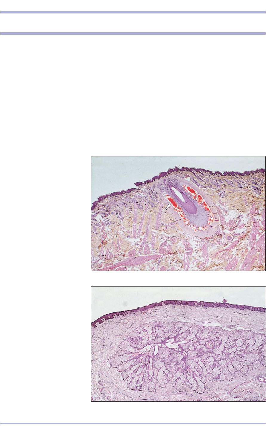
14.1 Eyelid (horse). Outer skin layer
of the eyelid with a tactile hair
(arrowed). H & E. ×7.5.
14.1
14.2 Eyelid (horse). (1) The
conjunctiva, stratified columnar
epithelium with mucus-secreting
cells. (2) Sebaceous glands in the
lamina propria. (3) Tarsal plate.
H & E. ×25.
14.2
2
3
3
1
circular fold of conjunctiva enclosing a plate of car-
tilage (hyaline in ruminants and dog; elastic in the
horse, pig and cat; 14.3–14.5).
Lacrimal apparatus
The lacrimal glands are tubuloacinar: serous in the
cat, seromucous in the horse, ruminant, dog and pig.
(In the horse and ruminant they are predominantly
serous, and in the pig predominantly mucous.)
Lymph nodules are seen in the lamina propria (14.6
and 14.7). The pig and ox also have a deep gland
(Harderin gland) of the nictitating membrane.
Development of the eye
The eyes are recognizable in the early embryo as lat-
eral diverticulae of the diencephalon. As the diver-
ticulum approaches the surface ectoderm, it
invaginates to form the optic cup. The inner layer
becomes the light-sensitive retina; the outer layer
becomes the retinal pigment, pigmented ciliary and
anterior iris epithelium. The surface ectoderm over-
lying the optic cup thickens and invaginates to form
the lens. The reconstituted ectoderm and the local
mesenchyme form the cornea; the surrounding
mesoderm provides the connective tissue, blood ves-
sels and ocular muscles (14.8 and 4.9).
228
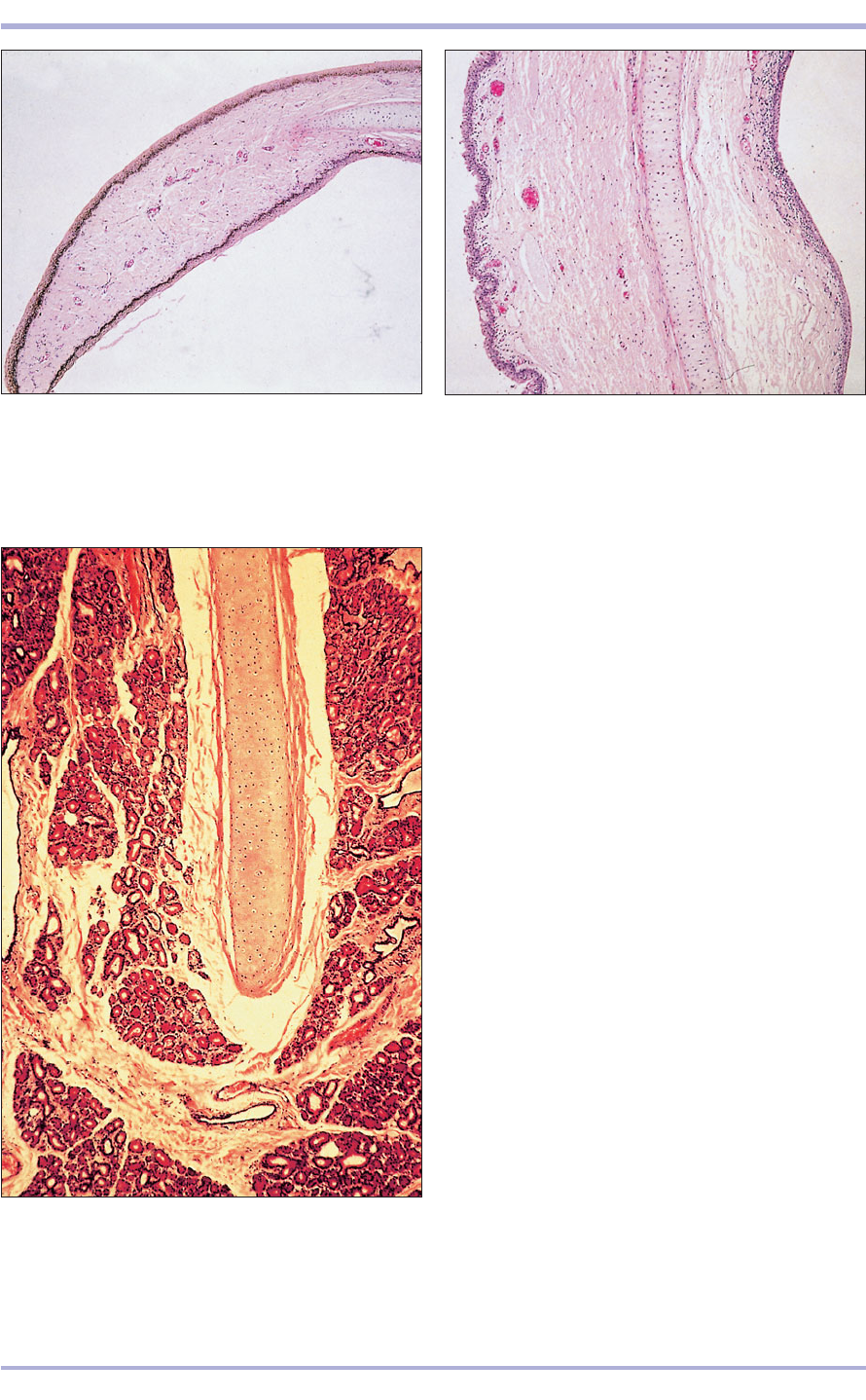
14.3 Nictitating membrane (membrana nictitans; horse).
(1) Anterior surface is covered by conjunctiva. (2) Elastic
cartilage plate in a central core of connective tissue.
(3) Posterior plate covered by conjunctiva. H & E. ×7.5.
14.3
14.4 Nictitating membrane (horse). (1) Anterior
conjunctival surface. (2) Elastic cartilage plate.
(3) Posterior conjunctival surface. (4) Lamina propria.
H & E. ×7.5.
14.4
14.5 Nictitating membrane (dog). (1) Hyaline cartilage.
(2) Seromucus-secreting glands in the lamina propria.
H & E. ×50.
14.5
2
3
4
1
3
2
2
1
1
2
3
Comparative Veterinary Histology with Clinical Correlates
Special Senses
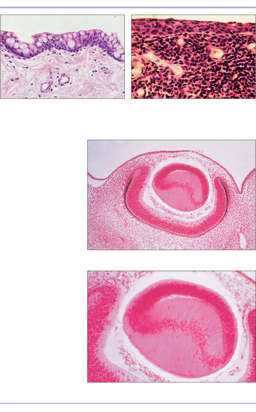
14.8 Developing eye in a 35-day
cat embryo. (1) Surface ectoderm.
(2) Developing lens. Optic cup has a
thick inner layer (3) and a thin outer
layer (4) with the pigment cells.
H & E. ×7.5.
14.8
14.9 Developing eye in a 35-day cat
embryo. (1) The lens fibres almost fill
the lens vescicle. (2) Loose vascular
mesenchyme. (3) Retina. H & E. ×25.
14.9
14.7 Nictitating membrane, posterior surface (dog).
(1) Lymphoid cells in the lamina propria. H & E. ×125.
14.7
14.6 Nictitating membrane (pig). The mucus-secreting
cells in the outer epithelium are stained pale pink.
H/PAS. ×100.
14.6
1
2
3
1
2
3
4
1
229
Structure of the eyeball
The eyeball (14.10) is spheroid and composed of
a lens and a wall that is divided into three layers:
an outer fibrous tunic (corneoscleral layer); a mid-
dle vascular tunic (uvea); and an inner retinal
tunic, which consists of a 10-layered photosensi-
tive retina and a bilayered non-photosensitive por-
tion that covers the ciliary body and posterior
surface of the iris.
The eye contains three fluid-filled regions: the
anterior chamber, bordered by the cornea, iris and
lens; the posterior chamber, located between the iris,
lens, zonular fibres and ciliary processes; and the
cavity of the vitreous humour, which lies behind the
lens (14.10). The lens is a biconvex transparent body
composed of epithelial cells within a homogeneous
outer capsule. The anterior epithelial cells are
cuboidal at the pole, and become elongated, pris-
matic and arranged in meridional rows at the equa-
tor where they form lens fibres (14.11). The aque-
ous humour circulates continuously, and is secreted
and absorbed by the blood vessels in the sclera at the
corneoscleral junction.
The corneoscleral layer is divided into a trans-
parent anterior segment, the cornea, and an opaque
posterior segment, the sclera. The avascular cornea
is transparent, covered with non-keratinized strat-
ified squamous epithelium resting on a basement
membrane (Bowman’s). The underlying stroma, the
substantia propria, consists of thin lamellae of col-
lagenous fibres and flattened fibroblasts running
parallel to the corneal surface in a mucoid ground
substance (14.12 and 14.13). The caudal limiting
membrane (Descemet’s) separates the connective tis-
sue of the substantia propria from the simple squa-
mous corneal endothelium (also called posterior
epithelium; 14.14).
The opaque sclera consists of interlacing bundles
of white fibrous tissue with a few elastic fibres. The
230
Comparative Veterinary Histology with Clinical Correlates
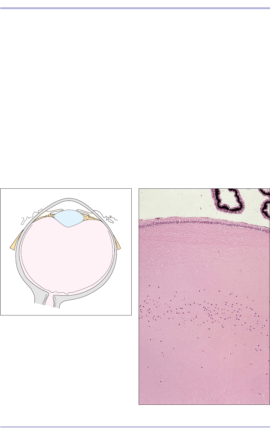
14.10 Diagram of the eye. (1) Cornea. (2) Anterior
chamber. (3) Iris. (4) Lens. (5) Posterior chamber.
(6) Ciliary body. (7) Ciliary processes. (8) Sclera.
(9) Choroid. (10) Retina. (11) Optic papilla.
(12) Optic nerve. (13) Vitreous humour.
14.10
1
2
3
4
5
6
7
8
9
10
11
12
13
14.11 Lens (dog). (1) Anterior epithelium. (2) Nuclei of
the lens fibres. H & E. ×62.5.
14.11
1
2
