Aughey E., Frye F.L. Comparative veterinary histology with clinical correlates
Подождите немного. Документ загружается.


241
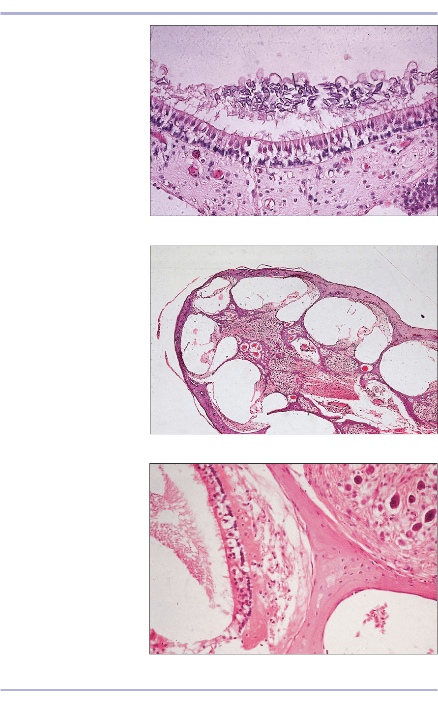
14.38 Inner ear. Macula (cat).
(1) Columnar epithelium of the
macula consists of neuroepithelial
cells and supporting cells continuous
with the lining epithelium of the
vestibule. (2) Otoliths. (3) Connective
tissue. H & E. ×160.
14.38
14.39 Inner ear. Cochlea (cat).
(1) Dorsal scala vestibuli. (2) Middle
cochlear duct. (3) Ventral scala
tympani. (4) Spiral ganglion.
H & E. ×7.5.
14.39
14.40 Inner ear. Spiral organ (of
Corti) (cat). Located on the floor of
the cochlear duct. (1) Osseous spiral.
(2) Spiral ganglion. (3) Basilar
membrane. (4) Sensory and
supporting cells. H & E. ×160.
14.40
1
2
3
4
1
1
4
2
3
1
4
3
1
2
3
Special Senses

14.41
Reptilian and amphibian
ears
The external pinna and ceruminous glands are
absent. Some terrestrial frogs and toads that uti-
lize audible calls in their courtship and territorial
behaviour have acute hearing. Many amphibians
possess external tympanic membranes; others lack
them entirely. However, some aquatic amphibians
have lateral line systems with which they detect
water-transmitted vibratory signals and hydrosta-
tic pressure changes (see 14.50).
Crocodilians are capable of hearing air-transmit-
ted sounds. They have slit-like auditory openings that
can be closed when they submerge themselves.
Most lizards have ears with tympanic membranes
that are located close to the integumentary surface
Avian ear
The external ear lacks a pinna and ceruminous
glands. The auditory ossicles are fused to form a
cartilaginous rod, the columella, which extends
from the tympanic membrane to the oval window.
The membranous labyrinth is essentially similar to
that of the mammal (14.42). The saccule, however,
differs in that it contains two maculae. The
cochlear duct also possesses a terminal expansion
that is peculiar to birds, the lagena, and is sepa-
rated from the dorsal scala vestibuli by the tegmen-
tum vasculosum, a thin connective tissue
membrane integrated with a highly folded vascu-
lar epithelium.
242
Comparative Veterinary Histology with Clinical Correlates
14.42 Avian inner ear. (1) Spiral ganglion. (2) Osseous
labyrinth. (3) Membraneous labyrinth. (4) Neuroepithelial
sensory cells. H.& E. ×125.
14.42
Clinical correlates
Diseases of the ear in domestic animals are prob-
ably not commonly investigated by pathologists,
although the three broad categories otitis
externa, otitis media and defects of hearing may
be considered. Otitis externa (14.41), or inflam-
mation of the external ear canal, may be a prob-
lem restricted to the waxy integument of this site
or it may be part of a widespread skin condition.
Usually only chronic cases of this very common
condition, in which the severe thickening of the
walls of the ear canal may raise clinical suspicion
of tumour development, are submitted for histo-
pathological examination.
14.41 Otitis externa (dog) The surface epithelium
(centre) is hyperplastic and there is dermal fibrosis. The
patchy appearance of the dermis is caused by a mixed,
mainly mononuclear, inflammatory infiltrate. The
sebaceous glands are hyperplastic and there is cystic
dilatation of the ceruminous glands, some of which are
seen filled with eosinophilic cerumen. H & E. ×20.
1
2
2
4
3

of the skull. In some species the tympanic membrane
is flush with the skin surface. In others it lies within
a shallow depression or deeper auditory meatus.
Snakes, chelonians and the tuatara lack external
auditory structures. The philosophical question of
whether their sense of ‘hearing’ is limited to substrate-
transmitted vibrations is a topic of spirited debate.
The inner ear is responsible for the spacial and
postural orientation of the animal within its envi-
ronment. The morphological and histological fea-
tures of the amphibian and reptilian inner ear are
sufficently similar to the mammalian and avian ear
that further discussion is not warranted here.
243
Organ of smell
(olfactory organ)
The nose, in addition to its role in the respiratory
system, also functions as the olfactory organ. In the
nasal cavity, brown–yellow olfactory epithelium
occurs together with pinkish respiratory epithelium.
It is a pseudostratified columnar epithelium with
olfactory (sensory) cells, basal cells and support-
ing cells. Tubular mucoserous glands (Bowman’s)
lie in the lamina propria and secrete onto the sur-
face through simple ducts lined with cuboidal cells
(14.43 and 14.44).
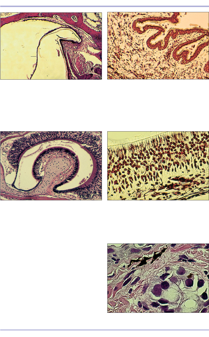
14.43 Olfactory organ (horse).
(1) The olfactory epithelium is
pseudostratified ciliated columnar
with many rows of nuclei; the more
superficial are of the sustentacular
cells; the deeper nuclei are of the
olfactory nerve cells. (2) Vascular
connective tissue with mucous
glands. H & E. ×160.
14.43
14.44 Olfactory organ (horse). The
mucous cells stain deep blue. Alcian
blue. ×100.
14.44
2
2
1
Special Senses

Other specialized
sense organs
Besides sight, hearing, taste, touch and the per-
ception of pain, heat and cold, many of the lower
vertebrates possess highly specialized organs that
augment their awareness of their external en-
vironment.
Parietal eye
In addition to their paired lateral eyes, many lizards
and the primitive tuatara (Sphenodon punctatus)
have a parietal eye. This photosensitive organ con-
sists of a scale-like and cell-poor cornea, a cellular
lens, a central chamber filled with clear fluid and a
few macrophages, and a cup-shaped pigmented
visual epithelium that is analagous to the retina and
choroid (14.45). The parietal eye is partially
responsible for regulating basking and other
thermoregulating behavioural activities, and it can
warn of the approach of potential predators whose
shadows are detected by the upward directed eye-
like structure.
Facial and labial pit organs
Rattlesnakes, water moccasins, copperhead snakes
and other pit vipers possess paired facial pit organs
(14.46) with which they sense very small differences
in the background thermal environment. This abil-
ity to discriminate slight temperature variations aids
in prey detection both before and after the prey
animals have been envenomated.
Non-venomous snakes of the family Boidae
(boas, pythons and anacondas that are ambush
predators) possess labial pit organs (14.47), which
help them locate warm-blooded prey. These
branched pit-like depressions are lined with lightly
keratinized squamous epithelium through which
numerous dendritic sensory neurons penetrate.
Vomeronasal organ
Most snakes and lizards have well-developed
vomeronasal (Jacobson’s) organs (14.48), which
assist in sampling and discriminating chemosensory
stimulatory particles such as prey-related scent or
pheromones. The vomeronasal organ consists of a
mushroom-shaped rounded column with a carti-
laginous core. This column is surrounded by a cup-
shaped spherical cavity. The luminal surfaces of the
column and cavity are covered with ciliated colum-
nar or pseudostratified columnar epithelium
through which myriad numbers of tiny dendritic
nerves pass between adjacent cell membranes. These
nerve endings project out into the lumen (14.49).
Lateral line organ
Most fish and many amphibians (particularly
aquatic frogs, newts and salamanders) have lateral
line systems (14.50) that are sensitive to slight
changes in hydrostatic pressure and water-borne
vibration. Tall cuboidal to fully columnar cells form
vibration- and pressure-sensitive neuromasts that
receive and transmit impulses from the aquatic envi-
ronment to the animal’s central nervous system.
244
Comparative Veterinary Histology with Clinical Correlates
14.45 Whole mount section of the
parietal eye of a green iguana (Iguana
iguana). At the top of the image is a
relatively thick, but avascular, cornea
(1). A cellular lens (2), composed of tall
columnar cells packed closely and parallel
to each other, lies beneath the cornea.
The lumen (3) of the central chamber
contains clear fluid and is surrounded at
the sides and back by heavily pigmented
photosensitive retinal cells (4) and
unpigmented ganglion cells (5). A giant
melanin-packed macrophage (6) can be
seen at the bottom of the capsule. The
parietal nerve exits the rear of the
parietal eye and courses through the
parietal foramen. A thin connective tissue
capsule (7) envelops the parietal eye
where it is surrounded by calvarial bone.
H & E. ×125.
14.45
1
3
2
4
5
6
7

14.46 Facial pit organ of a western diamondback
rattlesnake (Crotalus atrox) is surrounded on its inner
surfaces by a cup-shaped bony depression. A thin, lightly
keratinized diaphragm-like membrane (1) divides the
posterior of the pit chambers into two compartments.
Within the membrane are embedded numerous dendritic
neuron endings. H & E. ×20.
245
14.46
14.47 Labial pit organ of a reticulated python (Python
reticulatus). These infrared superficial depressions over
much of the external surface of upper lips of some boas
and pythons consist of parallel branched or unbranched
shallow passages lined by a thin layer of lightly
keratinized squamous epithelium. A rich network of dark
staining, fine dendritic nerve endings penetrate to just
beneath the epithelium. Bodian’s silver stain. ×125.
14.47
14.48 Whole mount sagittal section of the vomeronasal
(Jacobson’s) organ of a small skink (Scinella lateralis). The
raised mushroom-shaped protruberance is supported by a
core of hyaline cartilage and is surrounded by a narrow
cavity the surfaces of which are covered on all sides by
ciliated simple columnar epithelium. H & E. ×125.
14.48
14.49 Section through the superficial surface of the
vomeronasal organ of a green iguana (Iguana iguana).
Note the myriad number of thin dendritic nerve endings
that course between and penetrate the epithelium to the
lumenal surface. Bodian’s silver stain. ×250.
14.49
14.50 Cross-section of the dermis and the pressure-
sensitive lateral line of an aquatic salamander (Amphiuma
tridactyla). The clear central cavity is lined with large,
plump columnar glandular secretory cells. H & E. ×250.
14.50
1
Special Senses

Electric organ
Some teleost fish, such as the electric eel and elec-
tric catfish [and at least one family of elasmo-
branch ray, such as the torpedo (Torpedo spp.)],
possess specialized muscles arranged into discrete
electric organs (electroplax) that produce and
detect powerful electric impulses. Paired electric
lobes on the medulla oblongata are the motor cen-
tres for the integration of electroplax activity.
When these specialized muscular organs suddenly
discharge their electrical potential, prey fish and
predators are subjected to pulses of high amper-
age electrical current that can be incapacitating or
fatal. Some electric eels produce pulses of direct
current that measure 600 V.
Swim bladder
Most but not all teleost fish possess a specialized
elongated gas- (or oil-) filled organ: the swim blad-
der. This is a major hydrostatic organ that helps
these fish maintain their buoyancy and orientation
within a column of water. In some bottom-feeding
species the swim bladder is much reduced or may
be absent. In fast swimming fish it is elongated and,
thus, enhances streamlining. The swim bladder is
formed from thin sheets of dense fibrocollagenous
connective tissue laid at acute angles to each other.
This arrangement aids in maintaining its shape and
reducing deformation. In some fish, skeletal mus-
cles insert into its outermost surface. In other species
the swim bladder is only attached to the body wall
along its dorsal surface. The swim bladder is lined
by a thin, much flattened, non-keratinized squa-
mous epithelium.
Several bacterial, viral and protozoan diseases
are characterized by inflammation of the swim blad-
der. Thus, when performing a necropsy on a fish,
it is important to examine this organ for haemor-
rhage(s), oedematous thickening, discolouration or
other abnormality.
246
Comparative Veterinary Histology with Clinical Correlates

The lymphatic system has a dual function: the lym-
phatic vessels drain interstitial tissue, returning fluid to
the bloodstream; and lymphoid tissue produces phago-
cytes and the immunologically competent cells, which
are the body’s most important defence mechanism
against invasion by pathogens. Lymphoid tissue is
widely distributed in the body and comprises: lymph
nodes and associated lymphatic vessels; spleen; thy-
mus; local aggregations of lymphoid tissue in the
mucosa of the digestive, respiratory and urogenital
tracts; and any local tissue aggregation of lymphocytes.
Lymphoid tissue consists predominantly of lym-
phocytes. These and a variable number of plasma
cells, macrophages and other cells are supported by
a delicate network of reticular fibres that fill the
spaces between the trabeculae. Diffuse lymphatic tis-
sue and lymphatic nodules are the components of
most lymphatic organs, and also appear in the con-
nective tissue of the digestive, respiratory, urinary and
247
15. LYMPHATIC SYSTEM
15.1 Lymph node (dog). (1) Connective tissue capsule.
Cortex with (2) lymphatic follicles and (3) paracortex.
Masson’s trichrome. ×20.
15.1
1
2
3
15.2 Lymph node (sheep). (1) Connective tissue capsule.
(2) Connective tissue trabeculae. (3) Lymphatic tissue.
H & E. ×62.5.
15.2
1
2
3
3
3
15.3 Lymph node (cat). The reticular fibres form a black
network and support the cells of the lymphatic tissue.
Gordon and Sweet. ×100.
15.3
reproductive organs, among other locations. The for-
mer is characterized by a moderate concentration of
scattered lymphocytes; the latter comprises an aggre-
gation of mostly small, densely packed lymphocytes.
Lymph nodes
A lymph node contains diffuse and nodular lym-
phatic tissue and lymphatic sinuses that are organized
into a cortical and medullary region. A capsule of
connective tissue sends fine vascular fibrous trabec-
ulae into the substance of the node. Afferent lym-
phatic vessels enter the capsule and drain into the
subcapsular sinus. From there the lymph drains into
a labyrinth of sinuses extending along the trabecu-
lae, eventually emptying into the efferent lymphatics
in the hilus. In the cortex, circular aggregations of
lymphocytes form follicles or nodules (15.1–15.4).
15.4 Lymph node (dog). (1) Connective tissue capsule
with some fat cells. (2) Subcapsular sinus. (3) Cortical
follicle. (4) Parafollicular tissue. (5) Sinusoid. H & E. ×125.
15.4
1
2
3
4
5
5

248
15.7 Lymph node. Paracortex (pig). (1) Connective tissue
capsule. (2) Connective tissue trabeculae. (3) Subcapsular
sinus. (4) Thymus-derived T lymphocytes. (5) Sinusoid.
Alcian blue. ×125.
15.7
15.5 Lymph node. Cortex (dog). A single follicle is
present. The central zone is the pale staining reactive
germinal centre. There is an outer rim of closely packed
small lymphocytes. H & E. ×125.
15.5
15.6 Lymph node. Cortex (dog). The germinal centre
is composed of lymphoblasts, reticular cells and
macrophages. H & E. ×125.
15.6
The primary follicle is a solid packed, evenly distrib-
uted mass of cells. The secondary follicle has an outer
rim of densely packed small lymphocytes and a pale
staining, loosely packed germinal centre, with a mixed
population of lymphoblasts, dendritic reticular cells
and macrophages (15.1 and 15.4<15.6). The sec-
ondary follicle responds actively to antigen stimulus,
and B lymphocytes (originating in the bone marrow)
are present, as are macrophages. Thymus-derived T
lymphocytes between the follicles form the paracor-
tex (15.7).
In the medulla there is a looser aggregation of cells.
Sinusoidal spaces are lined with endothelial cells on
a reticular framework with attached macrophages.
Rosette-like clusters of lymphocytes, plasma cells,
3
5
1
2
4
macrophages and leucocytes form the medullary
cords (15.8 and 15.9). The sinuses are fenestrated and
lymphocytes and macrophages have free access.
Monoclonal antibodies are used to identify sub-
sets of lymphocytes and other cells in lymphoid tis-
sue. The cell surface glycoproteins CD4 and CD8 are
expressed on exclusive populations of mature T lym-
phocytes in the parafollicular and deep cortex of
lymph nodes (15.10–15.12). The anti-CD21 mono-
clonal antibody recognizes the follicular dendritic
cells and the cell processes (15.13). Haemolymph
nodes, dark red aggregations of lymphoid tissue the
function of which is unknown, are present in rumi-
nants. The sinusoids are filled with blood instead of
lymph (15.14).
15.8 Lymph node. Medulla (dog). (1) Loose aggregation
of lymphoid tissue. (2) Open meshwork of sinusoids.
H & E. ×250.
15.8
2
2
1
Comparative Veterinary Histology with Clinical Correlates

15.12
15.12 Lymph node. Cortex. Anti CD8/CD4 monoclonal
antibody. This subset of positive cells are rarely present in
the follicle germinal centre. Avidin/biotin method. ×100.
249
15.9 Lymph node. Medulla (dog). (1) Sinusoid lined by
macrophages. Also, clusters of small lymphocytes
(arrowed). Alcian blue. ×125.
15.9
15.10 Lymph node (cat). Anti-CD4 monoclonal antibody.
The avidin/biotin method detects an exclusive population of
T lymphocytes in the parafollicular and deep cortex of the
lymph node; brown reaction. Avidin/biotin method. ×62.5.
15.10
15.11 Lymph node (cat). Anti-CD4 monoclonal antibody.
The avidin/biotin method detects an exclusive population of
T lymphocytes in the parafollicular and deep cortex of the
lymph node; brown reaction. Avidin/biotin method. ×125.
15.11
15.13 Lymph node. Cortex. Anti-C.D.21 monoclonal
antibody. This method recognizes the follicular dendritic
cells and the cell processes; stained red. Avidin/biotin
method. ×62.5.
15.13
15.14 Haemal lymph node (ox). (1) Connective tissue
capsule. (2) Blood filled sinusoids. H & E. ×20.
15.14
1
2
2
1
1
Lymphatic System

250
15.15 Spleen (horse).
(1) Fibromuscular capsule.
(2) Fibromuscular trabeculae.
(3) Splenic pulp. H & E. ×12.5.
15.15
15.16 Spleen (horse). (1) White pulp.
(2) Red pulp. (3) Trabecula. H & E.
×62.5.
15.16
Spleen
The spleen, the largest organ in the lymphatic sys-
tem, is usually situated in the cranial part of the
abdominal cavity on the left of the stomach (in
ruminants on the left lateral wall of the reticulum).
It contains the largest collection of reticulo-
endothelial cells in the body. It has no afferent lym-
phatics. The capsule consists of smooth muscle,
collagen and elastic fibres with fibrocytes, and
extends into the gland to form the supporting
framework for the parenchyma, which is divided
into red and white splenic pulp (15.15).
White pulp contains lymphatic follicles (which
may be primary or secondary), together with dense
accumulations of T lymphocytes arranged around
arteries to form the periarterial lymphatic sheaths.
Splenic corpuscles, arterioles with a cuff of T lym-
phocytes, occupy an eccentric position in the white
pulp (15.16 and 15.17). The arteriolar branches leave
the white pulp and enter the red pulp as straight peni-
cilli. Some acquire a coat of reticular fibres and
become ellipsoids (well developed in the cat) and
empty into the splenic sinusoids of the red pulp
(15.18). These sinusoids are wide channels lined with
endothelial cells with gaps occupied by macrophages.
Foreign material is recognized and removed as part
of the immune response; senescent erythrocytes are
also removed from the circulation (15.19).
Red pulp, so called because of the large number
of erythrocytes it contains in the reticular frame-
work, is a loose arrangement of blood filled, fen-
estrated sinusoids, opening into venules and
draining into the splenic vein to leave at the hilus.
The region between the red and white pulp is the
marginal zone and is phagocytic (15.20). The spleen
thus functions as part of the immune system and
part of the mononuclear phagocyte system.
2
3
1
1
2
3
Comparative Veterinary Histology with Clinical Correlates
