Aughey E., Frye F.L. Comparative veterinary histology with clinical correlates
Подождите немного. Документ загружается.

201
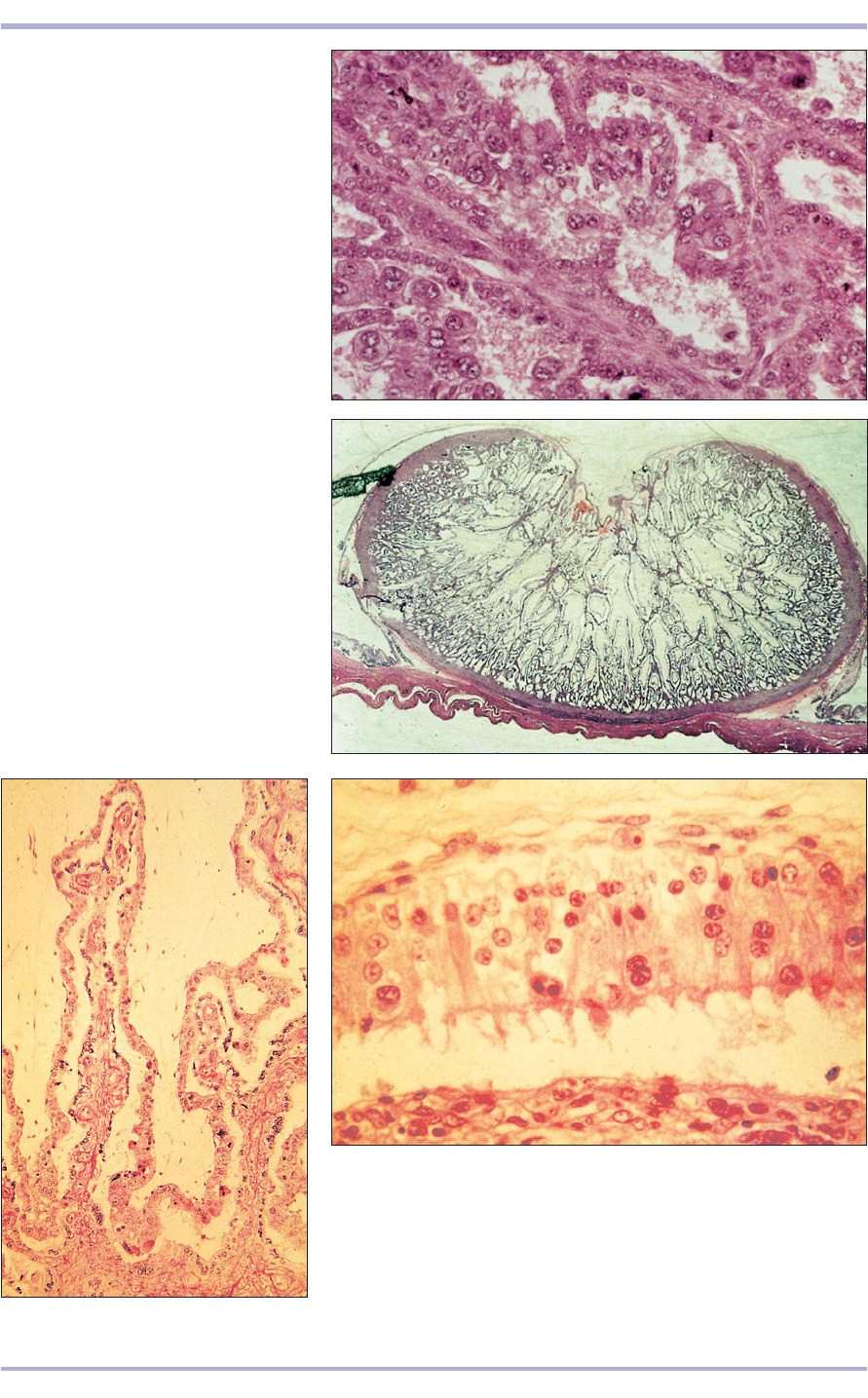
12.55 Epitheliochorial placenta (cow).
(1) Dark strands of maternal connective
tissue. (2) Maternal epithelium.
(3) Trophectoderm with binucleate giant
cells. (4) Mesenchyme. H & E. ×125.
12.55
12.56 Epitheliochorial placenta.
Placentome (sheep). Note the concave
appearance of the placentome. (1) The
chorioallantoic membrane projecting into
the caruncle. (2) Endometrium of the
caruncle surrounding the chorioallantois.
(3) Myometrium. (4) Intercaruncular area.
H & E. ×7.5.
12.56
12.57
12.58 Epitheliochorial placenta. Intercaruncular area (sheep).
(1) Absorptive trophectoderm with giant cells. (2) Uterine milk.
(3) Endometrium, note the apparent absence of epithelium. H & E. ×160.
12.57 Epitheliochorial placenta. Placentome (sheep). (1) Mesenchyme
of the chorioallantois. (2) Trophectoderm wth giant cells. (3) Maternal
epithelium. (4) Maternal connective tissue. H & E. ×62.5.
12.58
1
2
3
1
2
1
2
3
4
2
1
4
3
2
2
3
4
1
Female Reproductive System
Carnivores
Carnivores have an endotheliochorial, zonary decid-
uate placenta, and the following gestation periods:
bitch, 58–65 days; and queen, 60 days.
The trophoblast in the central zone of the chori-
ovitelline placenta becomes two-layered, with an
outer syncytial layer, the syncytiotrophoblast, and
an inner cellular layer, the cytotrophoblast. Cords
of trophoblast block the mouths of the uterine
glands and secrete lytic enzymes, destroying the
maternal tissue. With the development of the more
invasive chorioallantois, the maternal tissue is fur-
ther eroded and the trophoblast is apposed to the
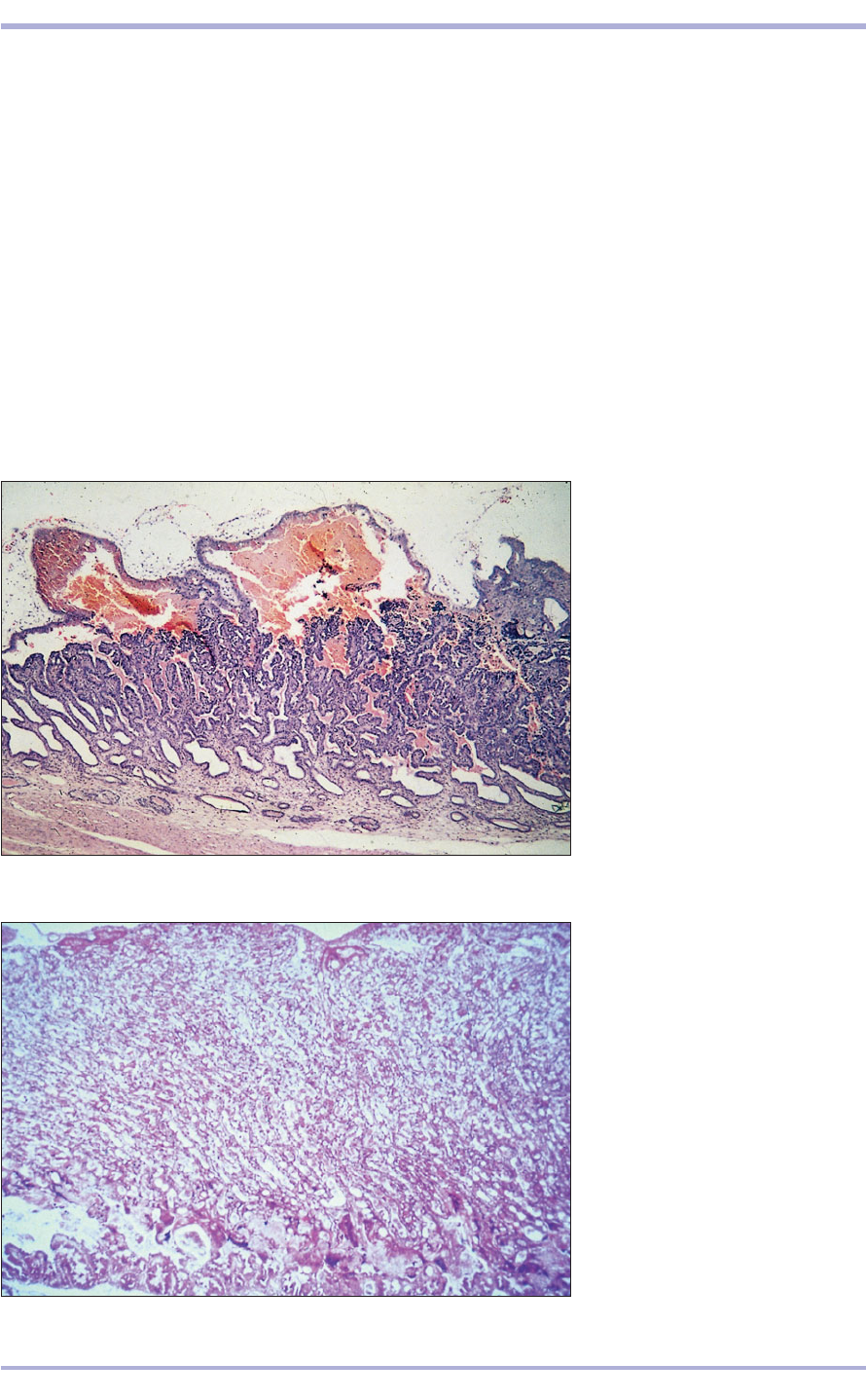
endothelium of the maternal blood vessels (12.59
and 12.60).
Decidual cells, probably fetal in origin, are also
present. Haematomata are present on the margin of
the zonary band (bitch) and centrally dispersed
(queen). These are pools of maternal blood sur-
rounded by absorptive trophoblast, where maternal
erythrocytes have been destroyed; these are the
haemophagous zones. A green deposit of uteroverdin
occurs in the bitch and a brown deposit in the queen.
Outside the zonary band there is simple apposition
of chorion and endometrium with no loss of mater-
nal tissue at parturition, as opposed to the zonary
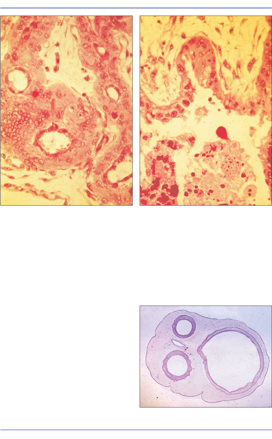
band where damage is considerable (12.61, 12.62).
202
Comparative Veterinary Histology with Clinical Correlates
12.59 Endotheliochorial placenta.
Cat placental band. (1) Absorptive
trophectoderm. (2) Haematoma,
pools of maternal blood.
(3) Interlocking zone of
fetal/maternal tissue.
(4) Endometrium. (5) Myometrium.
H & E. 7.5.
12.59
12.60 Endotheliochorial placenta.
Placental band in the bitch. The
lamellae are more regular than in the
cat. (1) Chorioallantoic membrane.
(2) Junctional zone marks the deep
penetration of fetal tissue. H & E. ×25.
12.60
1
2
1
2
4
5
3
Umbilical cord
The umbilical cord is the communications link
between the fetus and the placenta. The umbilical
arteries leave the fetus and carry deoxygenated
203
Female Reproductive System
12.61 Endotheliochorial placenta (bitch).
(1) Chorioallantoic mesenchyme. (2) Trophectoderm.
(3) Decidual cell. (4) Maternal blood vessels lined by
thickened endothelium. H & E. ×200.
12.61
12.62 Endotheliochorial placenta (bitch). This represents
the junctional zone, the limit of the fetal invasion.
(1) Chorioallantoic mesenchyme. (2) Trophectoderm.
(3) Maternal tissue debris. H & E. ×200.
12.62
12.63 Umbilical cord (foal). (1) Umbilical arteries.
(2) Umbilical vein. (3) Allantoic duct. H & E. ×7.5.
12.63
1
2
3
1
1
2
3
1
3
4
2
1
blood to the placenta. The umbilical veins carry
nutrient blood to the fetus (these commonly fuse to
form a single vein). The umbilical vesicle is the rem-
nant of the yolk sac and the small canal is the lumen
of the allantoic duct (12.63).
12.65
12.64
Clinical correlates
Mammary glands
Inflammation of the mammary glands, termed
mastitis, can affect any mammal and is most
common in the lactating mammary gland, where
the moist, nutrient-rich secretions provide an
ideal environment for growth of microorganisms.
A variety of mammary tumours are recognized
and several classification schemes exist that
divide the tumours according to the cell types and
patterns of growth present. For example, a mixed
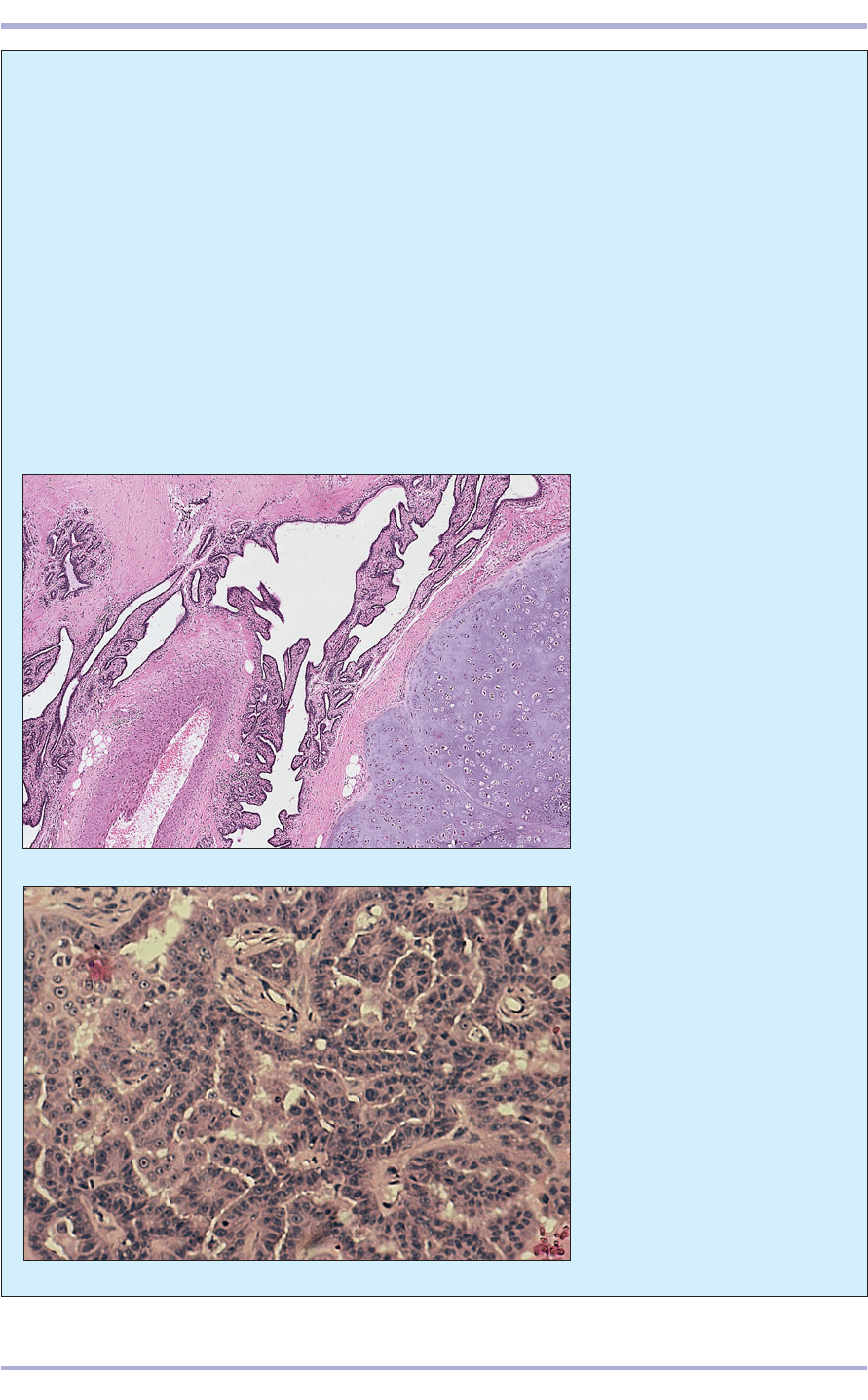
mammary tumour (12.64) is derived from more
than a single germ layer and includes epithelium,
myoepithelium, cartilage and possibly bone. In
the bitch, early ovariohysterectomy is known to
reduce significantly the incidence of future mam-
mary tumour development. True behavioural
malignancy is recognized in a minority of canine
mammary tumours, but is present in a higher
proportion of mammary tumours in the cat
(12.65, 12.66). Rats of both sexes have extensive
mammary-type glandular tissue and may present
with masses of this origin anywhere from the
neck to the inguinal region. Most are benign
fibroadenomas. Mammary tumours are also rec-
ognized in the rabbit (12.67).
204
Comparative Veterinary Histology with Clinical Correlates
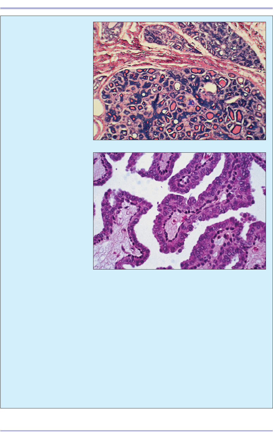
12.65 Feline mammary papillary
cystadenocarcinoma. The cells in
this malignant tumour form
multiple frond-like papillae that
extend into cystic spaces. H & E.
×125.
12.64 Mixed mammary tumour
from an elderly bitch. In addition to
glandular epithelial and stromal
components, cartilage (1) is
formed. The glandular lumina are
dilated and the epithelium is
arranged in a single layer. This
tumour is benign. H & E. ×62.5.
1
12.67
12.66
205
Female Reproductive System
12.66 Feline ductal
adenocarcinoma. The thin-walled
ductal structures are filled with
eosinophilic proteinaceous
secretion and are divided by
fibrocollagenous connective
tissue. Invasion into surrounding
tissue and lymphatic metastasis
are common with this type of
tumour. H & E. ×62.5.
12.67 Mammary adenoma from
an adult female rabbit. This high-
power view shows frond-like
papillary projections with fibrous
cores overlain by well-
differentiated columnar
epithelium. H & E. ×250.
Female reproductive system
Developmental disorders of the genital system
occur in all species of domestic animals, but are
uncommon. These disorders are caused by
abnormalities of genetic origin or by aberrant
hormonal influences. Often, precise mechanisms
have not been defined, but specific syndromes,
such as freemartinism in cattle, are recognized.
A freemartin is a genetically female calf twinned
with a male. If, as is common, anastomoses form
between the placental circulations, then factors
passed between the twins lead to abnormalities
in the female reproductive system, including
inhibition of ovarian development or testis-like
differentiation within the ovary and the absence
of parts of the tubular tract. Effects on the male
twin are minimal.
A spectrum of cystic changes are recognized
in the mammalian ovary. Cystic ovarian disease
in cows is important as a cause of reproductive
failure and hence economic loss. Tumours that
arise from tissues which are specifically ovarian
can be divided into three broad categories:
tumours of the surface coelomic epithelium,
those of the gonadal stroma and those of the
germ cells. Tumours of the surface epithelium are
significant only in the bitch as papillary and
12.69
12.68
cystic adenomas and rarely papillary adenocar-
cinomas. Tumours that arise from the gonadal
stroma include granulosa (12.68) and thecal
(12.69) cell tumours. These neoplasms can pro-
duce hormones and are rarely malignant in any
species. Germ cell tumours include dysgermino-
mas (12.70) and teratomas (12.71). The dysger-
minoma is morphologically similar to primordial
germ cells and resembles its testicular homo-
logue, the seminoma. In teratomas the totipo-
tential germ cells have undergone somatic
differentiation and a variety of tissues of differ-
ent germ lines are present within the tumour.
Obstructive and inflammatory conditions are
recognized in the uterine (Fallopian) tubes. A range
of inflammatory, hyperplastic or cystic changes
(12.72) that are under hormonal influence can
affect the uterus itself. Hyperplastic change, which
is usually focal within the uterus, does not appear
to be preneoplastic in domestic animal species, but
is an important precancerous indicator in humans.
Uterine neoplasia is uncommon in most domestic
species, although obviously many female domes-
tic animals are neutered. In the rabbit, however,
uterine adenocarcinoma occurs in a large per-
centage of adult females (12.73).
206
Comparative Veterinary Histology with Clinical Correlates
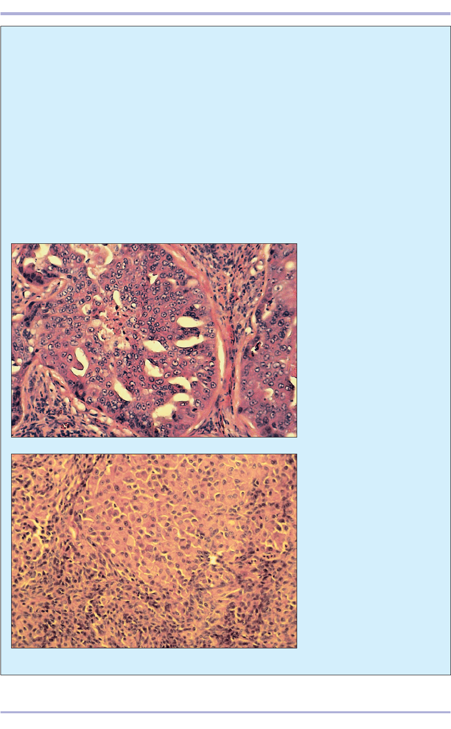
12.69 Canine ovarian thecoma in
a 12-year-old Springer Spaniel
bitch. The tumour cells are large
and polyhedral, contain finely
granular eosinophilic cytoplasm,
and are arranged in solid lobules
separated from each other by a
fine fibrovascular stroma. H & E.
×125.
12.68 Canine ovarian granulosa
cell tumour. This is the most
common gonadostromal tumour
in all species. Histological
appearances vary. This section
shows a lobular mass with clefting
between the tumour cells. H & E.
×125.
12.73
12.72
12.71
12.70
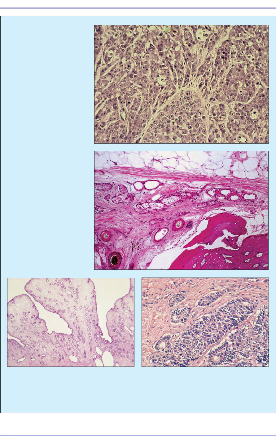
12.71 Teratoma from a 2-year-old
bitch. Several germ lines are
represented. Within this section,
(1) dense bone, (2) well-developed
hair follicles and (3) sebaceous
glands, and (4) adipose and
(5) collagenous connective tissue
can be seen. H & E. ×62.5.
207
Female Reproductive System
12.70 Canine ovarian
dysgerminoma. The tumour is
composed of lobular sheets of
large cells with central nuclei and
prominent nucleoli that resemble
the testicular seminoma. Giant
cells may be present. H & E. ×125.
12.72 Cystic endometrial hyperplasia in a 13-year-old
cat. Localized papillary outgrowths and cyst formation
are present within the uterine lining. Hydrometra or
mucometra may develop concurrently. Progestagen
administration is the most common cause of this
condition. H & E. ×44.
3
5
4
3
1
2
12.73 Uterine adenocarcinoma in a domestic rabbit.
The myometrium is infiltrated by intersecting cords of
neoplastic acini and distorted duct-like structures with
hollow lumens filled with pink staining proteinaceous
fluid. H & E. ×44.
Avian female
reproductive system
Ovary
In the majority of birds only the left ovary and
oviduct are functional. The ovary consists of an outer
cortex that envelops a vascular medulla. The cortex
is covered with cuboidal epithelium continuous with
the mesothelium of the peritoneum. The underlying
tunica albuginea is a thin layer of dense connective
tissue. The stroma is very vascular loose connective
tissue with sinusoidal blood vessels and follicles of
varying sizes, postovulatory follicles and atretic fol-
licles. Large follicles (the avian egg is megalethical or
large yolked) are suspended from the surface of the
ovary by stalks of cortical tissue (12.74 and 12.75).
Each follicle consists of a growing yolk-laden oocyte
with a rounded nucleus. The oocyte is surrounded
by several layers: the theca externa, theca interna,
membrana granulosa and perivitelline membrane
(12.76 and 12.77).
208
Comparative Veterinary Histology with Clinical Correlates
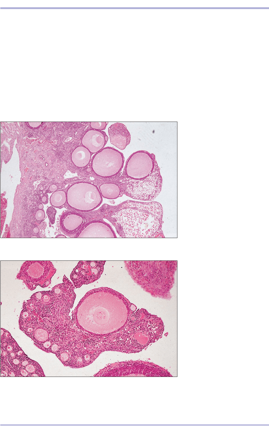
12.74 Ovary (bird). (1) The follicles
consist of the megalethic ovum
surrounded by a single layer of
granulosa cells. (2) Vascular ovarian
stroma. H & E. ×25.
12.74
12.75 Ovary (bird). (1) The range of
follicle sizes depends on the size of
the ovum. (2) Vascular stroma. H & E.
×62.5.
12.75
2
1
1
1
1
1
1
2
209
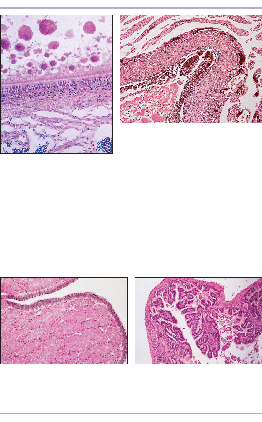
12.76 Ovary (bird). (1) Yolk granules. (2) Plasma
membrane of the egg. (3) Granulosa cells.
(4) Vascular ovarian stroma. H & E. ×200.
12.76
12.77 Ovary. Atretic follicle (bird). (1) Remnants of the yolk
filled ovum. (2) Disrupted granulosa layer. (3) Theca formed
from ovarian stroma. H & E. ×100.
12.77
Oviduct
In the domestic fowl the functional left oviduct con-
sists of five regions: infundibulum, magnum, isth-
mus, uterus or shell gland, and vagina. The wall of
the oviduct consists of a mucosa made up from
pseudostratified epithelium and a glandular lamina
propria. Longitudinal folds in the mucosa extend
spirally down the length of the oviduct but vary in
height and thickness. The muscularis is smooth
muscle with inner circular and outer longitudinal
layers increasing gradually in thickness. Loose con-
nective tissue forms the serosa.
The infundibulum engulfs the shed oocyte and,
after fertilization, lays down the first layer of albu-
men. The greatest proportion of albumen is pro-
duced by the next and longest part of the duct: the
magnum. The mucosal glands of the magnum are
lined with columnar cells packed with eosinophilic
granules before the arrival of an egg and depleted
after its passage (12.78 and 12.79). The shell mem-
branes are formed in the next short, narrow
12.78 Oviduct. Magnum (bird). (1) The lining epithelium is
pseudostratified columnar ciliated. (2) The lamina propria is
filled with simple tubular glands lined by columnar cells
packed with eosinophilic granules. H & E. ×100.
12.78
12.79 Oviduct. Magnum (bird). After passage of the egg,
the glands are empty and the deep mucosal folds project
into the lumen. H & E. ×20.
12.79
2
1
4
1
2
3
1
2
3
Female Reproductive System
region, the isthmus. Here the mucosal glands are
lined with poorly staining vacuolated cells (12.80)
that do not exhibit such marked secretory phases
as those seen in the magnum. The mucosal folds
of the isthmus are elongated and lie in leaf-shaped
folds. Once received by the ‘uterus’ or shell gland
(12.81), the egg remains in this region for about
20 hours, during which calcification of the shell
and formation of the cuticle take place (12.82 and
12.83). Watery fluid is also added to the albumen.
The uterine mucosa forms flat, leaf-shaped, longi-
tudinal folds. The epithelium is a continuous layer
of columnar cells with alternating basal and api-
cal nuclei, and these have been named basal and
apical cells. The basal cells have a restricted apical
surface; the apical cells are ciliated. The tubular
glands of the uterus are lined with cells that con-
tain pale staining granules both before and during
the phase of shell formation, but which are sub-
sequently depleted.
The vagina is short and narrow and has a well-
developed muscularis. Short simple tubular glands,
the sperm host glands, are found near the junction
of the vagina with the shell gland and lie within the
mucosal tissue (12.84). As their name suggests, their
function is to store sperm after insemination. The
mucosal folds are long and slender at this point and
bear short secondary folds.
The surface is lined with pseudostratified colum-
nar epithelium and mucous cells. The vagina opens
into the cloacal urodeum.
210
Comparative Veterinary Histology with Clinical Correlates
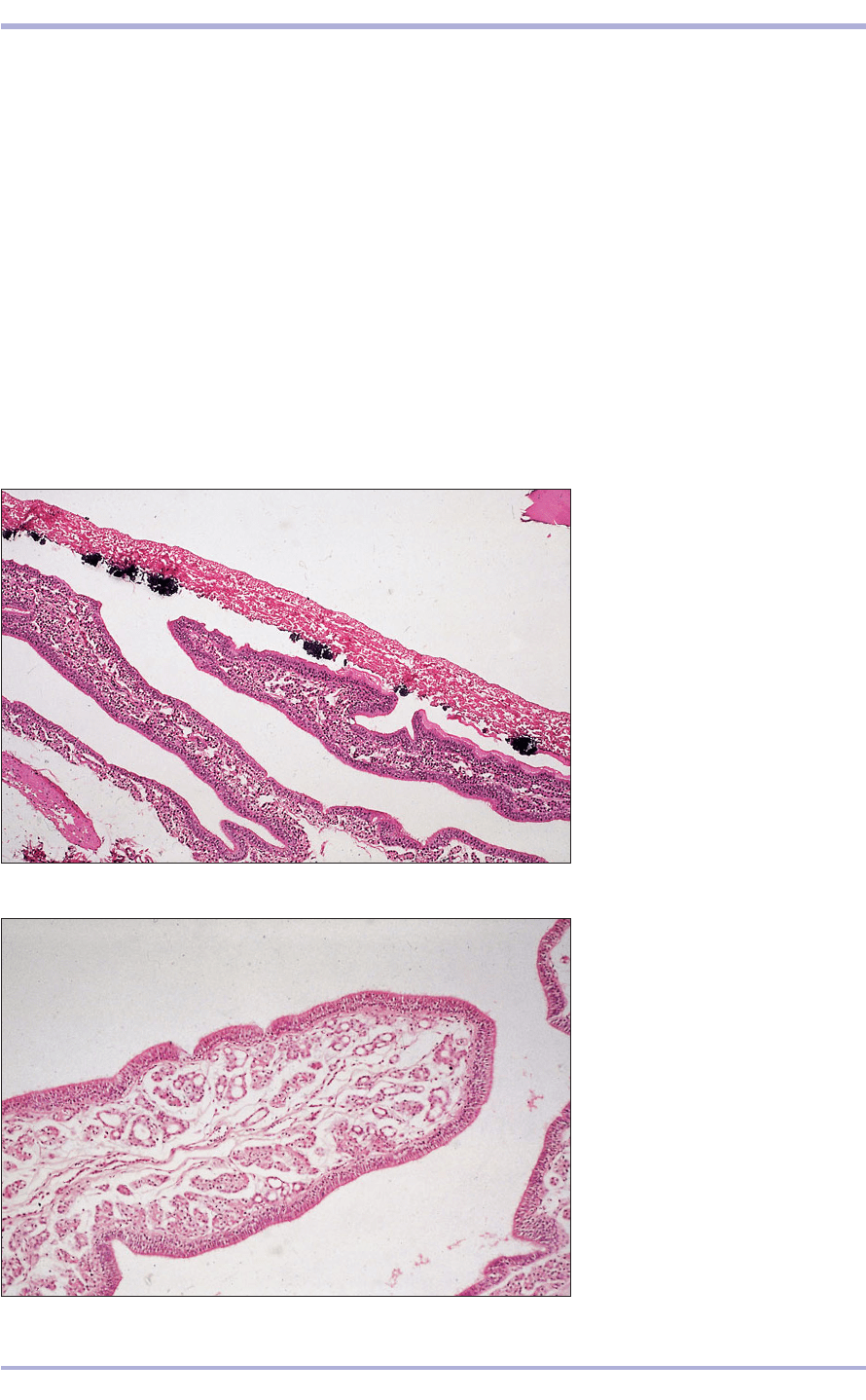
12.81 Oviduct. Shell gland/uterus
(bird). (1) The lining epithelium is
pseudostratified columnar with two
distinct rows of nuclei. (2) The
mucosal glands in the long folds
appear empty. H & E. ×62.5.
12.81
12.80 Oviduct. Shell gland/uterus
(bird). (1) Shell membranes, the dark
blue areas, are sites of calcification.
(2) The long mucosal folds lie parallel
to the developing shell. H & E. ×62.5.
12.80
1
2
2
2
2
1
