Aughey E., Frye F.L. Comparative veterinary histology with clinical correlates
Подождите немного. Документ загружается.

141
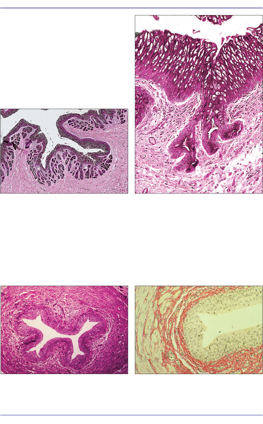
9.14 Ureter (dog). (1) Urethelium lines the lumen of the
ureter. (2) Vascular lamina propria. (3) Muscularis externa.
H & E. ×62.5.
9.14
9.15 Ureter (dog). The elastic fibres are stained reddish-
orange. Van Giesen. ×62.5.
9.15
Ureter
The ureter leaves the kidney at the pelvis and runs
to the bladder. The mucosa is formed into plait-
like longitudinal folds, and elastic fibres allow
stretching. The urethelium consists of at least five
to six cell layers, and in the horse simple tubu-
loalveolar mucous glands are present in the lam-
ina propria. There is no submucosa. The muscu-
laris externa is ill-defined with connective tissue
between the bundles of smooth muscle (9.14 and
9.15). The outer coat may be loose connective tis-
sue adventitia or a serosa, depending on the part
of the ureter that is examined.
2
1
3
Renal pelvis
This is the funnel-like dilatation at the cranial end
of the ureter. It is usually within the renal sinus, but
in certain conditions a large part may be extrarenal.
It is lined with urethelium resting on a loose con-
nective tissue lamina propria. In the horse there are
numerous mucus-secreting glands (9.12 and 9.13).
The muscularis is three ill-defined layers of smooth
muscle. The tunica adventitia is loose connective
tissue.
9.12 Kidney. Renal pelvis (horse). The renal pelvis is lined
by urethelium; simple mucus-secreting glands are present
in the lamina propria. H/PAS. ×62.5.
9.12
9.13 Ureter (horse). (1) Urethelium. (2) Simple mucus-
secreting tubular glands. (3) Vascular lamina propria.
H/PAS. ×250.
9.13
1
2
3
Urinary System
142
Urethra
The male urethra serves a genital function and is
discussed in Chapter 11. The female urethra is
short, running from the bladder to the external ure-
thral orifice, and has a purely urinary function. The
mucosa is folded longitudinally and the epithelium
varies from urethelium at the bladder to stratified
squamous at the urethral orifice. (See 11.14–11.17
and 12.26.)
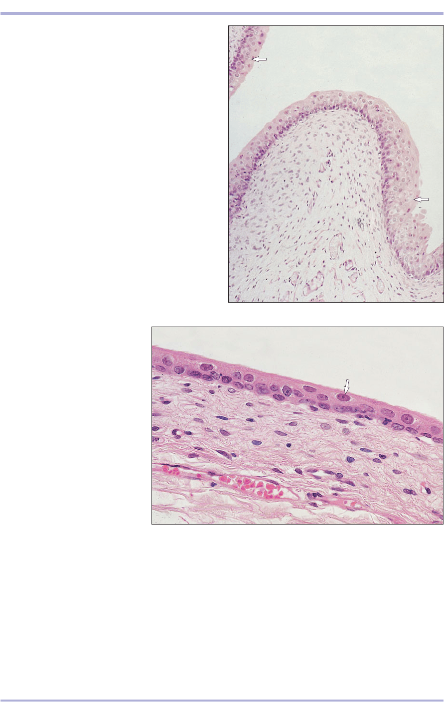
9.16 Urinary bladder (dog). The bladder is relaxed and
the surface cells are rounded adjacent to the lumen
(arrowed). H & E. ×125.
9.16
9.17 Urinary bladder (dog). The
bladder is stretched and the surface
cells are flattened (arrowed). H & E.
×250.
9.17
Urinary bladder
The urinary bladder is lined by urethelium, the num-
ber of cell layers depending upon whether the blad-
der is stretched or unstretched. There are elastic
fibres in the lamina propria. The muscularis is the
same as in the ureter, and the outer layer may simi-
larly be adventitia or serosa. Parasympathetic gan-
glia and nerve receptors are present (9.16 and 9.17).
Comparative Veterinary Histology with Clinical Correlates
143
Avian urinary system
Each kidney consists of three pyramidal divisions:
cranial, middle and caudal. These are not compa-
rable to the lobes of the mammalian kidney. Each
division receives a branch of the renal artery, a
branch of the great renal vein, and in the renal
portal vein this is a branch of the internal iliac.
The avian renal division is composed of a number
of indistinct lobes made up of lobules, the struc-
tural unit of the kidney. Lobules that drain into a
single branch of the ureter constitute a lobe. Each
lobule is pear-shaped; the wider part is cortical tis-
sue and the tapering part is medullary tissue. In
histological sections the appearance is of the larger
cortical areas surrounding cone-shaped islands of
medullary tissue, called medullary tracts. The
interlobular veins are wedged between the lobules.
There is no renal pelvis or urinary bladder.
There are two types of uriniferous tubules: the
mammalian (metanephric) tubule extends into the
medullary tissue and the shorter ‘reptilian’
(mesonephric), or cortical, tubule lacks a loop (of
Henle). Both types begin with a renal corpuscle.
The reptilian renal corpuscule is smaller than the
mammalian, but more numerous, and has a
prominent central mass of mesangial cells. The
PCT is about half of the tubule and is connected
by a short intermediate segment to the DCT. In
the medullary tubule the intermediate segment is
the loop descending into the medullary tissue. A
juxtaglomerular complex is present as in the
mammal. The DCTs are joined by collecting
tubules, lined with mucus-secreting cuboidal to
low columnar epithelium, to the perilobular col-
lecting ducts. These fuse with other ducts to form
larger ducts and lead to a secondary branch of the
ureter. Five or six secondary branches fuse to form
a primary ureteral branch (9.18 and 9.19).
The ureters are lined with a mucus-secreting,
pseudostratified epithelium supported by a cellu-
lar lamina propria with variable amounts of diffuse
lymphoid tissue. The thick muscularis consists of
an inner longitudinal and an outer circular layer of
smooth muscle (9.20). The ureter drains into the
middle compartment of the cloaca: the urodeum.
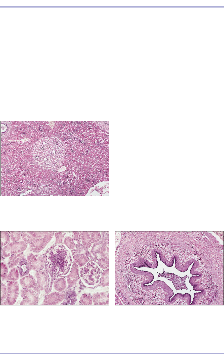
9.18 Kidney (bird). (1) The central, pale staining
medullary area is surrounded by (2) the much denser
staining cortical area. (3) Lobar duct. (4) Renal vein.
H & E. ×25.
9.18
9.19 Kidney cortex (bird). (1) The renal corpuscule has a
central mass of epithelial cells and mesangial cells.
(2) Proximal convoluted tubule. (3) Distal convoluted
tubule. (4) Small collecting tubule with low columnar
mucus-secreting epithelium. H & E. ×125.
9.19
9.20 Ureter (bird). (1) Pseudostratified mucus-secreting
epithelium lines the lumen. (2) Cellular lamina propria
with groups of lymphocytes (arrowed). (3) Muscularis
externa. H/PAS. ×125.
9.20
1
2
2
3
1
1
2
3
4
3
4
2
Urinary System
144
Reptilian urinary system
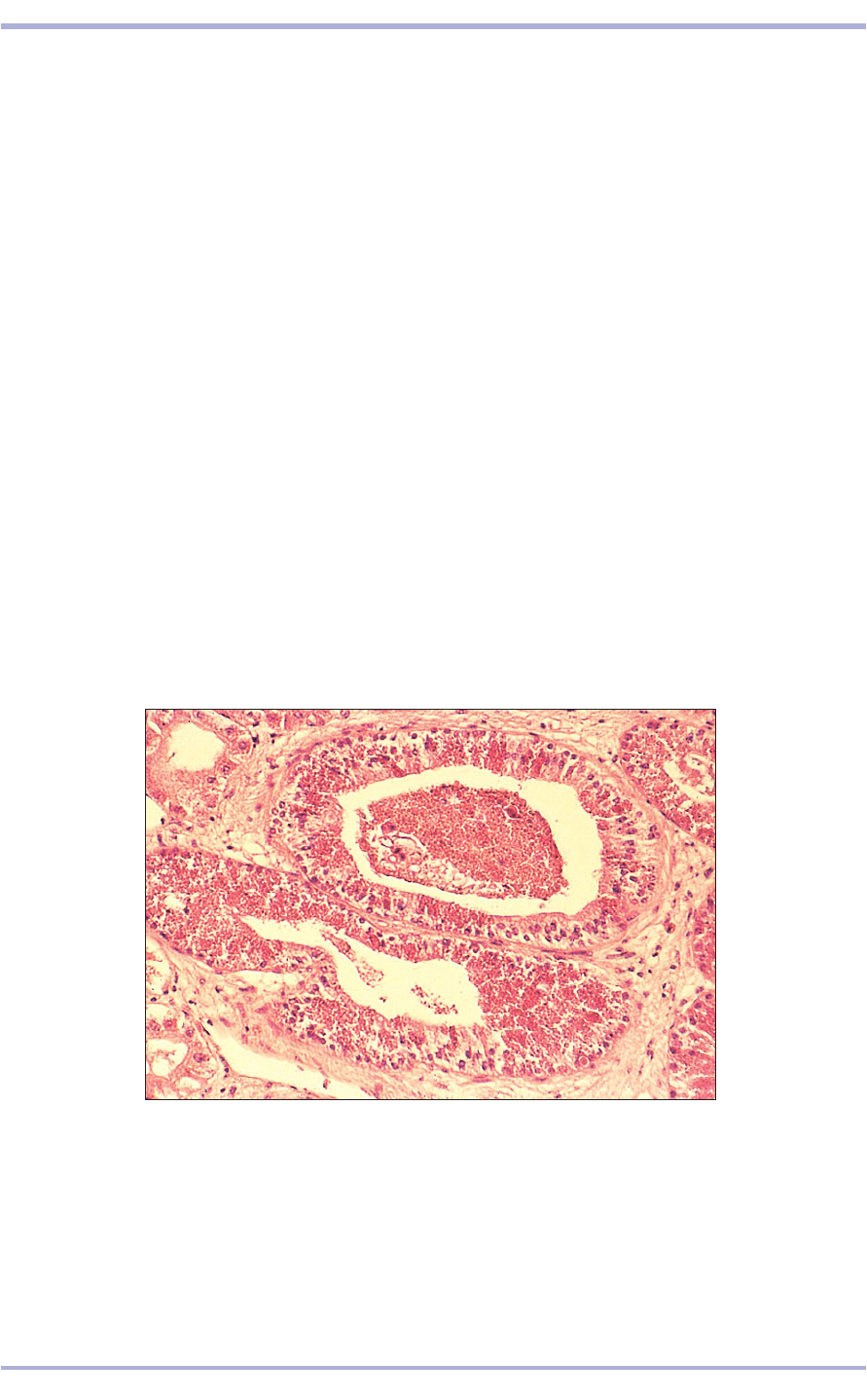
The renal tissues of reptiles are superficially very
similar to those of birds and mammals, but there
are some notable differences. In adult males of some
species of squamate reptiles (snakes and lizards)
there is an obvious and seasonal alteration in the
size, shape, staining characteristics and cytoplasmic
granularity of the cells comprising the DCTs. This
change is termed the ‘sexual segment’ and is seen in
sexually mature male crotalids (pit vipers), teiid and
some iguanid lizards, to name but a few (9.21). The
change is so striking that the sex of the animal from
which the renal tissue was obtained is immediately
apparent to the histologist. The epithelial cells of
the DCTs of mature males become markedly hyper-
trophied and become packed with small, round and
highly eosinophilic cytoplasmic granules that are
extruded into the urinary wastes. They are believed
to contribute a pheromone or similar chemical cue
to the urates that are passed during periods of sex-
ual courtship and mating activity. The sexual seg-
ment does not occur in females.
The histological characteristics of the amphibian
and reptilian ureter and urinary bladder are similar
to those of mammals. The lumen is lined by transi-
tional epithelium (urethelium) that is several layers
thick when the bladder is empty but becomes flatter
and thinner when the bladder is distended. There is
no submucosa; the bladder wall contains wispy
strands of smooth muscle. Not all reptiles possess
urinary bladders. Instead, the urinary wastes are
passed from the twin ureters into the urodeum por-
tion of the cloaca and thence out through the cloa-
cal vent. In some reptiles, the urine is retained for a
variable length of time, during which water is
actively removed and ‘recycled’ before the now con-
centrated, urate-rich wastes are discharged as a pasty
white, relatively water-insoluble microcrystalline
substance.
9.21 The kidneys of some species of sexually mature male snakes and lizards
possess a characteristic ‘sexual segment granulation’ which is discerned by a
marked hypertrophy and eosinophilic granularity of the distal convoluted
tubules. This makes the kidneys of sexually mature males readily
distinguishable from the kidneys of sexually mature females of the same
species. Illustrated is a section of kidney from a mature male timber
rattlesnake (Crotalus horridus). H & E. ×125.
9.21
Comparative Veterinary Histology with Clinical Correlates

145
Amphibian urinary
system
There is a substantial change in the renal histology
and function before, during and after metamorpho-
sis in most amphibians. The pronephric kidney of
early larval amphibians changes to become fully
metanephric in the adult stage of most amphibians.
However, in the caecilian amphibians, which are
elongated, legless creatures, the kidneys are
opisthonephric.
Some amphibians without tails (frogs and toads)
excrete most of their nitrogenous wastes as toxic
ammonia. Others excrete urea, yet others produce
urate salts of uric acid similar to those produced by
terrestrial reptiles. The mode of nitrogen excretion
is dictated mainly by the living habits; aquatic forms
tend to be more ammonotelic, whereas terrestrial
amphibians tend towards ureotelism and uricotelism.
In addition, season of the year and hydration affect
the means by which these animals excrete their
nitrogenous wastes.
The kidneys are one of the major sites of
haematopoiesis in larval amphibians. During and
after metamorphosis, the kidneys lose most of this
ability and, as a result, assume a more familiar his-
tological pattern consisting of nephrons that super-
ficially resemble those of reptilian and avian species
(9.21).
There is no loop interposed between the PCT
and DCT and, therefore, the degree of concentra-
tion of the glomerular filtrate is variably limited.
Fish urinary system
Most fish excrete toxic ammonia-rich urinary wastes
that are converted via nitrification by the bacteria
Nitrosomonas spp. to nitrite and then by
Nitrobacter spp. to nitrate, a less toxic ionic prod-
uct that can be metabolized by aquatic plants. Thus
the potential toxicity of water containing urinary
wastes is prevented by the action of these two essen-
tial microbial organisms and aquatic flora which, in
turn, yield oxygen via photosynthetic pathways.
The kidneys of fish are divided into a cranial
pronephric (head) kidney and caudal mesonephric
(tail) kidney. The cranial portion is the major site of
erythropoiesis. Erythropoietic tissue occupies the
interstitial spaces between adjacent glomeruli and
renal tubules. The histology of fish kidneys varies
widely between species and between marine and
freshwater fish: the kidneys of some marine teleosts
lack glomeruli. Structurally, the renal tissue of fresh-
water fish is readily recognizable as kidney at low
magnification, but the intervening erythropoietic
component may seem to be a cellular inflammatory
infiltrate to histologists unfamiliar with fish kidneys.
The caudal portion functions in conventional
renal manner as a site of proteinaceous waste fil-
tration and removal. The glomerulus is easily rec-
ognizable by the tuft of capillaries and its parietal
and visceral capsule. The renal tubules are com-
posed of cuboidal to low columnar epithelial cells,
similar to those seen in other vertebrates. In teleosts
the kidney also plays a role in osmoregulation of
sodium and chloride. The gills also participate in
osmoregulation.
Clinical correlates
In all animals the main role of the kidney is the
homeostatic control of extracellular fluid com-
position. This involves maintenance of normal
concentrations of salt and water in the body, con-
trol of acid–base balance and excretion of waste
products.
To function normally, the kidney requires ade-
quate perfusion with blood, sufficient functional
renal tissue and unimpeded urinary outflow.
Failure of kidney function can therefore be
related to inadequate perfusion (pre-renal), to
inadequate processing in the kidney (renal) or to
blockage of urinary outflow (post-renal).
Each of the four main contributing tissues in
Urinary System
9.22
Comparative Veterinary Histology with Clinical Correlates
the kidney, the blood vessels, glomeruli, tubules
and interstitium can be a primary target of dis-
ease.
In animals, renal disease is often subclinical.
Clinical disease may be divided into acute and
chronic renal failure. Acute renal failure involves
a sudden onset of oliguria or anuria and azo-
taemia and is often the result of acute glomeru-
lar, interstitial damage or acute tubular necrosis.
This form of renal disease is often reversible.
Once the kidney is fully developed, new
nephrons (functional units) are not produced and
chronic renal failure with progressive destruction
of functional tissue, regardless of initiating cause,
leads to a syndrome of salt and water imbalance,
acid–base disturbance and accumulation of
wastes. Chronic renal failure results in irre-
versible changes that produce shrunken, fibrosed
‘end-stage’ kidneys.
A wide variety of developmental, circulatory,
metabolic, inflammatory and neoplastic condi-
146
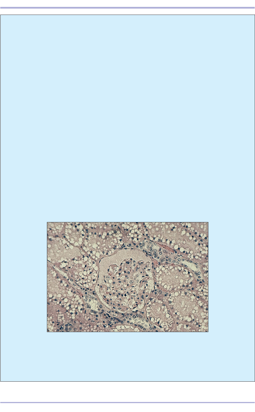
9.22 Feline infectious fibrinoperitonitis is caused by a feline coronavirus
and can produce an immune mediated glomerulonephritis with severe
proteinaemia. The glomerular space and tubular lumina contain
eosinophilic proteinaceous material. The numerous intracytoplasmic lipid
vacuoles seen in the tubular epithelial cells are normal in the feline kidney.
H & E. ×250.
tions can affect the kidneys. Familial nephropa-
thies are recognized in several dog breeds and
renal cysts are quite common in pigs and cattle.
Glomerulonephritis, often of immune origin, is a
common cause of chronic renal failure in both
dogs and cats (9.22–9.25). As glomerular dam-
age leads to significant protein loss this can result
in the development of nephrotic syndrome, char-
acterized by hypoalbuminaemia, generalized
oedema and hypercholesterolaemia. Primary
renal neoplasms are uncommon in domestic ani-
mals, with renal carcinoma the most commonly
recognized tumour in dogs, sheep and cattle,
while in pigs, nephroblastomas (true embryonal
tumours which arise in primitive nephrogenic tis-
sue) are more frequently seen, especially in
younger animals.
Renal tumours are common in salmonid fish
and some amphibians, particularly leopard
frogs (Rana pipiens) in which a specific virally
induced adenocarcinoma (of Lucke) is found.
9.24
9.23
9.25
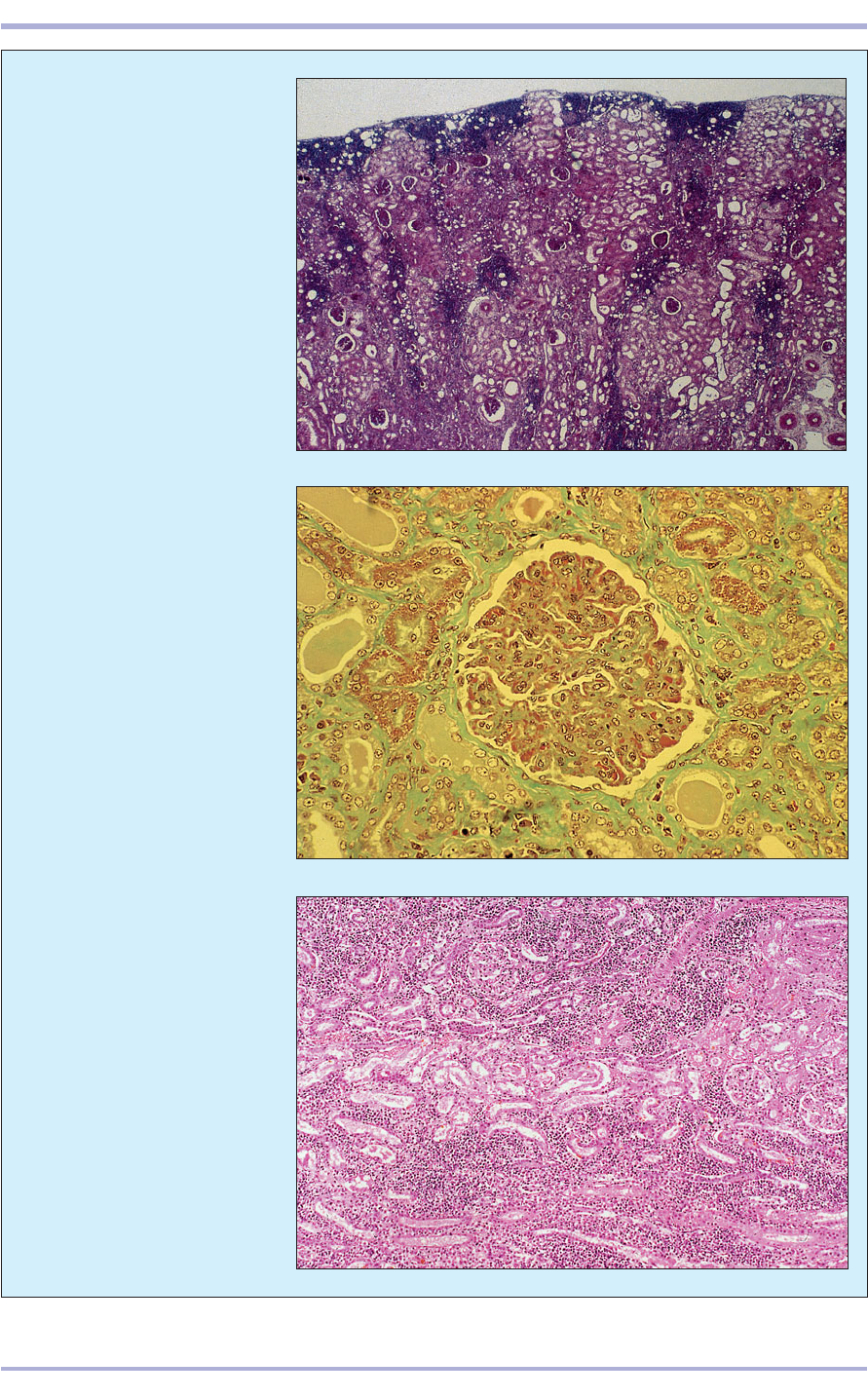
9.24 Chronic glomerulonephritis
(dog). This high power micrograph
shows thickening of the
glomerular capillary loops,
fine interstitial fibrosis and
accumulation of proteinaceous
fluid in the tubules. This disease `
is usually of immune origin and
is a common precursor to the
‘end-stage’ kidney of renal failure.
Masson’s Trichrome & Orange
G. ×250.
Urinary System
147
9.25 Acute interstitial nephritis
(dog). This micrograph is of a
kidney section from a dog with
acute interstitial nephritis. Large
numbers of inflammatory cells,
mostly lymphocytes, are present
between the tubules and around
glomeruli. The disease in this dog
was caused by infection with
Leptospira canicola which can
cause death through renal failure.
H&E. ×62.5.
9.23 Severe chronic interstitial
nephritis in an adult male
neutered cat. A dense multifocal
accumulation of mononuclear,
mostly lymphoid, inflammatory
cells is present in the interstitial
tissue, especially in the
subcapsular regions. The capsular
outline is irregular and there is
interstitial fibrosis with distortion
of the tubules. H & E. ×25.
9.26
9.27
9.29
9.28
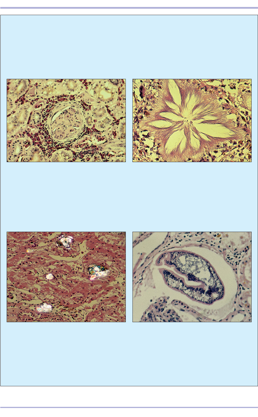
9.28 Cholesterol nephrosis. Illustrated is a section
of kidney from a Galapagos tortoise (Geochelone
elephantopus), which displays multifocal deposits
of cholesterol crystals that are obstructing some
glomeruli and renal tubules. This condition is seen
in captive herbivorous reptiles that have been fed
abnormal diets such as commercial dog and cat
food. H & E, photographed with cross-polarized
illumination. ×48.5.
Comparative Veterinary Histology with Clinical Correlates
148
9.29 Renal trematodiasis. The renal pelvis of this
Argentine horned frog (Ceratophrys ornata) is dilated
and contains a large fluke. H & E. ×98.
9.26 Purulent interstitial glomerulonephritis in a
kingsnake (Lampropeltis getulus). The glomerular
capsule and glomerular tuft are thickened. The renal
interstitial connective tissue is infiltrated by heterophil
granulocytic and mixed small mononuclear leucocytes.
H & E. ×180.
9.27 Renal gout. Illustrated is a section from the
kidney of a boa constrictor that had been treated
with several aminoglycoside antibiotic drugs without
receiving adequate parenteral fluid therapy. A large
‘star-burst’-shaped tophus has replaced the glomerulus
and the periglomerular interstitial connective tissue is
infiltrated by mixed small mononuclear leucocytes.
H & E. ×180.
Histopathologically, similar renal tumours
have been described in the Argentine horned
frog (Ceratophrys ornata). Renal tubular ade-
noma and carcinoma are relatively frequently
recognized in captive snakes and lizards.
Purulent interstitial glomerulonephritis in a
kingsnake (9.26), renal gout in a boa constrictor
(9.27), cholesterol nephrosis in a Galapagos tor-
toise (9.28) and renal trematodiasis in an Argen-
tine horned frog (9.29) are shown.

10.1
Endocrine tissue
Endocrine tissue is derived from epithelioid
parenchymal cells and may form discrete glands,
such as hypophysis cerebri (pituitary), thyroid,
parathyroid, adrenal and epiphysis cerebri (pineal).
Groups of endocrine cells are also active in the inter-
stitial cells of the testes, the granulosa and luteal
cells of the ovary, the pancreatic islets (of
Langerhans) which are responsible for insulin pro-
duction and release (see 8.92), the juxtaglomeru-
lar apparatus of the kidney and individual
amine–precursor–uptake–decarboxylation (APUD)
cells acting locally (paracrine). Endocrine cells
secrete chemical messengers called hormones
directly into a blood or a lymphatic vessel or tis-
sue fluid to influence the activity of target organs.
Endocrine tissue is characteristically ductless.
149
10. ENDOCRINE SYSTEM
Clinical correlates
Diabetes mellitus, one of the most common
endocrinopathies of the dog, may be asssociated
with degenerative change and fibrosis in the pan-
creas (10.1). However, the diagnosis of this con-
10.1 Diabetus mellitus (dog). The section of pancreas shown here was
taken from an 11-year-old, neutered female English Springer Spaniel with
a 2 year history of diabetus mellitus. There are pale eosinophilic areas of
replacement fibrosis and the remaining epithelial cells of the exocrine
pancreas, with their dark basal nuclei and strongly eosinophilic cytoplasm,
can be seen. No islet tissue is discernible. H & E. ×62.5.
dition rests on clinical testing rather than biopsy,
as the pancreas of non-diabetic elderly dogs may
appear similar and, conversely, some diabetic
dogs will have a histologically normal pancreas.
Hypophysis cerebri
(pituitary gland)
The hypophysis cerebri consists of a glandular lobe,
the adenohypophysis, and a fibrous lobe, the neu-
rohypophysis (10.2 and 10.3).
Adenohypophysis
The adenohypophysis is derived from an outpocket-
ing of the ectoderm of the dorsal portion of the oral
cavity, the hypophyseal (Rathke’s) pouch. It has three
sub-divisions: the pars distalis, the pars intermedia
and the pars tuberalis.
Pars distalis
The pars distalis is the major constituent of the glan-
dular pituitary. The dense connective tissue capsule is
continuous with a fine network of reticular fibres sup-
porting the cords and clusters of parenchymal cells
and the capillary/sinusoidal blood vessels. There are
150
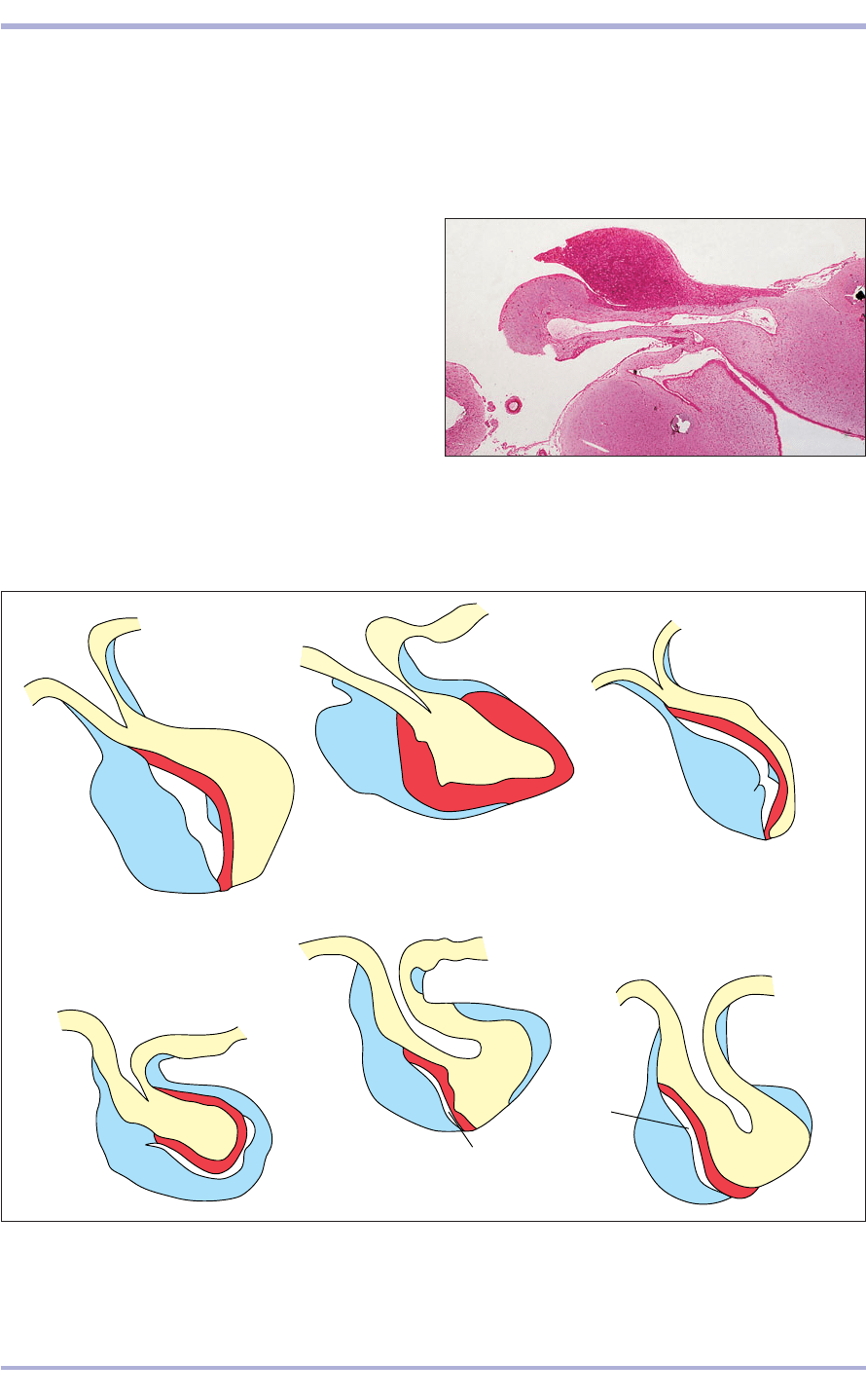
10.3 Diagram of the hypophysis cerebri (pituitary) in some domestic species. (1) Pars distalis. (2) Pars intermedia.
(3) Pars nervosa. (4) Pars tuberalis. (5) Hypophyseal cleft (residual lumen of Rathke’s pouch). (6) Infundibular recess
(third ventricle). (7) Infundibular stalk.
10.3
6
6
7
7
7
7
7
7
7
7
7
7
7
7
6
6
6
6
4
4
4
4
4
4
4
4
4
1
1
2
2
3
3
3
3
3
5
5
5
5
5
2
2
2
2
1
1
1
1
1
1
1
4
4
Horse
Pig
Cat
Dog
Small Ruminant (sheep)
Large Ruminant (ox)
3
10.2 Hypophysis cerebri (cat). (1) Pars distalis. (2) Pars
intermedia. (3) Pars nervosa. (4) Pars tuberalis.
(5) Residual lumen (Rathke’s pouch). (6) Infundibular
recess (third ventricle). (7) Infundibular stalk. H & E. ×5.
10.2
3
6
7
4
1
2
5
two broad categories of cells based on staining affin-
ity: the chromophobes and the chromophils
(10.4–10.8). The chromophobe cytoplasm has a few
granules that are non-reactive to dyes. These may be
reserve cells or exhausted degranulated cells. The
Comparative Veterinary Histology with Clinical Correlates
