Aughey E., Frye F.L. Comparative veterinary histology with clinical correlates
Подождите немного. Документ загружается.

111
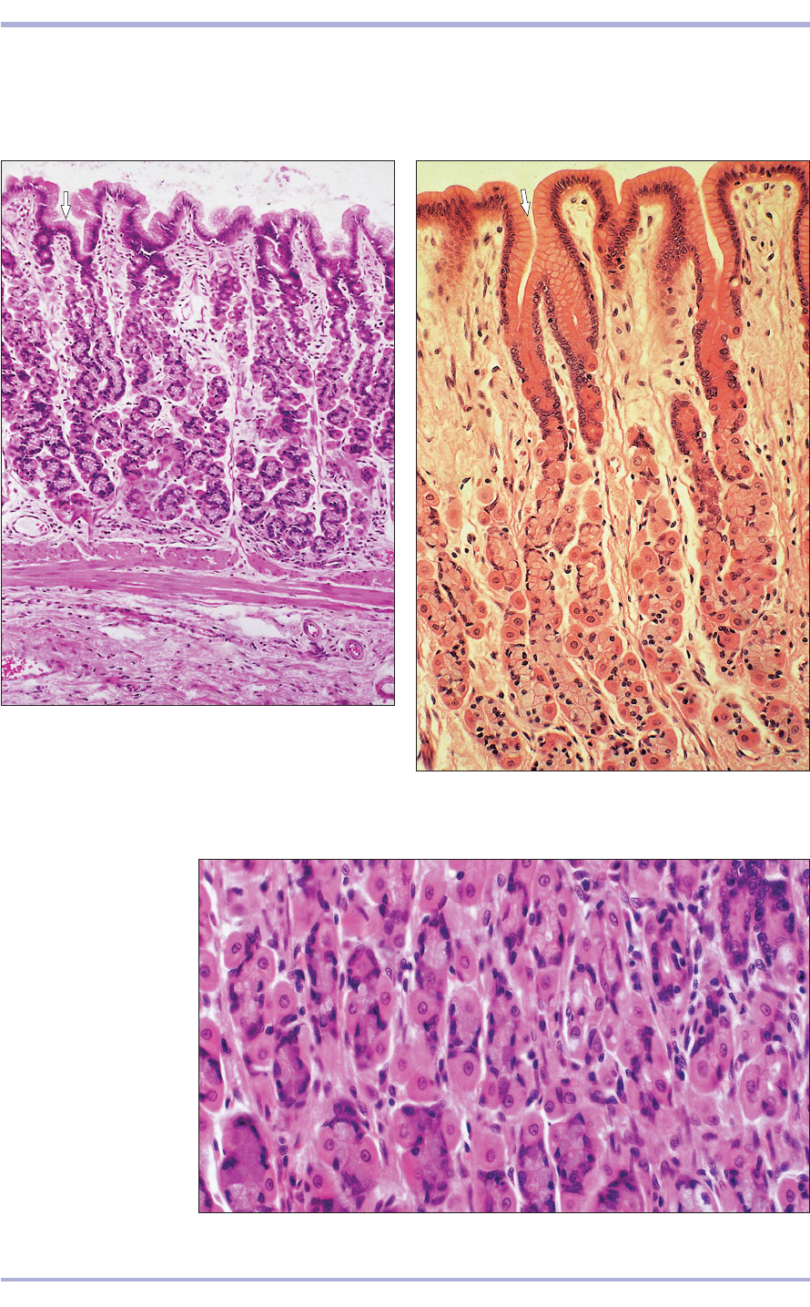
8.42 Fundic gland.
Stomach (cat). (1) Parietal
cell. (2) Zymogen cell.
H & E. ×250.
8.42
8.40 Fundic glands. Stomach (dog). Simple columnar
mucus-secreting epithelium extends into the gastric pits
(arrowed). (1) Parietal cell. (2) Zymogen cell.
(3) Muscularis mucosae. (4) Submucosa. H & E. ×125.
8.40
8.41 Fundic glands. Stomach (dog). Simple columnar
mucus-secreting epithelium extends into the gastric pits
(arrowed). (1) Parietal cell. (2) Zymogen cell. H & E. ×160.
8.41
1
2
1
2
1
1
2
3
4
recognized: the cardia, the fundus and the pylorus. Glands are sparse
with few cells in the cardia, but are abundant and cellular in the fun-
dus (8.37–8.42).
Digestive System
There are five cell types in the gastric glands:
• Stem cells at the neck of the gland divide and
replace the surface epithelium.
• Mucous neck cells at the neck of the gland secrete
mucus.
• Parietal (oxyntic) cells are large polyhedral cells
with a central nucleus and eosinophilic cytoplasm.
They secrete hydrochloric acid into canaliculi and
elaborate invaginations of the plasma membrane.
• Chief (zymogenic, peptic) cells secrete the enzyme
pepsinogen; this is converted into pepsin by the
gastric acid. In common with all enzyme-secret-
ing cells, the rounded basal nucleus is surrounded
with basophilic cytoplasm (rough endoplasmic
reticulum). The apical cytoplasm is eosinophilic
(stored secretory granules).
• Enteroendocrine cells are a diffuse population
that are identified with specialized silver stains
and are also known as argentaffin and argyrophil
cells. Chemical messengers (serotonin, gastrin,
somatostatin and enteroglucagon) are secreted
locally to control digestion. These cells are
regarded as part of the ‘amine–precursor–
uptake–decarboxylation’ (APUD) cell system,
which is characterized by the ability to take up
and process biogenic amines. However, not all
of these cells process amines and the term ‘dif-
fuse neuroendocrine system’ is more accurate.
The pyloric glands are mucus secreting (8.43 and
8.44).
The lamina propria is loose cellular connective
tissue with lymphatic cells present as a local popu-
lation and part of the gut-associated lymphoid tis-
sue (GALT). The muscularis mucosae is composed
of several layers of smooth muscle fibres. The sub-
mucosa is aglandular loose connective tissue with
parasympathetic nerve plexi (8.37–8.44). The mus-
cularis externa consists of three layers of smooth
muscle: oblique, circular and longitudinal. The
myenteric parasympathetic nerve plexus (Meiss-
ner’s) lies between the muscle layers (8.45). The
outer layer, the serosa, is vascular connective tis-
sue covered with mesothelial cells continuous with
the visceral peritoneum.
112
Comparative Veterinary Histology with Clinical Correlates
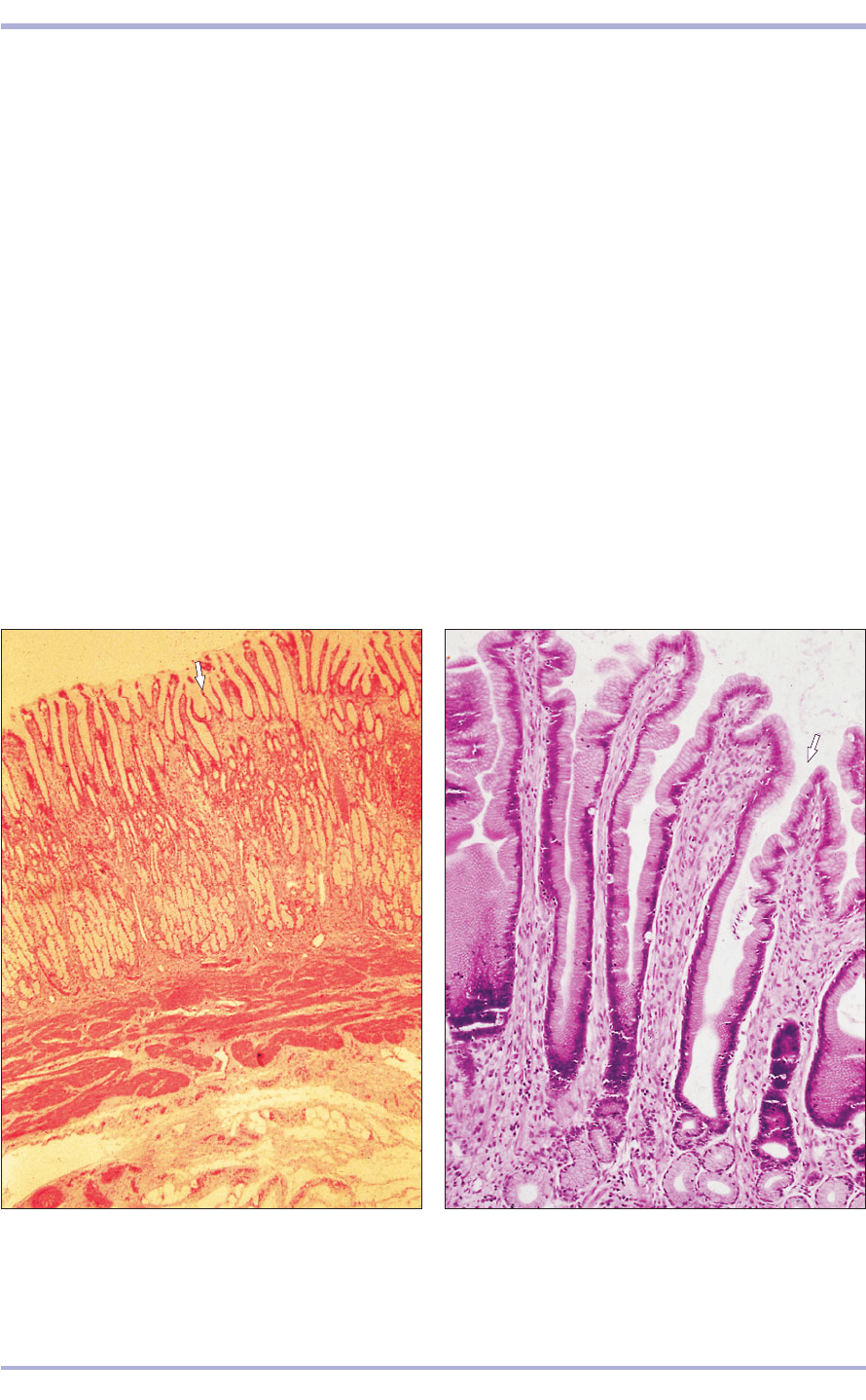
8.43 Pyloric glands. Stomach (pig). Simple columnar
mucus-secreting epithelium extends into the gastric pits
(arrowed). (1) Simple tubular mucus-secreting glands.
(2) Muscularis mucosae. (3) Submucosa. H & E. ×25.
8.43
8.44 Pyloric glands. Stomach (dog). Simple columnar
mucus-secreting epithelium extends into the gastric pits
(arrowed). Simple columnar epithelium lines the gland
tubule seen here cut in transverse section. H & E. ×100.
8.44
1
1
2
3
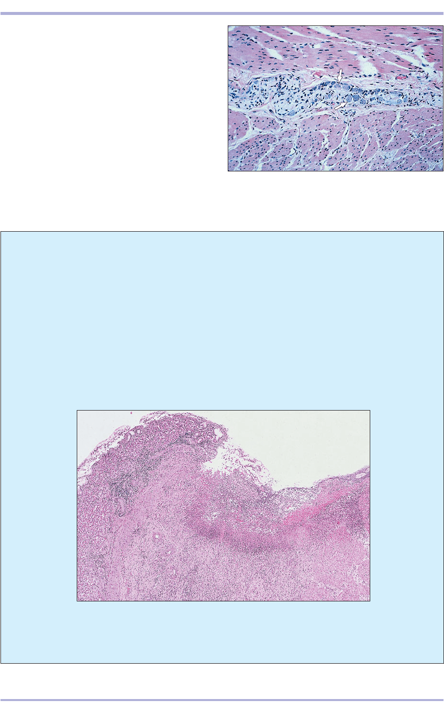
8.46 Peptic ulceration (dog). Loss of mucosal epithelium is seen, with
eosinophilic necrotic debris within the defect. Granulation tissue is
developing in the base of the ulcer. H & E. ×32.
113
8.45 Myenteric nerve plexus. Stomach (horse).
Parasympathetic neuron cell bodies (arrowed) lie in
the connective tissue between the smooth muscle layers.
H & E. ×125.
8.45
Clinical correlates
Gastric lesions may be associated with many
conditions which have signs that also affect
other body systems or other levels of the gas-
trointestinal tract. The aetiopathogenesis of
peptic ulceration (8.46) is incompletely under-
stood, but it results from an imbalance between
the damaging effects of gastric acid and pepsin
and the protective mechanisms of the gastric
mucosa. Administration of non-steroidal anti-
inflammatory drugs is known to predispose to
gastric ulceration by inhibiting prostaglandin
metabolism and damaging the gastric epithe-
lium. Systemic disturbances, such as endotox-
aemia or uraemia, may produce gastric lesions
and complex factors associated with stress can
also be implicated. Gastric ulceration can pro-
duce abdominal pain, vomition or haemateme-
sis, melaena and anaemia.
8.46
Digestive System
114
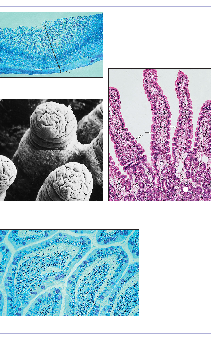
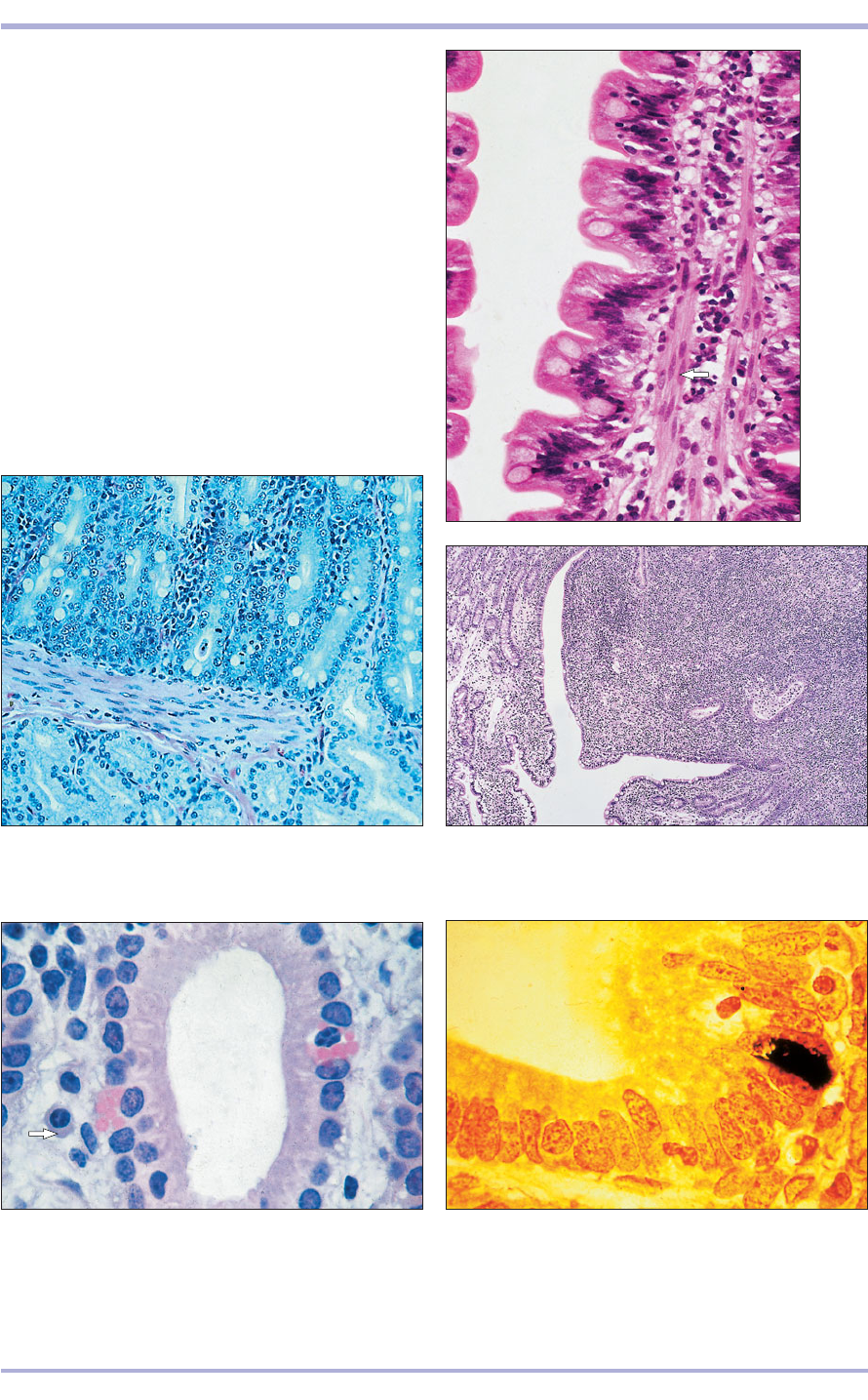
8.49 Duodenum (dog). (1) Villus covered by simple
columnar epithelium. (2) Lamina propria forms the
core of the villus; the contractile crypts are arrowed.
(3) Mucosal glands. H & E. ×62.5.
8.49
8.50 Duodenum (horse). (1) Goblet
cells in the epithelium are stained
deep pink. (2) Lamina propria.
Haematoxylin/PAS. ×125.
8.50
Small intestine
The small intestine consists of the duodenum,
jejunum and ileum (8.47–8.55). The function of the
mucosa is absorption. Finger-like projections of the
intestinal villi are long and thin in carnivores and
short and thick in ruminants. They increase the sur-
face area for absorption. The core of each villus is
1
2
3
1
1
1
2
2
2
8.47 Duodenum (dog). (1) Mucosa. (2) Submucosa.
(3) Muscularis externa. (4) Serosa. H & E. ×25.
8.47
1
2
3
4
8.48 Duodenal villi (dog). The finger-like villi project
into the lumen of the duodenum. Scanning electron
micrograph. ×100.
8.48
Comparative Veterinary Histology with Clinical Correlates
3
115
8.51 Duodenum (horse). (1) Simple columnar epithelium
with goblet cells. (2) Lamina propria with smooth muscle
fibres (arrowed). (3) Contractile crypts. H & E. ×250.
8.51
8.52 Duodenum (dog). (1) Mucosal glands. (2) Muscularis
mucosae. (3) Submucosal glands. H & E. ×160.
8.52
8.53 Duodenum (dog). The lamina propria is filled with
lymphatic tissue and lymphocytes are seen migrating
through the epithelium (a Peyer’s patch). H.& E. ×12.5.
8.53
8.54 Globular leucocyte (horse). The globular leucocyte
(function unknown) is in the epithelium of the intestinal
gland. A plasma cell is present in the lamina propria
(arrowed). H & E. ×500.
8.54
8.55 Enteroendocrine argentaffin cell (cat). Argentaffin
cell stains black in the intestinal gland epithelium.
Methanamine silver/safranin. ×500.
8.55
1
2
3
1
1
3
3
2
formed by the lamina propria, which is vascular, cel-
lular and reticular, with local aggregations of lym-
phoid cells. The tall columnar cells that line the
intestine have a striated border containing mucus-
secreting goblet cells; these increase in number
with distance from the stomach. At the bases of
the villi, the epithelium dips into the lamina pro-
pria to form mucosal intestinal glands (the crypts
of Lieberkühn). The cells lining the crypts are colum-
nar, secreting mucus, enzymes and local hormones,
and are the stem cells that are active in the repair
and replacement of the epithelium. Paneth cells,
Digestive System
which contain secretory granules that contain pep-
sidase, may also be present in horses and ruminants.
The muscularis mucosae consists of two layers of
smooth muscle, inner circular and outer longitudi-
nal, and separates the crypts from the underlying
mucosa. A strip of muscle extends into each villus
from the muscularis mucosae; a lacteal (lymphatic
that transports chyle) is also present. Indentations on
the villi, called contractile crypts, are created by the
contraction of the central strip of the muscle. Mucus-
and seromucus-secreting submucosal glands are
found in the horse in 6–7 m of the intestine, 3–5 m
in the pig, 4 m in the cow and 60–70 cm in sheep.
The muscularis externa consists of two layers of
smooth muscle dispersed in a gentle spiral, appearing
as an inner circular and outer longitudinal layer. As
in the stomach the myenteric parasympathetic nerve
plexus (Meissner’s) can be found between the layers.
The serosa consists of loose connective tissue,
and the mesothelium is continuous with the visceral
peritoneum.
Large intestine
There are no villi in the large intestine (caecum,
colon, rectum and rectal canal). Goblet cells are
abundant in the surface epithelium and in the
mucosal glands, which are simple regular tubules.
Lymphoid tissue is present in the lamina propria, as
are eosinophil leucocytes associated with parasitic
infestations. Lymphocytes are present in the epithe-
lium when immunoglobulin, which bathes the
epithelial cell surface as a defence against luminal
antigen, is released. There are no submucosal glands.
A muscularis mucosae is present, and the muscu-
laris externa consists of an inner circular and outer
longitudinal layer of smooth muscle. The outer layer,
or taenia coli, is arranged in bands and is charac-
teristic of the colon of the horse and pig. In the
horse, elastic fibres replace muscle fibres. The serosa
is continuous with the peritoneum (8.56 and 8.57).
The rectum is lined with simple columnar epithe-
lium. The mucosal glands decrease in number and
may disappear entirely as the anus is approached,
where there is an abrupt change to a stratified squa-
mous epithelium. The muscularis externa is thicker
here and becomes striated at the anal sphincter. Part
of the rectum is covered by a serosa and the rest by
adventitia. Tubuloacinar anal glands are present at
the cutaneous<rectal junction where they secrete
lipids in carnivores and mucus in the pig. In carni-
vores, circumanal, sebaceous-secreting glands are
found in the anal canals. Anal sacs, opened by small
tubular alveolar glands and lined with stratified squa-
mous epithelium, open into the perianal region (8.58).
GALT is part of the immune system. Both T and
B lymphocytes, as well as macrophages and
eosinophils, are present. The tissue may be so pro-
fuse that the enterocytes are stretched over a bulging
mass. Lymphocytes may migrate through the epithe-
lium (see 8.53).
116
Comparative Veterinary Histology with Clinical Correlates
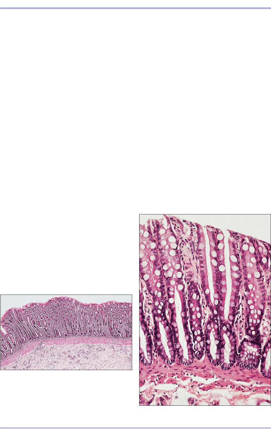
8.56 Colon (horse). (1) Mucosa. (2) Muscularis mucosae.
(3) Submucosa. H & E. ×9.7.
8.56
8.57 Colon (horse). (1) Simple columnar epithelium with
goblet cells. (2) Intestinal mucosal glands. H & E. ×125.
8.57
2
1
3
2
1
Alimentary system of
reptiles and amphibians
When amphibians metamorphose from larvae to
adults, significant changes take place in form and
function. Many larval amphibians are facultative
(or obligatory) herbivores, the alimentary tracts of
which are elongated and often tightly coiled (par-
ticularly in frog and toad larvae). During the latter
stages of metamorphosis, postlarval amphibians
usually cease eating and, therefore, must subsist on
their tails and other sources of readily catabolized
tissue. As adults, most amphibians are carnivorous.
The lingual apparatus of many amphibians and
reptiles is modified for the apprehension of prey:
some contain glandular acini that secrete sticky
mucus (8.59 and 8.60); others are characterized by
numerous papillary projections at the lingual tip to
which food particles stick and are then brought into
the mouth. When not being used, the tongue of
snakes retracts into a lingual sheath that is lined by
117
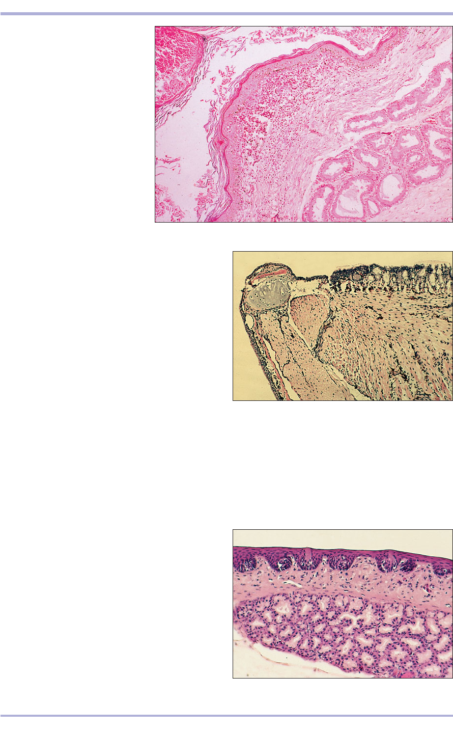
8.58 Anal sac (dog). (1) Stratified
squamous keratinized epithelium
lines the sac. (2) Tubuloalveolar
glands in the lamina propria.
H & E. ×12.5.
8.58
8.59 Tongue of a poison-arrow frog (Dendrobates spp.).
The dorsal lingual surface (1) is covered by an unusual
and complex epithelium composed of small, dark-staining
cuboidal cells and acini of sticky mucin-secreting
columnar cells that maintain a coating of adhesive mucus.
The muscle fibres (2) are primarily arranged in a
longitudinal direction and are attached at the front of
the mandible. This facilitates the tongue being rapidly
protruded and retracted in order to catch small
invertebrates. H & E. ×12.5.
8.59
1
1
2
2
8.60 The tongue of some lizards overlies a sublingual
salivary gland (1), as is illustrated by this longitudinal
section of the tongue of a small skink (Scincella lateralis).
The dorsal surface is covered by a non-keratinized
stratified squamous epithelium (2) in which cup-shaped
taste receptors are embedded. Some lizards (for example,
many iguanines) possess tongues with a terminal tip
composed of papillary projections that are kept moist and
sticky with mucus secreted by goblet cells and several
salivary glands. H & E. ×62.5.
8.60
2
1
Digestive System
mucus-secreting glands. It does not contain glands
but is lubricated when it comes into contact with
the luminal surface of the lingual sheath. The
tongues of other reptiles contain taste receptors that
are similar to taste buds found in the tongues of
mammals.
The dental histology of amphibians and reptiles
is similar to that in mammals, although the teeth of
these animals are periodically and continually shed
throughout life. Chelonians (turtles, tortoises and
terrapins) lack teeth entirely. Their premaxillae,
maxillae and mandibles are covered with hard and
horny keratinous surfaces, called ramithecae, with
which these animals cut their food items.
The salivary glands of amphibians and reptiles
are similar to those found in mammals. They may
be either entirely serous, entirely mucus-secreting
or a mixture of the two.
In venomous snakes and helodermatid lizards (the
Gila monster lizard, Heloderma suspectum, and the
Mexican beaded lizard, Heloderma horridum) some
salivary glands are greatly modified into structures
(see Chapter 2) that secrete extremely toxic secre-
tions that help these animals capture their prey and
defend themselves. In venomous snakes the secre-
tions from these glands are conducted to the hollow
needle-like fangs through coiled venom ducts. The
passage of venom through these ducts is aided by
the contraction of the temporal and masseter skele-
tal muscles that surround the glands and myoep-
ithelial cells that surround the ducts (see Chapter 2).
The fangs are replaced periodically throughout a
snake’s life. They are formed with a separate hollow
channel (8.61). Some nominally non-venomous
snakes, especially many colubrids, possess modified
salivary (Duvernoy’s) glands (8.62), the secretions
118
Comparative Veterinary Histology with Clinical Correlates
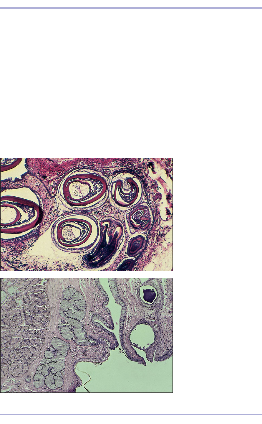
8.61 The fangs of venomous snakes
are continually being renewed.
Illustrated are several teeth
primordia of a juvenile rattlesnake
(Crotalus spp.), forming modified
fangs with a central enamel-lined
channel through which venom is
conducted. H & E. ×12.5.
8.61
8.62 Some non-venomous snakes
possess modified (Duvernoy’s)
maxillary and premaxillary salivary
glands connected to short ducts that
empty into the oral cavity. Current
studies indicate that the secretions
from some of these glands manifest
venom-like bioactivity on the lower
vertebrate prey of these snakes.
Also, mild clinical envenomation
of sensitive humans bitten by these
snakes has been reported. Illustrated
are two lobules of gland from a
watersnake (Natrix cyclopion).
H & E. ×62.5.
8.62
119
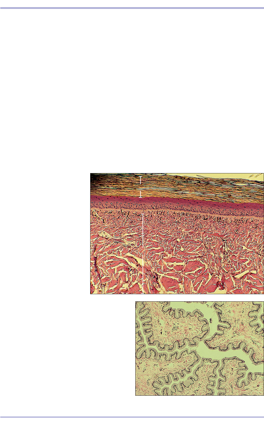
8.63 The oesophageal lumen of
many chelonians, such as this green
sea turtle (Chelonia mydas), is heavily
keratinized and lacks mucus-secreting
goblet cells. These characteristics
reflect the scabrous diet of these
marine animals. (1) Stratum corneum.
(2) Stratum lucidum. (3) Stratum
granulosum. (4) Stratum spinosum.
(5) Stratum basale. (6) Muscularis
externa. H & E. ×12.5.
8.63
8.64 The oesophagus of most snakes and many lizards is
characterized by its extensive plaiting which permits the
oesophagus to stretch to accommodate enormous prey.
Illustrated is a cross-section of the oesophagus of a
kingsnake (Lampropeltis triangulum). Because of the
necessity for abundant lubrication during the swallowing
of furry, feathered or scaly prey, the oesophageal lumen
is lined by a mucous epithelium composed of simple
non-keratinized columnar cells bearing basal nuclei.
H & E. ×62.5.
8.64
of which induce a toxic reaction when injected into
particularly sensitive prey and humans.
Generally, the alimentary system of the lower ver-
tebrates is similar to that found in mammals, but
major variations exist in species that are highly
adapted to a particular diet. Folivorous (leaf eating)
reptilian herbivores utilize hindgut rather than
foregut fermentation to accomplish the processing
of cellulose and other complex carbohydrates.
Modifications that aid in this process are an
expanded sacculated colon, which is similar in func-
tion to the sacculus rotundus of lagamorphs (rab-
bits and hares) and some herbivorous rodents, and
to the massive caecum and colon of equids. In all of
these organs, the surface of the luminal lining is aug-
mented by numerous mucosal villous projections,
which greatly increase the area available for micro-
bial digestion and nutrient absorption. Thus, the
sacculated colon of reptilian folivores serves the
same purpose as the large rumen complex of rumi-
nants, even though it is part of the hindgut rather
than the foregut.
The anterior alimentary tracts of various reptiles
are modified. The oropharynx and oesophagus of
some sea turtles have a heavily keratinized lining
(8.63) that helps to protect the lumen from trauma
when scabrous food items such as rocky and silica-
rich coral are swallowed. The egg-eating snake
(Dasypeltis scabra) ingests eggs with calcareous
shells. As the egg enters the cranial oesophagus, the
snake contracts its throat and thereby compresses
the egg against multiple horny ridges that extend
from the ventral region of the cervical vertebrae.
After the eggshell is slit, the snake swallows the fluid
and/or embryonic contents and regurgitates the shell
fragments en masse. Most snakes and many lizards
possess an oesophagus with walls formed into mul-
tiple longitudinal plaits that permit the swallow-
ing of enormous meals (8.64), many times the
diameter of their necks. Other reptiles, such as most
1
3
4
5
6
2
Digestive System
crocodilians, have thick-walled muscular stomachs
in which their prey are macerated with the aid of
ingested stones.
The gastric mucosa of reptiles is similar to that
found in mammals, except that only chief and clear
cells are present; parietal cells are lacking (8.65 and
8.66). The small intestine lacks Brünner’s glands (see
Chapter 3). The serosa covering most or all of the
coelomic viscera of many diurnal lizards is heavily
pigmented (8.67). Lymphoid patches or aggregates
are scattered throughout the length of the alimen-
tary tract. Discrete lymph nodes are not present.
Many lizards and some snakes possess salt-
secreting glands through which hyperosmolar solu-
tions containing sodium, potassium and chloride
ions are secreted. In many lizards these glands are
situated in the nasal cavity. Some sea snakes possess
sublingual salt glands. In some crocodilians, par-
ticularly crocodiles that inhabit salt marshes and
travel between oceanic islands, salt-secreting glands
are located on the dorsal surface of the tongue. All
of these aforementioned glands permit the non-
renal secretion of electrolytes without the appre-
ciable loss of water.
120
Comparative Veterinary Histology with Clinical Correlates
8.67 The colon of some lizards, particularly folivorous
species, is a highly modified sacculated organ divided into
multiple chambers that are functionally analogous to the
hindgut of lagamorphs and some (herbivorous/folivorous)
rodents, and the forestomachs of ruminants. Digestion is
enhanced because the villous surface of the colon is
covered by a highly absorptive columnar mucosa across
which nutrients processed from cellulose-digesting micro-
organisms are assimilated. The elongated villi that cover
the surface are supported, and stiffened, by thin cores of
smooth muscle. Illustrated is the sacculated colon of a
green iguana (Iguana iguana). H & E. ×12.5.
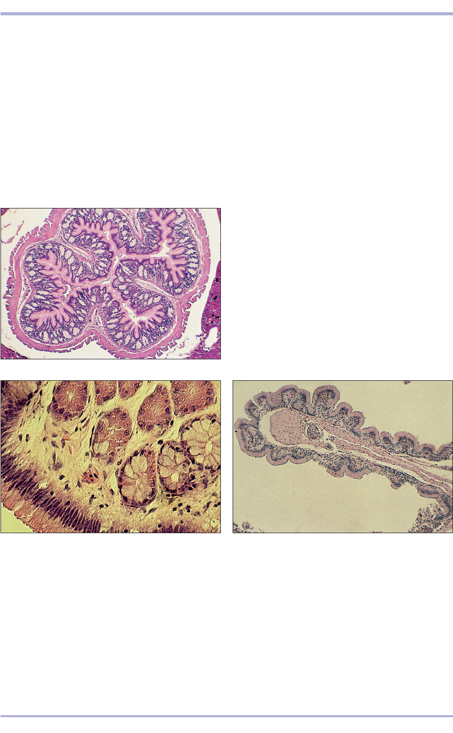
8.65 Whole mount cross-section of the fundic stomach of
a small skink (Scincella lateralis). A very thin serosa covers
the outermost visceral surface. The gastric wall is
composed of an outer external longitudinal muscularis
externa (1), a circular muscularis externa, the muscularis
mucosa), and immediately beneath is the glandular
mucosa (2) which is composed of pink staining granular
chief cells and clear cells. The lumen is lined by tall mucus-
secreting columnar cells. Parietal cells, present in
mammalian gastric mucosae, are lacking in amphibians
and reptiles. The outermost surface of the stomach is
covered by a delicate serosa (3) formed of non-keratinized
squamous cells. H & E. ×12.5.
8.65
8.66 Gastric mucosa of a boa constrictor (Boa
constrictor). The lumen is covered by tall columnar
epithelium. The gastric glands consist of only granular,
cuboidal, pink staining chief cells (1) with large vesicular
nuclei, and pale staining clear cells (2) whose nuclei are
dark and basal. Some gastric pits are lined by both cell
types. H & E. ×250.
8.66
8.67
2
2
1
1
3
