Aughey E., Frye F.L. Comparative veterinary histology with clinical correlates
Подождите немного. Документ загружается.

101
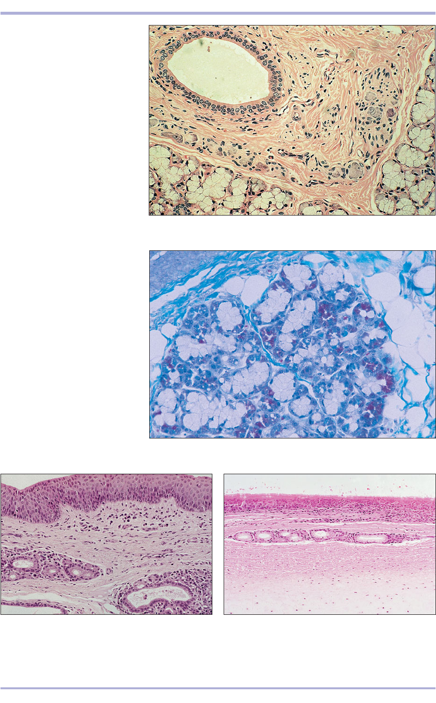
8.10 Mixed salivary gland (dog).
(1) Interlobular duct lined by a
stratified columnar epithelium.
(2) Connective tissue stroma.
(3) Parasympathetic ganglion.
(4) Seromucous acini. H & E. ×125.
8.10
8.11 Mixed salivary gland (dog).
(1) Pale staining triangular mucous
cells with a basal nucleus. (2) Deeply
staining serous cells with a round
nucleus. Gomori’s trichrome. ×125.
8.11
8.13 Soft palate (ox). (1) Respiratory epithelium.
(2) Lymph nodule. (3) Bone. H & E. ×62.5.
8.13
8.12 Hard palate (ox). (1) Stratified squamous
epithelium. (2) Lamina propria. (3) Mucus-secreting
glands in the lamina propria. H & E. ×125.
8.12
1
2
3
1
3
3
1
1
2
2
2
2
3
4
1
Digestive System
Teeth
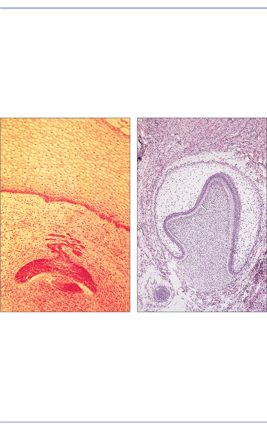
In the embryo teeth develop in the ectoderm as den-
tal papillae within the enamel organs (8.14). The
mesoderm invaginates each enamel organ into a bell
shape, with an inner enamel epithelium of
ameloblasts laying down enamel continuous with
the outer enamel epithelium and enclosing the stel-
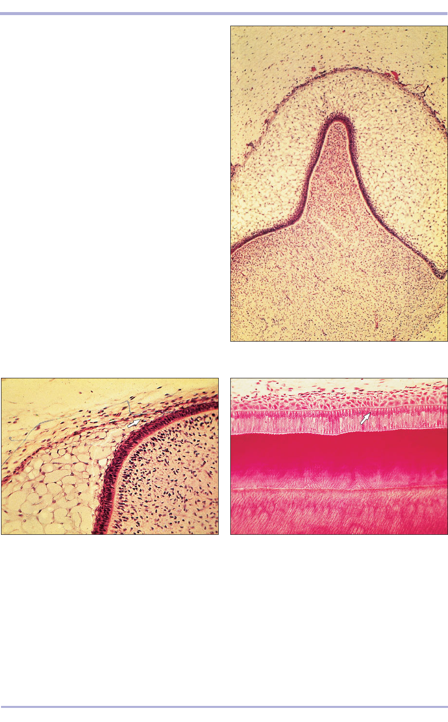
late reticulum (8.15). The mesenchymal cells of the
papilla differentiate to become odontoblasts, the
dentine-forming cells (8.16) and cementoblasts, and
secrete cementum in a similar pattern to that of
bone. Enamel and dentine are involved with the cre-
ation of the crown; dentine and cementum are
involved in the root. The root is formed by an
extension of the enamel organ at the junction of the
inner and outer enamel epithelium: the root tubule
(8.17 and 8.18). The tooth is held in the develop-
ing mandible and maxilla by the periodontal mem-
brane of collagen fibres embedded in the cementum.
Temporary teeth develop first. The permanent teeth
are secondary offshoots on the lingual side of the
102
Comparative Veterinary Histology with Clinical Correlates
8.14 Developing tooth (cat embryo). (1) Oral epithelium.
(2) Enamel organ surrounded by mesoderm. H & E. ×12.5.
8.14
8.15 Developing tooth (cat embryo). (1) Mesenchymal
papilla with a layer of odontoblasts (arrowed). (2) Inner
enamel epithelium, continuous with (3) the outer enamel
epithelium. (4) Stellate reticulum. H & E. ×62.5.
8.15
1
1
2
3
4
2
103
8.16 Developing tooth (cat embryo). (1) Mesenchymal
dental papilla. (2) Ameloblasts in the inner enamel
epithelium. (3) Outer enamel epithelium. (4) Stellate
reticulum. H & E. ×62.5.
8.16
8.17 Developing tooth (cat embryo). (1) Mesenchymal
dental papilla lined by odontoblasts. (2) Inner enamel
epithelium, ameloblasts, turns to become (3) outer
enamel epithelium at the point of root development
(arrowed). H & E. ×125.
8.17
3
1
2
1
2
3
4
8.18 Developing tooth (cat). The ameloblasts are
tall columnar cells with a basal nucleus (arrowed).
H & E. ×125.
8.18
Digestive System
104
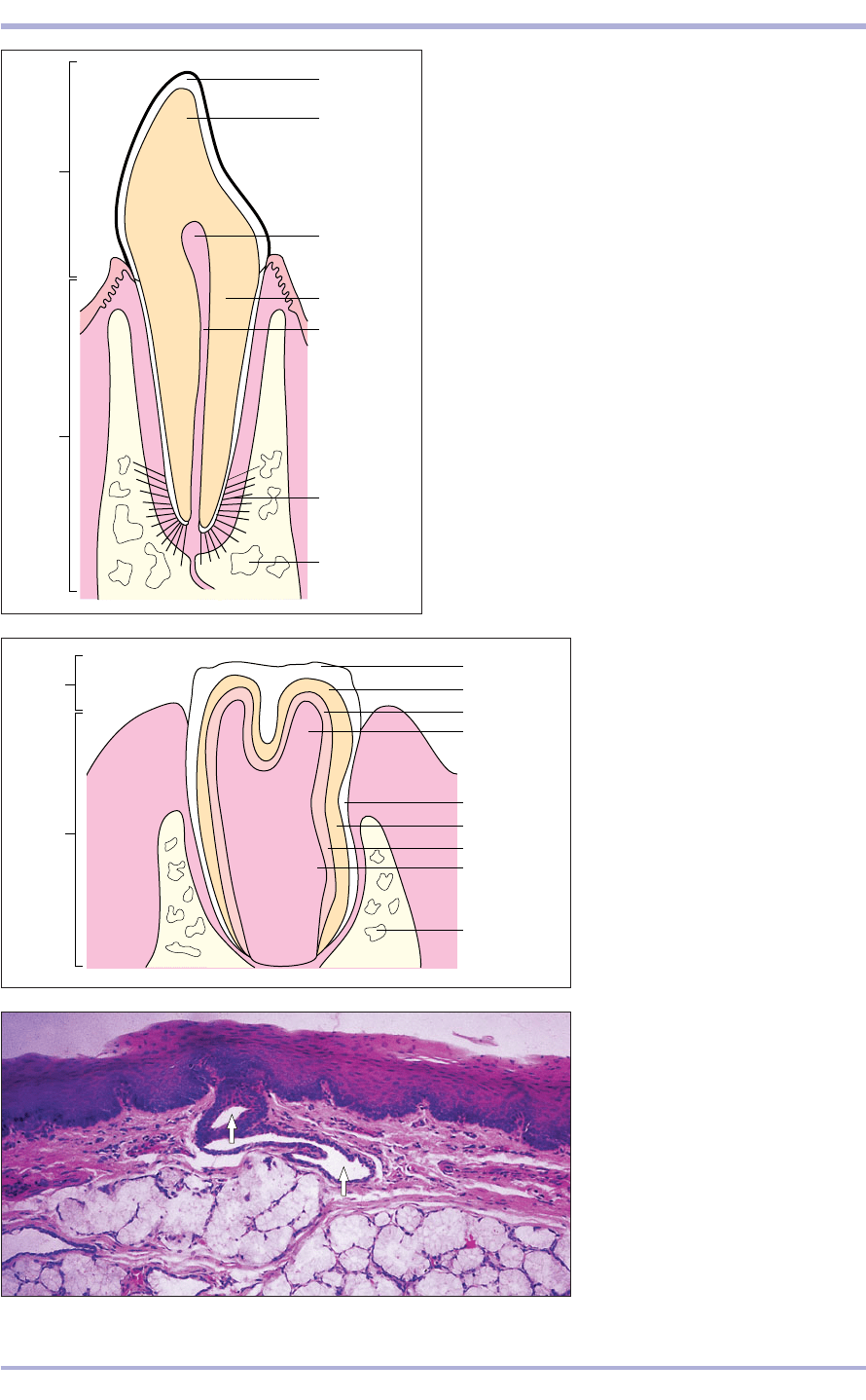
8.19 Diagram of an incisor.
8.19
Enamel
Dentine
Dentine
Pulp cavity
Pulp cavity
Periodontal
membrane
Alveolar
bone
Root
Crown
8.20 Diagram of a molar.
8.20
Enamel
Cementum
Dentine
Pulp cavity
Enamel
Cementum
Dentine
Pulp cavity
Maxillary/
mandibular
bone
Root
Crown
8.21 Oropharynx. (1) Stratified
squamous epithelium. (2) Mucus-
secreting glands open onto the
surface (arrowed). H & E. ×62.5.
8.21
temporary teeth. The tooth is divided into a crown
and a root (8.19). In the carnivore, teeth cease to
grow after eruption and the ameloblast layer is lost:
brachydont teeth. In the horse, ruminant and pig,
teeth are much longer and continue to grow for all
or part of adult life: hypsodont teeth. In these the
dental sac covers the whole of the tooth before
eruption and cementum covers the entire tooth, pre-
venting loss of the ameloblasts and allowing con-
tinuing deposition of enamel and cementum and
thus allowing for the wear and tear in these species
(8.20), which include rodents and lagomorphs.
Oropharynx
This is a short junctional area between the oral cav-
ity and the alimentary canal with some mucus-
secreting glands (8.21).
2
1
Comparative Veterinary Histology with Clinical Correlates
8.22
Alimentary canal
The alimentary canal (digestive tract) is a muscular
tube extending from the oropharynx to the anus,
comprising the oesophagus, stomach, and small and
large intestines. Two large glands, the liver and pan-
creas, are also derived from the embryonic alimen-
tary canal. Each part of the canal has a specific
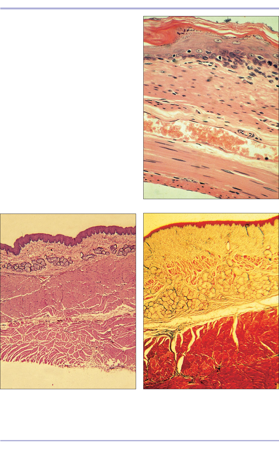
function and the histology reflects this (8.23). The
canal wall is derived from endoderm and splanchnic
mesoderm and consists of four layers:
• Tuna mucosa (mucous membrane), with epithe-
lial lining, supporting vascular lamina propria
and lymphatic cells. Mucosal glands, which are
derived from the epithelium, are variably present.
The outer muscularis mucosae (absent from the
mouth, pharynx, portions of the oesophagus and
rumen) is smooth muscle.
• Tela submucosa: a connective tissue layer with
lymphatic tissue and nerve plexi. Submucosal
glands may be present.
• Tunica muscularis: smooth muscle (except in the
oesophagus and the anus where the muscle is vol-
untary, striated muscle).
• Tunica adventitia/serosa (within the peritoneal
cavity) or adventitia (retroperitoneal).
105
Digestive System
8.23 Diagram.TS. Alimentary
canal.
8.23
Extratubular glands
(liver and pancreas)
Serosa/adventitia
Mucosa
Muscularis
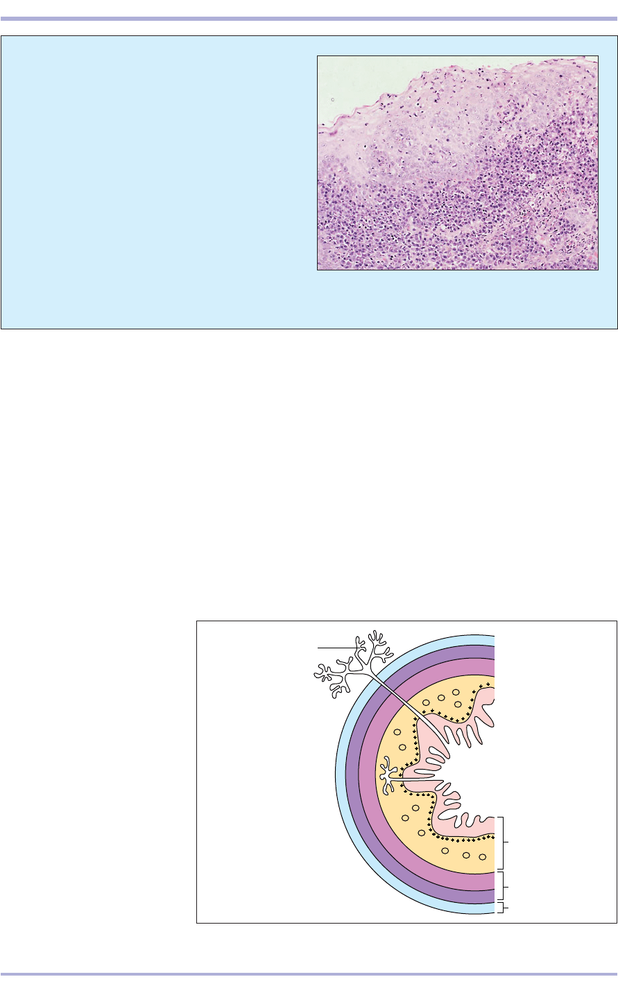
8.22 Plasmacytic gingivitis (dog). H & E. ×125.
Clinical correlates
Any level of the intestinal tract can be affected
by inflammation. Gingivitis, or inflammation of
the gums, is common in dogs and cats, often in
association with dental disease.
A gingival biopsy from a dog in which the gin-
gival epithelium is irregularly hyperplastic is
shown in 8.22. A dense inflammatory infiltrate
occupies the superficial submucosa and extends
into the mucosal epithelium. Plasma cells (mature
immunoglobulin-secreting cells) predominate in
the infiltrate. Their presence indicates a persistent
antigenic stimulus. The initiating disease may be
local or systemic.
Oesophagus
The oesophagus is lined with stratified squamous
epithelium, and both mucosal and submucosal
mucus- or seromucus-secreting glands may be pre-
sent. In ruminants the muscularis externa is skele-
tal muscle; in the pig and cat the distal part is
smooth muscle (8.24–8.27).
106
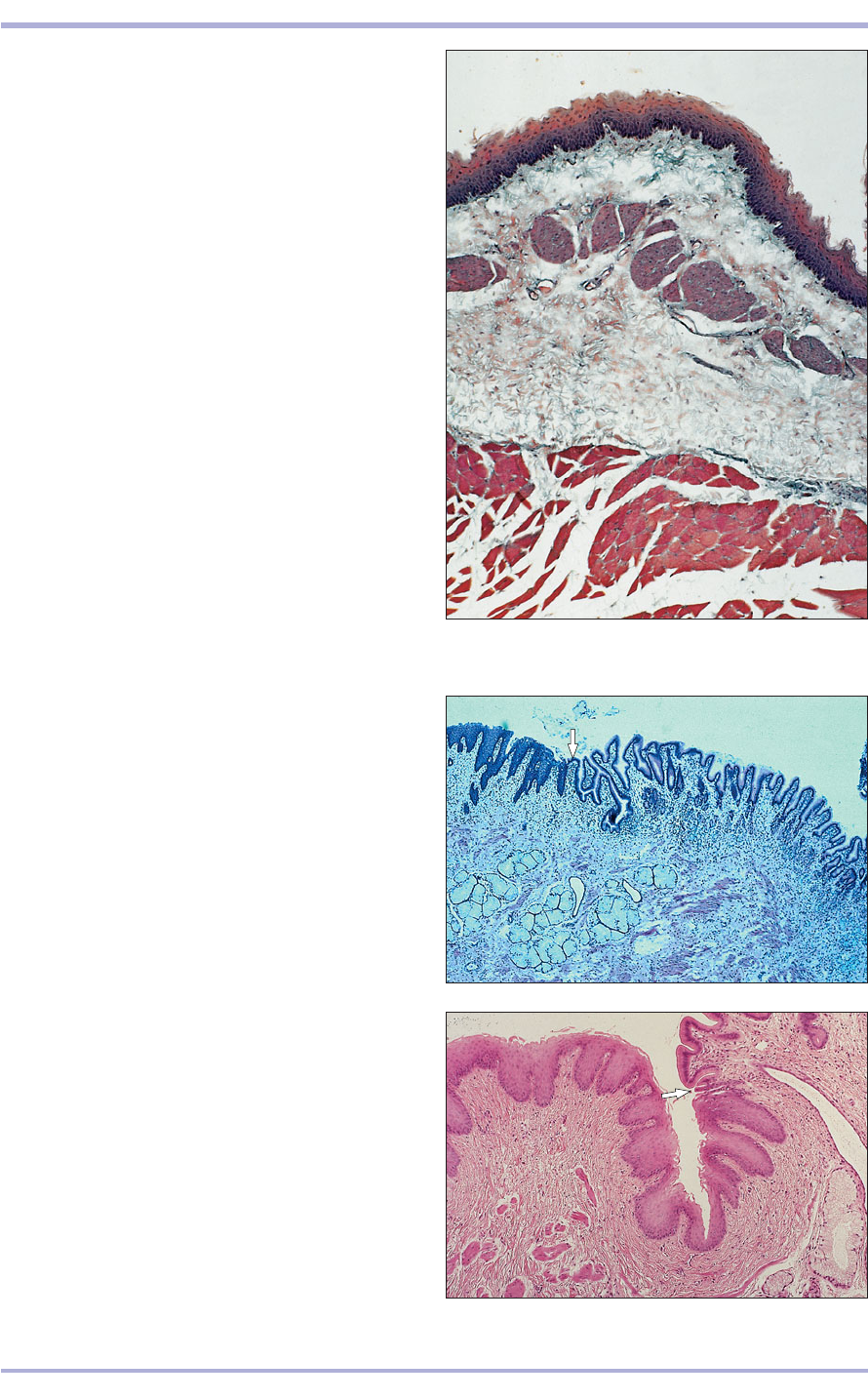
8.24 Oesophagus (cat). (1) Stratified squamous
keratinized epithelium. (2) Lamina propria. (3) LS vein.
(4) Inner layer of circular smooth muscle. (5) Outer layer
of longitudinal smooth muscle. H & E. LP.
8.24
8.25 Oesophagus (dog). (1) Stratified squamous
epithelium. (2) Mucus-secreting glands in the lamina
propria. (3) Inner layer of circular skeletal muscle. (4) Outer
layer of longitudinal skeletal muscle. H & E. ×12.5.
8.25
8.26 Oesophagus (cat). (1) Stratified squamous
epithelium. (2) Lamina propria. (3) Muscularis mucosae.
(4) Submucosal mucous glands. (5) Muscularis externa.
Masson’s trichrome. ×12.5.
8.26
2
3
4
1
5
4
1
2
3
2
3
4
1
5
Comparative Veterinary Histology with Clinical Correlates
Stomach
The stomach mucosa may be non-glandular or glan-
dular in domestic animals. In the simple stomach of
the dog and cat, the mucosa is glandular. In the pig
and horse, there is a non-glandular (oesophageal)
region and a glandular region (8.28 and 8.29). In the
ruminant the non-glandular forestomach has three
compartments: the rumen, the reticulum and the
107
Digestive System
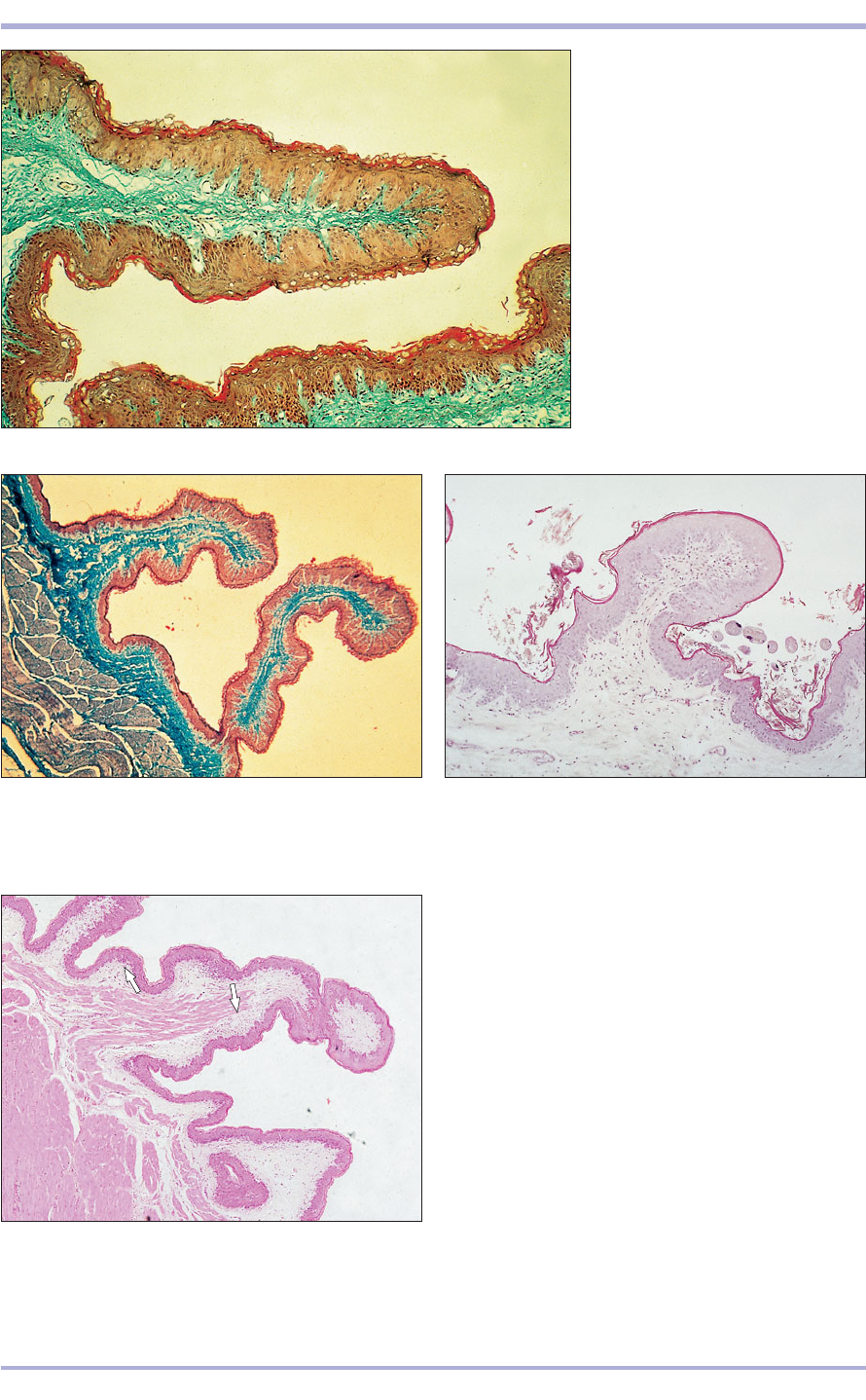
8.27 Oesophagus (dog). (1) Stratified squamous
epithelium. (2) Lamina propria. (3) Muscularis mucosae.
(4) Submucosal mucous glands. (5) Striated muscle of the
muscularis externa. H & E. ×62.5.
8.27
8.28 Oesophageal/stomach junction (horse). The
epithelium changes abruptly from stratified squamous to
simple columnar at the junction (arrowed). H & E. ×62.5.
8.28
8.29 Oesophageal/stomach junction (horse). The
epithelium changes abruptly from stratified squamous to
simple columnar at the junction (arrowed). H & E. ×100.
8.29
2
4
1
3
5
omasum; the glandular stomach is a separate com-
partment: the abomasum (8.30–8.35).
The non-glandular stomach is lined with a strat-
ified squamous epithelium with some keratiniza-
tion. In the ruminant clear vacuolated cells in the
epithelium give it a distinctive appearance and allow
the transfer of water, electrolytes and short-chain
fatty acids. Muscularis mucosae is present in the
omasum and reticulum, but absent from the rumen.
The glandular mucosa of the stomach is folded
and lined with a simple columnar mucus-secreting
epithelium. The gastric pits, the foveoli, are surface
depressions continuous with the simple tubular gas-
tric glands (8.36). Three histological regions are
108
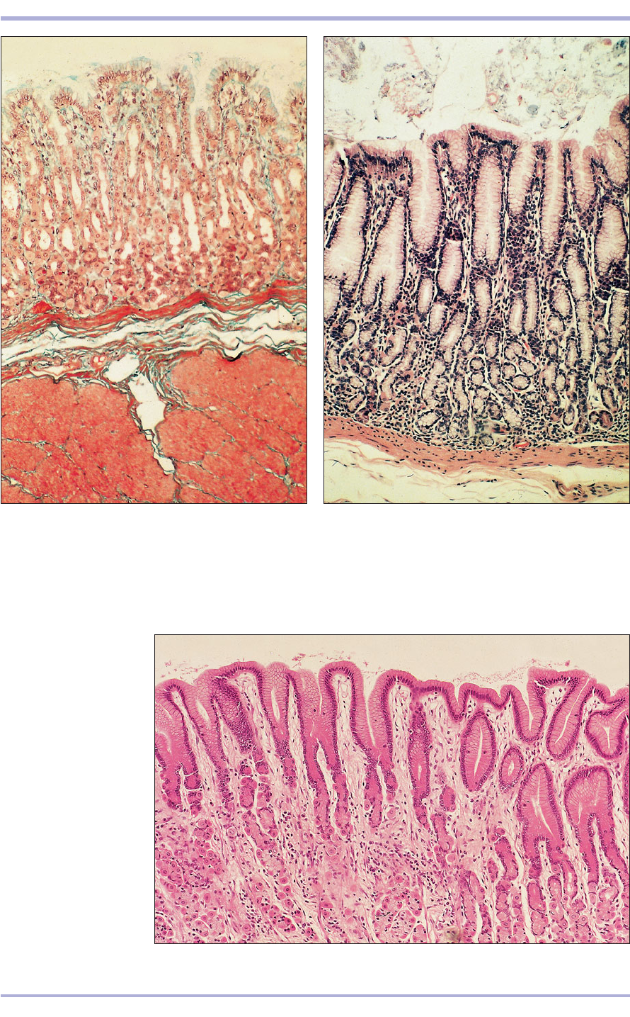
8.30 Rumen (sheep). (1) Stratified
squamous epithelium lines the
rumen. (2) Lamina propria. Masson’s
trichrome. ×12.5.
8.30
8.33 Omasum (sheep). The muscularis mucosae is present
in the long omasal folds (arrowed). H & E. ×12.5.
8.33
8.31 Rumen (goat). The lining epithelium is stratified
squamous. The lamina propria is loose connective tissue,
stained green. Gomori’s trichrome. ×12.5.
8.31
8.32 Reticular groove (goat). The mucosa is folded; this
allows stretching. The lining epithelium is stratified
squamous. H & E. ×12.5.
8.32
2
1
Comparative Veterinary Histology with Clinical Correlates
109
Digestive System
8.34 Abomasum. Fundic region (goat). (1) Mucosa.
(2) Muscularis mucosae. (3) Submucosa. (4) Muscularis
externa. Masson’s trichrome. ×25.
8.34
8.35 Abomasum. Pyloric region (goat). (1) The lining
epithelium is simple columnar and mucus secreting.
(2) Simple tubular mucus-secreting glands. (3) Muscularis
mucosae. (4) Submucosa. H & E. ×62.5.
8.35
8.36 Stomach (horse).
(1) Simple columnar
mucus-secreting
epithelium. (2) Gastric
pits. H & E. ×62.5.
8.36
2
2
2
1
1
2
3
4
1
3
2
4
8.37
110
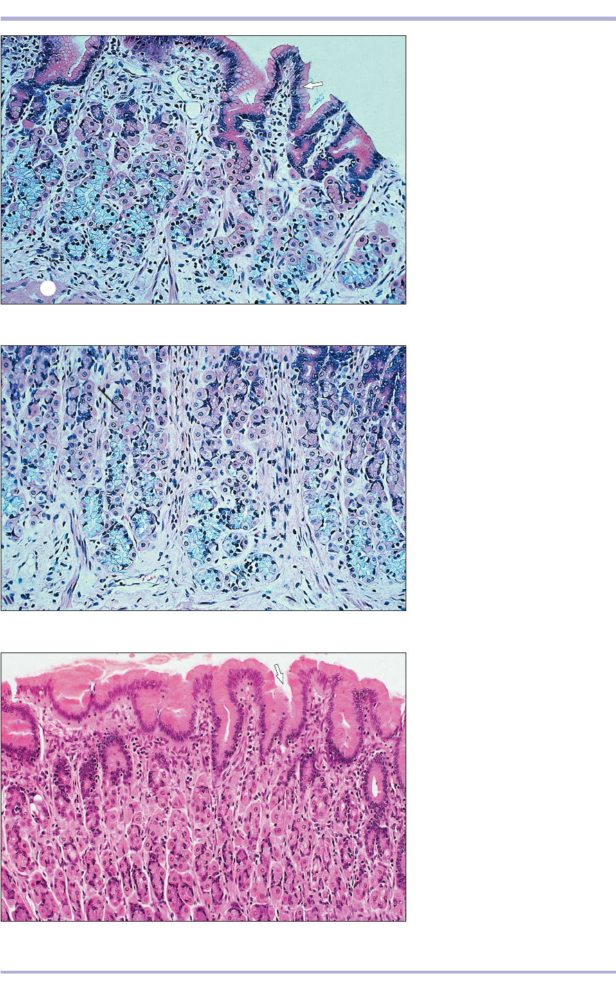
8.37 Cardiac glands. Stomach
(horse). Simple columnar mucus-
secreting epithelium lines the
stomach (arrowed). (1) Parietal
(oxyntic) cell, deep red staining.
(2) Zymogen (chief) cell, basophilic
staining. (3) Lamina propria.
(4) Muscularis mucosae. H & E. ×125.
8.38 Cardiac glands. Stomach
(horse). (1) Parietal cell. (2) Zymogen
cell. (3) Lamina propria. H & E. ×125.
8.38
8.39 Fundic glands. Stomach (horse).
Simple mucus-secreting epithelium
extends into the gastric pits
(arrowed). (1) Parietal cell.
(2) Zymogen cell. H & E. ×250.
8.39
1
2
1
2
3
1
3
2
4
Comparative Veterinary Histology with Clinical Correlates
3
1
2
2
4
