Aughey E., Frye F.L. Comparative veterinary histology with clinical correlates
Подождите немного. Документ загружается.

The mammalian circulatory system is a closed system
of tubes with an endothelial lining from the heart
through the arteries into the capillaries and back
through the veins.
Arteries
Arteries are the conducting channels conveying blood
from the heart to the capillary bed. There are three
types of artery: elastic, muscular (distributing) and
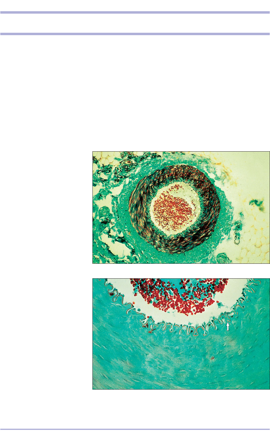
arteriole. The arterial wall has a common structure:
tunica intima (inner lining layer), tunica media (mid-
dle layer) and tunica adventitia (outer layer; 6.1 and
6.2). The tunica intima consists of elongated flattened
endothelial cells resting on loose areolar connective
tissue.
The tunica media of the elastic arteries has a
high proportion of concentric lamellae of fenes-
trated elastic fibres interspersed with smooth
71
6. CARDIOVASCULAR SYSTEM
6.1 Artery (dog). (1) Lumen.
(2) Tunica intima. (3) Tunica media.
(4) Tunica adventitia. Masson’s
trichrome. ×62.5.
6.1
6.2 Artery (dog). (1) Lumen.
(2) Tunica intima with the internal
elastic lamina (arrowed). (3) Tunica
media. Masson’s trichrome. ×125.
6.2
1
1
2
3
4
3
2
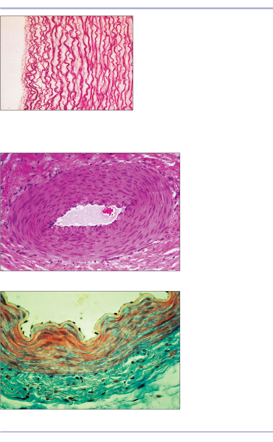
muscle fibres (6.3). These allow the vessels to
dilate. The recoil sends the blood onwards, creat-
ing the pulse in the major elastic artery, the aorta.
In muscular (distributing) arteries, the elastic
content is reduced and the smooth muscle increased.
Internal and external elastic lamina are present (6.2
and 6.4<6.6).
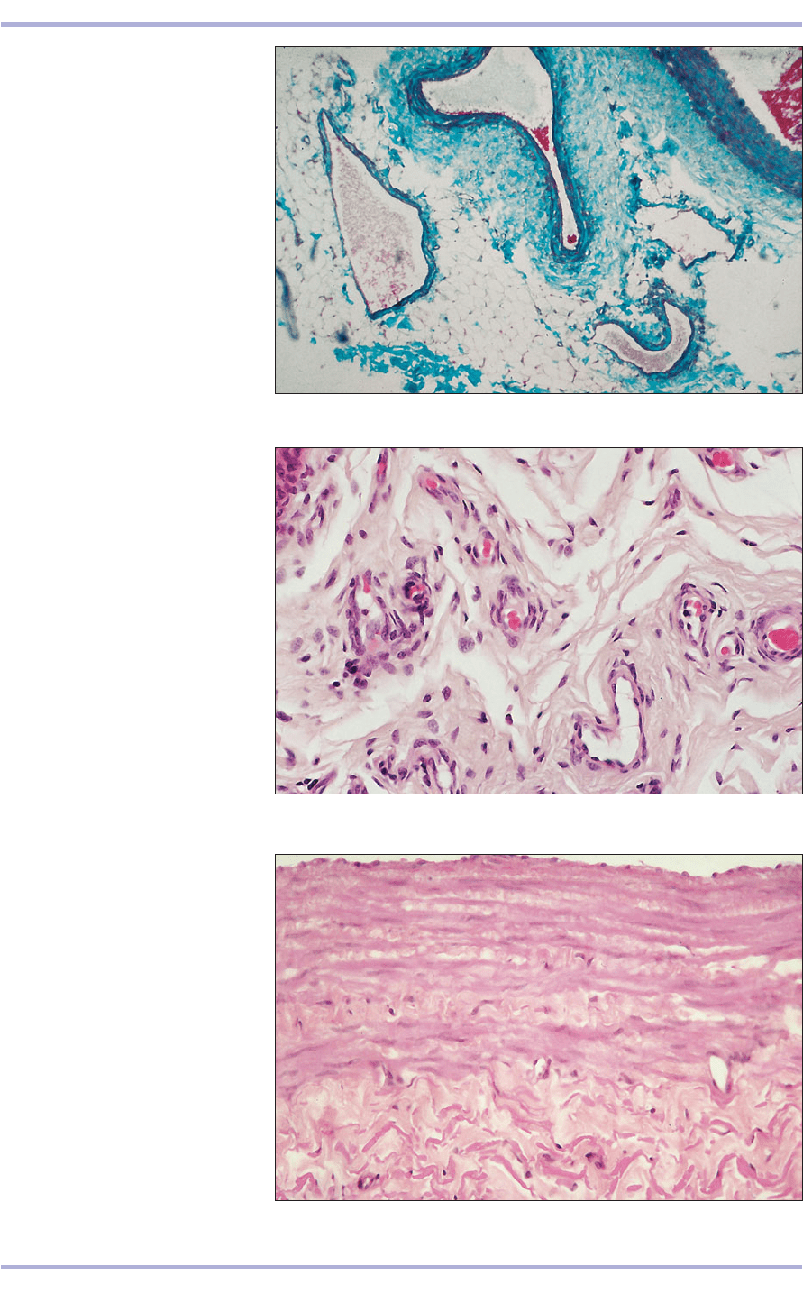
The arterioles (6.7) reduce the pressure of the
blood and supply the capillary bed. The tunica media
may consist of only one layer of smooth muscle cells,
and the luminal diameter is less than the thickness of
the wall (6.8). The tunica adventitia is composed of
collagen and elastic fibres, and contains the vasa
vasorum, the small nutrient arteries and veins in the
walls of the larger blood vessels.
72
6.3 Aorta (horse). Elastic artery stained to illustrate the
wavy elastic fibres. Weigert’s elastin. ×100.
6.3
6.4 Muscular artery (sheep).
(1) Lumen. (2) Tunica intima with the
internal elastic lamina. (3) Tunica
media. (4) Tunica adventitia. H & E.
×125.
6.4
6.5 Muscular artery (sheep).
(1) Lumen. (2) Tunica intima.
(3) Tunica media. (4) Tunica
adventitia. Masson’s
trichrome. ×250.
6.5
1
2
3
4
2
3
4
1
Comparative Veterinary Histology with Clinical Correlates
73
6.6 Artery and vein. Stomach (dog).
(1) Small artery. (2) Small vein.
(3) Lymphatic vessel. H & E. ×62.5.
6.6
6.7 Arteriole, vein and a Iymphatic
vessel in connective tissue. Tongue
(ox). (1) Arteriole. (2) Vein.
(3) Lymphatic vessel. H & E. ×62.5.
6.7
6.8 Arterioles in the wall of a small
artery (horse). (1) Arteriole. (2) Veins.
(3) Artery. H & E. ×160.
6.8
2
2
1
3
3
3
2
1
1
3
2
2
Cardiovascular System

Capillaries and venules
The capillary is the smallest unit of the vascular sys-
tem. The diameter of the lumen is no larger than an
erythrocyte and permits these cells to pass in sin-
gle file only (6.9). The capillary wall is two-layered:
a tunica intima of one or two squamous endothe-
lial cells resting on a basal lamina, and a fine tunica
adventitia of collagen and elastic fibres. The
endothelial cells usually form a continuous layer,
but fenestrated capillaries occur in the renal
glomerulus, the endocrine glands, intestinal villi and
the choroid plexus where gaps are present between
adjoining cells closed by a membrane diaphragm.
The wall also contains pericytes: undifferentiated
cells believed capable of becoming fibroblasts or
muscle cells. Venules collect the blood from the cap-
illaries. Their lumina are wider than those of the
arterioles. The tunica intima (there is no tunica
media and adventitia) in each venule consists of a
continuous layer of endothelial cells and areolar
connective tissue. Pericytes are also present.
74
Comparative Veterinary Histology with Clinical Correlates
6.9 Capillaries (sheep) in the connective tissue of the cervix (arrowed). Masson’s
trichrome. ×125.
6.9
Sinusoids
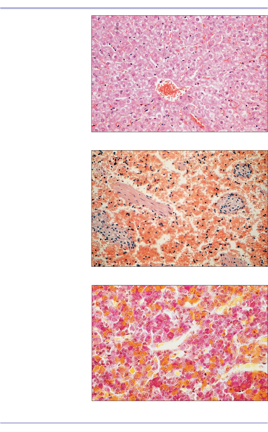
Sinusoids are found in the liver, spleen, bone mar-
row and adenohypophysis. Their wide lumina are
lined by endothelial cells interspersed with fixed
macrophages of the mononuclear phagocyte scav-
enging and defence system of the body (6.10<6.12).
Similar thin-walled venous sinuses are found in
endocrine glands.
75
6.10 Liver (sheep). (1) Central vein.
(2) Liver sinusoids. H & E. ×62.5.
6.10
6.11 Spleen (horse). Sinusoids filled
with erythrocytes. H & E. ×125.
6.11
6.12 Pars distalis of the
adenohypophysis (cat). (1) Sinusoids.
(2) Cords of hypophyseal cells.
Orange G. ×250.
6.12
1
2
1
2
2
2
Cardiovascular System
Veins
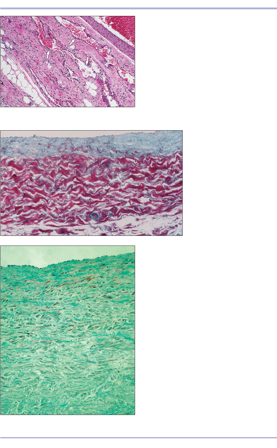
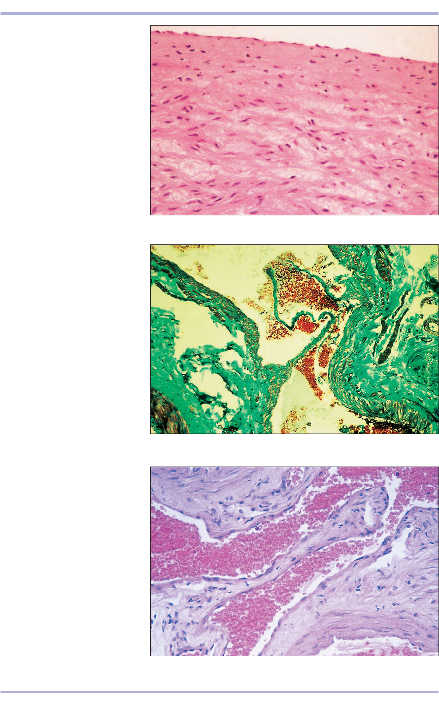
Veins are lined by a continuous layer of endothelial
cells and areolar connective tissue. The tunica
media, which is always narrow, contains a few cir-
cular smooth muscle fibres, some elastic fibres, but
no elastic lamina. The tunica adventitia consists of
longitudinal collagen fibres and, in the large veins,
some smooth muscle (6.13–6.16). The lumen con-
tains valves that are projections of the tunica intima;
these allow only unidirectional blood flow (6.17
and 6.18).
76
6.15 Caudal vena cava (horse). (1) Tunica intima.
(2) Tunica media. Masson’s trichrome. ×125.
6.15
6.14 Caudal vena cava (dog).
(1) Tunica intima. (2) Tunica media.
(3) Tunica adventitia with vasa
vasorum (arrowed). Gomori’s
trichrome. ×125.
6.14
6.13 Spermatic cord (horse). Venous plexus (arrowed).
H & E. ×62.5.
6.13
2
1
1
2
3
Comparative Veterinary Histology with Clinical Correlates
77
6.16 Cranial vena cava (horse).
(1) Tunica intima. (2) Tunica media.
H & E. ×125.
6.16
6.17 Femoral vein with valves (cat).
(1) Lumen. (2) Valve. (3) Tunica
intima. (4) Tunica media. (5) Tunica
adventitia. Masson’s trichrome. ×25.
6.17
6.18 Valve in the brachial vein (cat).
(1) Lumen filled with erythrocytes.
(2) Valve. (3) Tunica intima. H & E.
×125.
6.18
1
1
1
3
3
2
1
2
3
4
5
1
2
Cardiovascular System
Arteriovenous anastomoses
Heart
The cardiac wall consists of three layers: endo-
cardium (inner), myocardium (middle) and epi-
cardium (outer).
The endocardium contains continuous squa-
mous endothelial cells, vascular areolar connective
tissue and conducting fibres. The myocardium is
composed of cardiac muscle and also contains vas-
cular areolar connective tissue. The epicardium is
78
Comparative Veterinary Histology with Clinical Correlates
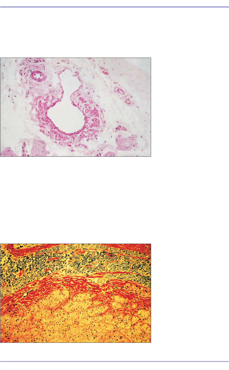
6.19 Arteriovenous anastomosis in
loose connective tissue (cat).
(1) Artery. (2) Vein. H & E. ×62.5.
6.19
thicker than the endocardium, and fat deposits in
the rather dense connective tissue and coronary
blood vessels are often found (6.20 and 6.21).
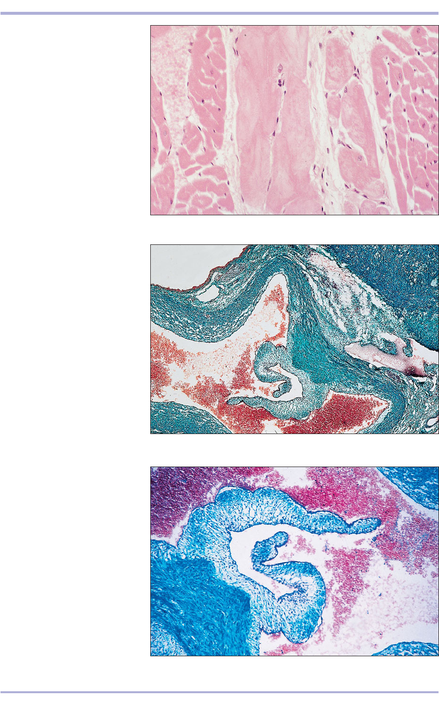
Fibrous rings support the heart valves (6.22). They
provide a means of insertion for the cardiac mus-
cle and may be referred to as the fibrous or cardiac
skeleton (6.23). In older horses the aortic rings
may become calcified to form the ossa cordis. Two
bones form in the aortic rings of cattle, the ossa
cordis.
6.20 Cardiac muscle (ox). (1) Cardiac
muscle. (2) Connective tissue of the
fibrous skeleton. (3) Atrioventricular
node. Masson’s trichrome. ×125.
6.20
3
1
1
2
2
1
2
Arteriovenous anastomoses are special areas of
the skin of the nose, lips and pads where the arte-
riole opens directly into a venule without going
through the capillary bed (6.19). This provides an
alternative channel of blood supply and regula-
tion of heat loss.
79
6.21 Cardiac muscle (horse).
(1) Cardiac muscle fibres.
(2) Conducting (Purkinje) fibres.
H & E. ×125.
6.21
6.22 Heart (dog). (1) Lumen of the
atrium. (2) Lumen of the pulmonary
artery. (3) Valve cusps. Masson’s
trichrome. ×62.5.
6.22
6.23 Heart (dog). (1) Valve cusps.
(2) Dense connective tissue part of
the fibrous skeleton of the heart.
Masson’s trichrome. ×62.5.
6.23
1
2
1
3
2
3
1
2
Cardiovascular System

6.24
Lymphatic vessels
Lymphatic vessels drain excess fluid from the tis-
sues and are made up of a thin layer of connective
tissue with an endothelial lining. Larger lymphatic
vessels, such as the thoracic duct, may have a few
smooth muscle fibres in the wall.
Amphibians and reptiles have perilymphatic and
endolymphatic systems that are particularly well
developed in some species. These lymph-filled
structures serve several functions. Perilymphatic
pathways encircle the auditory apparatus and may
participate in the transmission of sounds. The
endolymphatic system consists of receptor organs of
the inner ear as well as either bilateral separate or
fused thin-walled sacs. These communicate with the
skull via narrow ducts and serve as reservoirs for the
storage of calcium carbonate microcrystals. In some
amphibians, endolymphatic sacs form a ring-like
extension of the vertebral canal around the brain.
80
Comparative Veterinary Histology with Clinical Correlates
Clinical correlates
A range of cardiovascular diseases are important
in veterinary medicine. The heart itself can be
affected by congenital, degenerative, inflamma-
tory and neoplastic conditions with a variety of
underlying causes. Disease caused by congeni-
tal malformations is naturally recognized most
often in the young animal. Mitral valve dyspla-
sia in an 11-week-old Bearded Collie is shown
in 6.24. The heart is opened to show the left atri-
oventricular (mitral) valve. The valve leaflets are
thickened and malformed and the chordae tend-
inae are short, thick and partially fused. This con-
genital defect leads to mitral valve incompetence
and produces a holosystolic murmur centred
around the fifth intercostal space near the left
sternal border.
In adult animals primary cardiac disease is
most often encountered in the form of cardiomy-
opathy or degenerative change in the muscle of
the heart. In 6.25 and 6.26 special stains have
been used to highlight features of interest. Both
are from the same case, a 12-year-old dog with
myocardial degeneration. In 6.25 a Sirius Red
stain, which colours collagen red, demonstrates
the large amount of fibrosis replacing muscle
bundles, which are stained yellow. In 6.26 a
Masson’s trichrome stain highlights muscle fibres
undergoing degeneration. Myocardial degener-
ation and fibrosis may be found in cases of
dilated cardiomyopathy or as a non-specific
response of the cardiac muscle to injury or insult.
One of the most important diseases to affect
the blood vessels is haemangiosarcoma, a
malignant tumour of the endothelial cells that
line blood vessels. In dogs the spleen, right
atrium and skin are common primary sites and
metastasis can be very widespread. Other
species are also affected. A haemangiosarcoma
in a 6-year-old thoroughbred gelding is shown
in 6.27.
The smooth muscle in the tunica media of
blood vessels can be affected by the deposition
of calcium salts. This change is frequently
induced by oversupplementation of the diet of
herbivorous reptiles or amphibians with vita-
min D
3
either directly or by inclusion of com-
mercial dog, cat or primate food. Similarly
hypervitaminosis D
3
may be induced when sup-
plemented goldfish are fed to fish-eating reptiles
and amphibians. Early arteriosclerotic mineral-
ization of the tunica media in a large pulmonary
artery in an iguana is shown in 6.28.
6.24 Mitral valve dysplasia in an 11-week-old Bearded
Collie. Note the thickened, distorted chordae
tendinae.
