Aughey E., Frye F.L. Comparative veterinary histology with clinical correlates
Подождите немного. Документ загружается.

61
Blood
4.31
Clinical correlates
Disorders of the blood can be attributed to
abnormalities in production of blood cells from
the bone marrow or other central blood-form-
ing organs or to abnormal loss or consumption
of these cells. Anaemia, the deficiency of oxy-
gen-carrying erythrocytes, can be regenerative
(i.e. the bone marrow is able to respond by
releasing new cells, including immature cells
into the circulation) or non-regenerative (in
which this response does not occur). A blood
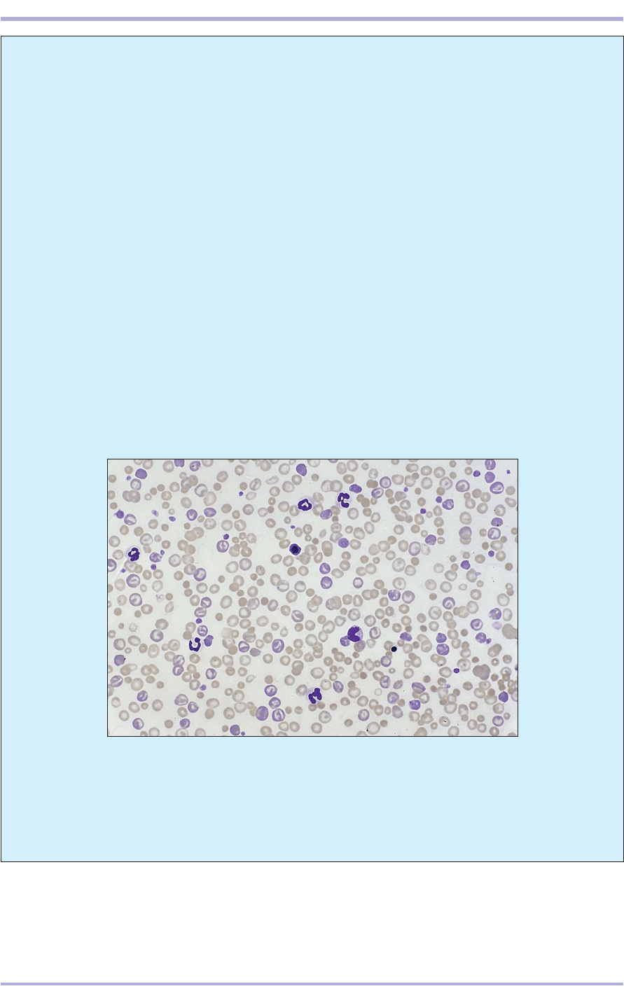
film that illustrates haemolytic anaemia in a dog
is shown in 4.31. In this regenerative anaemia
there is variation in cell size (anisocytosis) with
red cell precursors, such as the large, stippled
reticulocyte (1) and nucleated normoblast (2),
4.31 Haemolytic (regenerative) anaemia in a dog. Note the variation in
appearance of the red cells. Reticulocytes, nucleated normoblasts and
spherocytes are present. Giemsa. ×200.
visible. Some of the red cells have small, dense
dots (Howell–Jolly bodies), which represent
remnants of nuclear chromatin and are charac-
teristic of regenerative anaemias. Spherocytes,
red cells that have lost their biconcave shape
and become globular due to immunological
attack, are present (3). There is an increased
neutrophil count with some immature neu-
trophils (reactive neutrophilia with a left shift),
which typically accompanies haemolysis. These
findings are diagnostic of autoimmune
haemolytic anaemia, the most common type of
anaemia in the dog.
In immune-mediated anaemia, erythrocyte
destruction follows the attachment of antibody
to the cell membrane. In most cases the aetiology
is unknown.
62
Comparative Veterinary Histology with Clinical Correlates
4.32
4.33
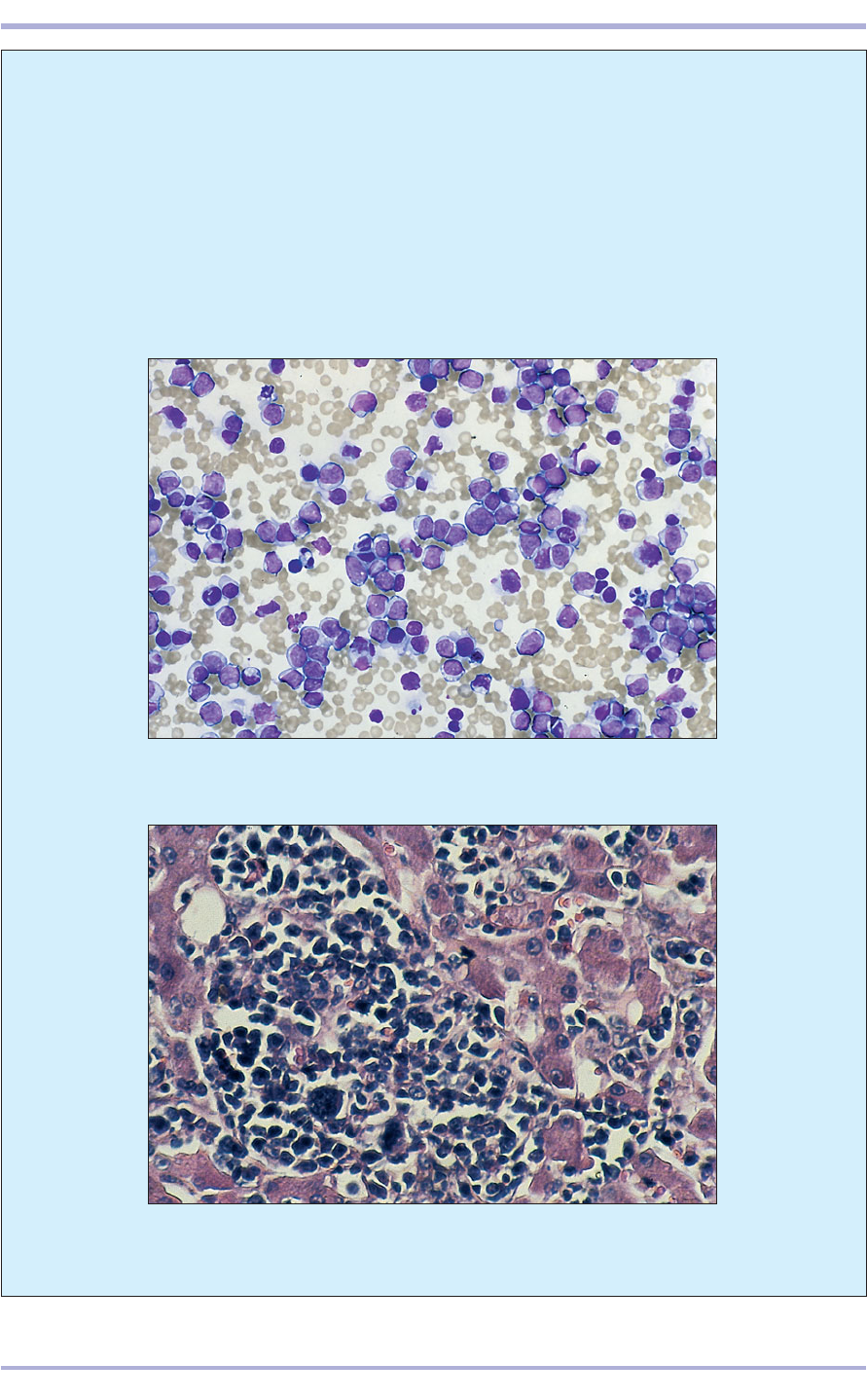
4.33 Megakaryocytic myelosis in the liver of a cat. Note the megakaryocyte
with the multilobed nuclei in the cellular infiltrate. H & E. ×255.
4.32 Acute lymphoblastic leukaemia (dog) showing large numbers of
malignant lymphoblasts. Giemsa. ×255.
The blood film in 4.32 was prepared from a
dog with acute lymphoblastic leukaemia. Very
high numbers of malignant lymphoblasts, which
are larger than normal lymphocytes and have
bluer cytoplasm, are seen. In some cases of
leukaemia, although the bone marrow is invaded
by neoplastic haemopoietic cells, no abnormal
cells are released into the peripheral blood. Such
cases may be termed aleukaemic leukaemias.
Non-regenerative anaemia is a common finding
with many leukaemias.
The liver of a cat in which the sinusoids (vas-
cular channels) are heavily infiltrated by large
neoplastic megakaryocytes with multilobed
nuclei is shown in 4.33. This is megakaryocytic
myelosis, a rare, chronic disease of dogs and
cats characterized clinically by bleeding and
thrombosis of the ear and tail tips.
63
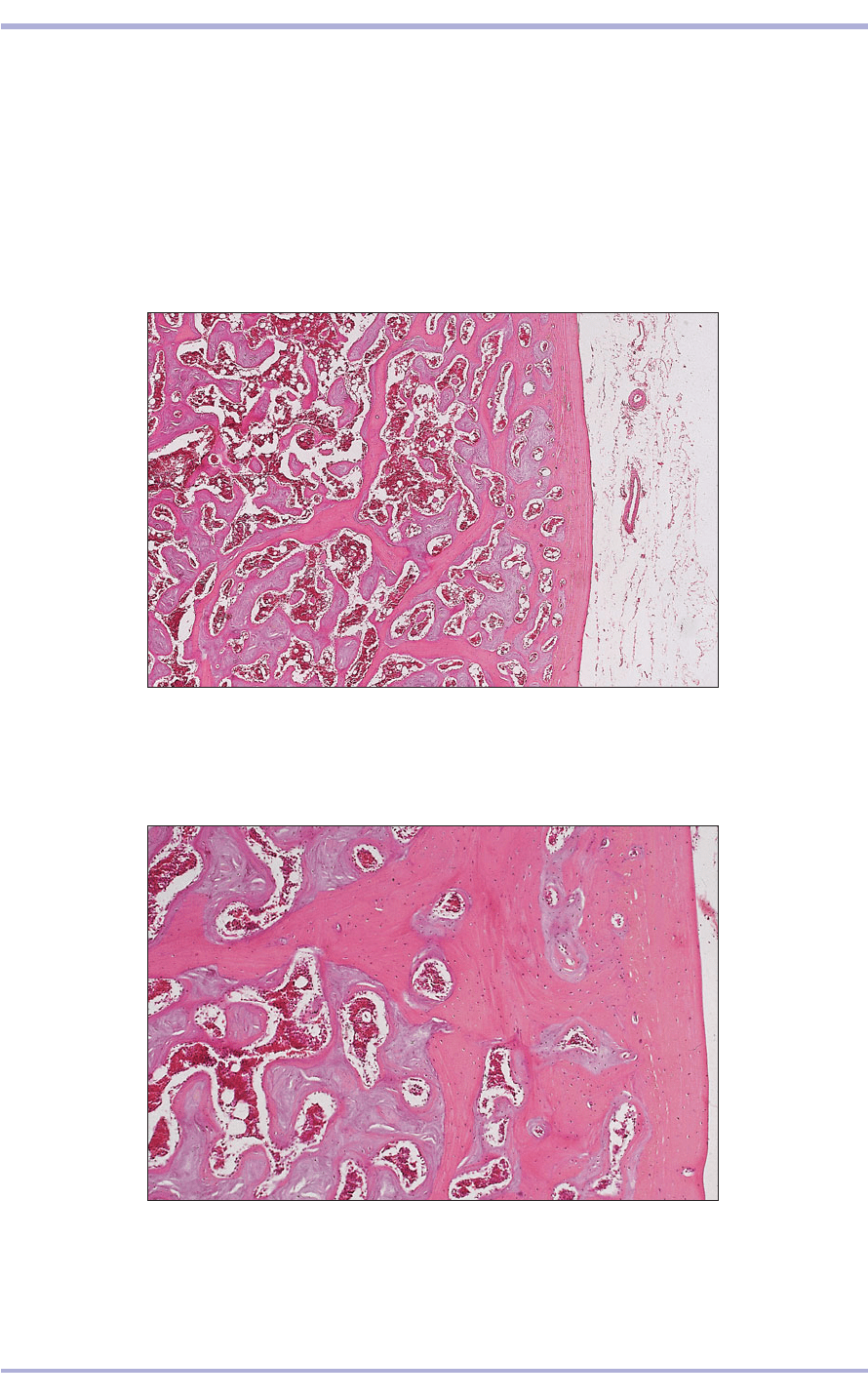
4.34 Head of the humerus (domestic hen). (1) Outer layer of eosinophilic
cortical bone. (2) The spiculated medullary bone is basophilic. (3) The marrow
cavity is lined by osteoblasts. H & E. ×62.5.
4.34
4.35 Head of the humerus (domestic hen). (1) The osteocytes in the
eosinophilic cortical bone form an osteon. (2) Osteocytes in the open lacunae
of the basophilic medullary bone. (3) Marrow cavity lined by osteoblasts.
H & E. ×125.
4.35
Avian bone
A unique feature of avian bone in egg-producing
birds is the accumulation of spiculated bone in the
medullary cavity, under the combined influence of
oestrogens and androgens. This medullary bone
is particularly labile and the stored calcium is uti-
lized in the formation of the calcareous egg shell.
The medullary bone is basophilic and decreases
during shell deposition and increases at other
times. The basophilia is caused by a change in the
density of the matrix and in the glycosaminogly-
cans that are present. During resorption, osteo-
clasts are active; during deposition, osteoblasts are
prominent on the surface of the bone spicules
(4.34 and 4.35).
2
3
1
1
2
3
Blood
64
Reptilian, amphibian and fish
bone and bone marrow
Reptiles do not possess pneumatized bone such as
that found in birds capable of flight. The box-like
carapace and plastron of chelonians are composed
of specialized bone that must be both strong and
relatively lightweight in order for this bone to pro-
tect efficiently the delicate internal structures.
Compressional stresses applied to the dorsal and
ventral surfaces are distributed widely and are then
borne upon buttress-like vertical supporting pillars
of bone that are at each end of the ‘bridge’ that joins
the carapace to the plastron on each side. The
strength of their shells is further enhanced by the
curved shape and by additional internal struts that
distribute compressive forces so that they are not
concentrated onto a single focus. The shell is com-
posed of parallel layers of inner and outer tables
of compact bone with an intervening layer of
spongy cancellous bone characterized by spaces
filled with bone marrow. In form and function the
bony shell resembles the calvaria that covers the
brain case of a mammalian skull.
The bone of amphibians and reptiles contains
numerous sites of haemopoiesis; long bones (see
3.29), ribs, skull and mandibles are locations in
which active blood cell formation normally occurs.
During severe blood loss and a few other condi-
tions, sites other than bone marrow are recruited
for extramedullary haemopoiesis; liver, spleen and
kidney are then most often involved.
In fish, the major organ of haemopoietic activity
is the cranial pole of each kidney (see Chapter 9),
with lesser amounts occurring in other extra-
medullary sites during times of severe anaemia,
infection or stress.
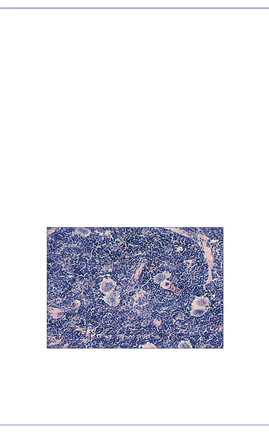
The presence of numerous megakaryocytes in
the splenic red pulp is a common finding in the
healthy house mouse, Mus musculus, whereas
extramedullary haemopoiesis is usually considered
an abnormal finding in most other animals (4.36).
4.36 Extramedullary haemopoiesis in the spleen of a domestic mouse (Mus
musculus). Note the multinucleated megakaryocytes which are normal in
murine splenic tissue. H & E. ×125.
4.36
Comparative Veterinary Histology with Clinical Correlates

Contractility, a fundamental property of cytoplasm,
is developed to a highly specialized degree in mus-
cle tissue. The elongated muscle cell is commonly
referred to as a muscle fibre, the plasma membrane
as the sarcolemma and the cytoplasm as the sar-
coplasm. There are basically two types of muscle:
smooth, visceral or involuntary muscle; and striated
muscle, which is further subdivided into skeletal
voluntary muscle and cardiac involuntary muscle.
Muscle contraction depends upon the proteins
actin and myosin in the sarcoplasm. In skeletal and
cardiac muscle the longitudinal arrangement of
these proteins is aligned in register to give cross-
striations, which are absent from smooth muscle.
Muscle types
Smooth muscle
Smooth muscle cells are elongated fusiform fibres
(2–50 +m long) with a single, centrally located
nucleus with several nucleoli. These fibres may
occur singly, as in the lamina propria of intestinal
villi, but are more commonly arranged in sheets or
layers in the walls of a wide range of tubes of the
alimentary, urogenital, respiratory and cardiovas-
cular systems. The fibres are packed in a staggered
fashion with the thickest nucleated portion of one
fibre juxtaposed to the thin tapered end of an
adjoining one. Individual smooth muscle fibres are
supported by reticular fibres, with collagen and
elastic fibres forming a supporting framework of
connective tissue carrying blood vessels and nerves.
Smooth muscle is under involuntary control; it is
innervated by the autonomic nervous system
(5.1–5.3).
65
5. MUSCLE
5.1 Smooth muscle. Uterus (cat). (1) Nucleus.
(2) Sarcoplasm. H & E. ×100.
5.1
5.2 Smooth muscle. Uterus (cat). (1) Nucleus.
(2) Sarcoplasm. (3) Longitudinal fibres. (4) Transverse
fibres. H & E. ×200.
5.2
5.3 Smooth muscle. Duodenum (dog). (1) Fusiform
smooth muscle fibre. H & E. ×200.
5.3
1
1
1
2
3
4
1
2
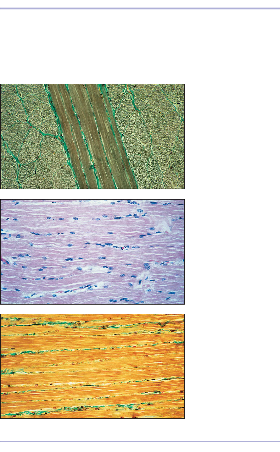
Striated (skeletal) muscle
Skeletal muscle fibres are multinucleated giant cells
that range in length from a few millimetres to sev-
eral centimetres. The nuclei lie immediately beneath
the sarcolemma, and the myofibrils give both a lon-
gitudinal and a cross-striation arrangement. The
contractile myofilaments are arranged in alternat-
ing isotropic I bands and anisotropic A bands. This
imparts the cross-striated effect.
Skeletal muscle fibres are bound into large bun-
dles by an outer connective tissue investment, the
epimysium. This dips into the muscle and invests
bundles of muscle fibres (fascicles) in connective
66
Comparative Veterinary Histology with Clinical Correlates
5.4 Striated muscle. Tongue (ox).
(1) Transverse section (TS) muscle
fibres. (2) Longitudinal section (LS)
muscle fibres. Masson’s trichrome.
×125.
5.4
5.5 Striated muscle. Tongue (cat).
(1) Nucleus. (2) Sarcoplasm.
(3) Endomysium. H & E. ×200.
5.5
5.6 LS striated muscle. Tongue (cat).
(1) Longitudinal striated muscle.
(2) Nuclei. (3) Endomysium stained
green. Masson’s trichrome. ×200.
5.6
1
3
2
3
3
2
1
21
67
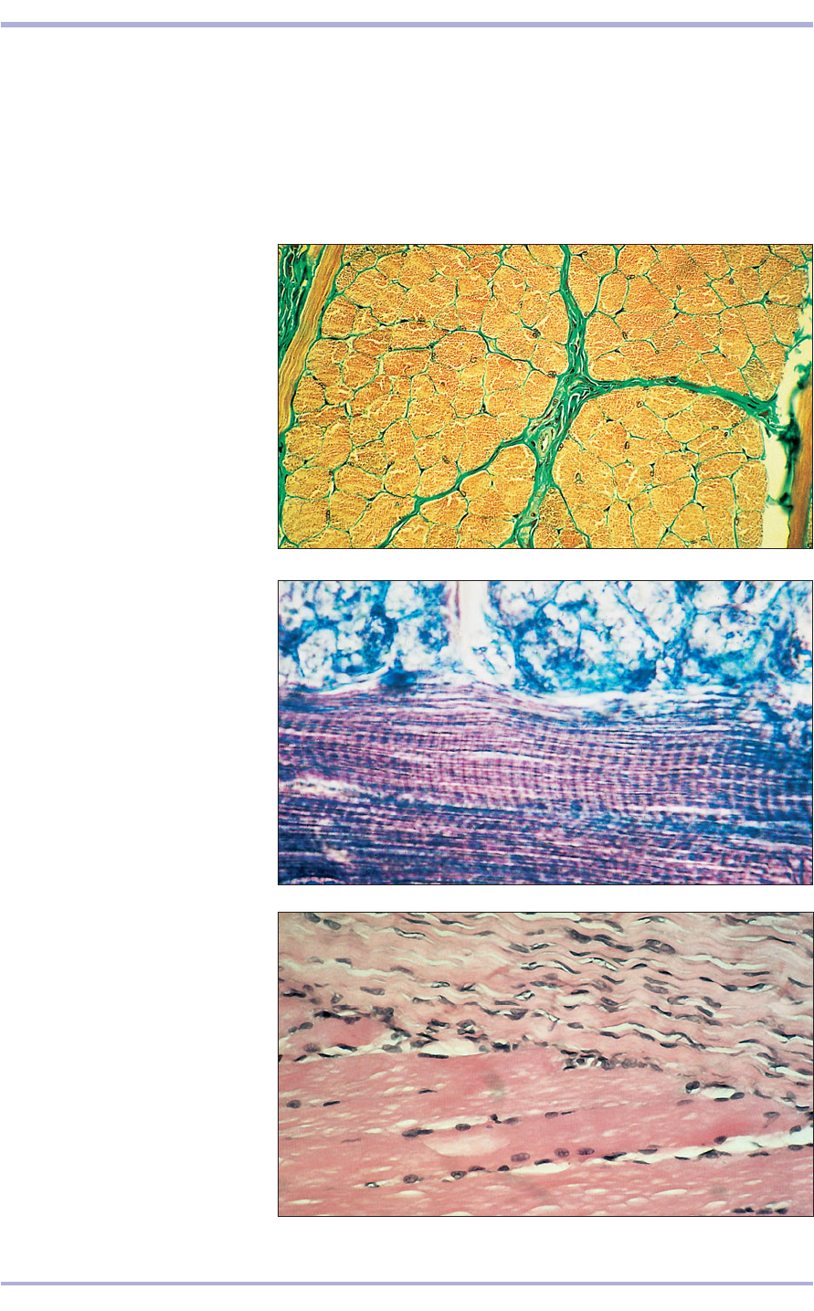
tissue, the perimysium. This in turn invests each
muscle fibre in a vascular loose connective tissue,
the endomysium. A fine network of reticular fibres
lies against the sarcolemma. The collagen fibres of
the tendon extend into the epimysium and allow
muscular contraction to effect movement (5.4–5.9).
Every skeletal muscle fibre receives an axon termi-
nal from a motor neuron at the myoneural junction,
the motor endplate (see 13.27). Muscle spindles,
which are attenuated skeletal muscle fibres, act as
stretch receptors that are innervated both by motor
and by sensory nerve terminals.
5.7 TS striated muscle. Tongue (cat).
(1) Transverse section of muscle
fibres. (2) Connective tissue
perimysium. Masson’s trichrome.
×62.5.
5.7
5.8 Striated muscle. Tongue (ox).
Muscle fibres showing cross-striations.
Masson’s trichrome. ×625.
5.8
5.9 Tendon/muscle junction (dog).
(1) Collagen fibres of the tendon.
(2) Striated muscle fibres. H & E.
×200.
5.9
1
2
1
2
Muscle
Cardiac muscle (myocardium)
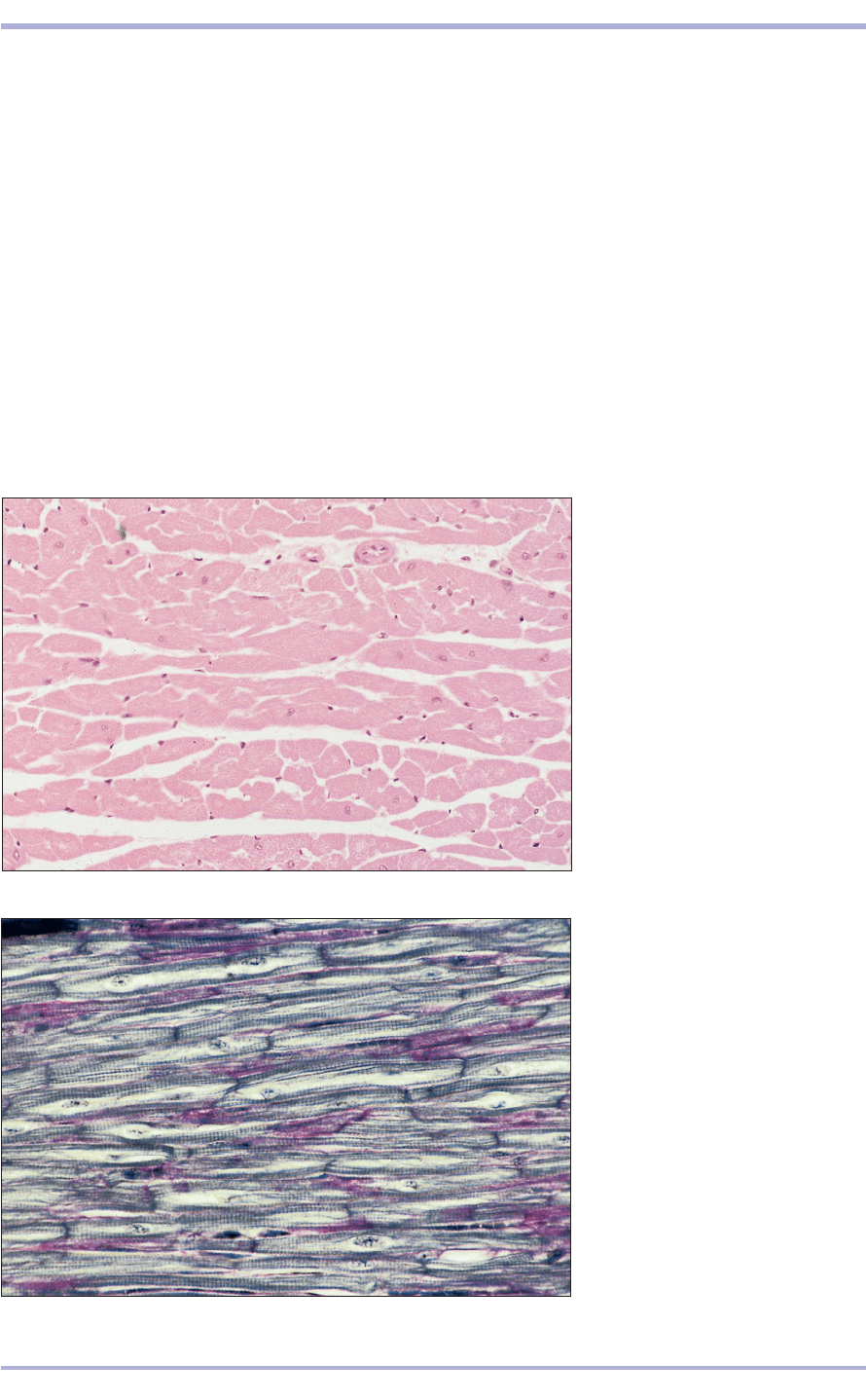
Cardiac muscle is exclusive to the myocardium, the
muscular wall of the heart. The fibres are smaller
than skeletal muscle fibres and branch repeatedly
(5.10). The nucleus lies in the centre of the fibre
(occasionally two nuclei are present) and the fibres
have strong areas of attachment at the intercalated
disc. These are visible as a dark cross-striation at
the end of one fibre and the beginning of the next,
and confer structural integrity on the heart muscle
to allow contraction to spread throughout the
myocardium (5.11 and 5.12). These are represented
at the ultrastructural level by tight junctions and
gap junctions. The cardiac conducting system is
composed of several specialized muscle fibres in the
sinoatrial and atrioventricular nodes. In the sinoa-
trial node (5.13, 5.14), small cardiac muscle fibres,
which are low in myofilaments, have an intrinsic
ability to contract at a species-specific rate and act
as the pacemaker for cardiac muscle contraction.
The atrial wave of depolarization converges on the
second node, the atrioventricular node, from where
specialized large muscle fibres spread throughout
the ventricular muscle and initiate contraction.
These specialized conducting fibres are binucleate
and the nucleus lies in a clear area of sarcoplasm
(5.15).
The various types of muscle tissue can be dis-
cerned, even in invertebrates low on the phyloge-
netic scale. For example, the striated muscle fibres
of a spider are structurally similar to those found
in mammals and the multiple heart-like pumping
chambers that circulate the haemolymph and
68
Comparative Veterinary Histology with Clinical Correlates
5.10 Cardiac muscle (horse).
(1) Nucleus of the muscle fibre.
(2) Sarcoplasm. H &.E. ×125.
5.10
5.11 Cardiac muscle (horse).
(1) Cardiac muscle fibre. (2) Vascular
connective tissue of the endomysium.
(3) Intercalated disc. Heidenhain’s
iron haematoxylin. ×625.
5.11
1
2
3
3
1
1
2
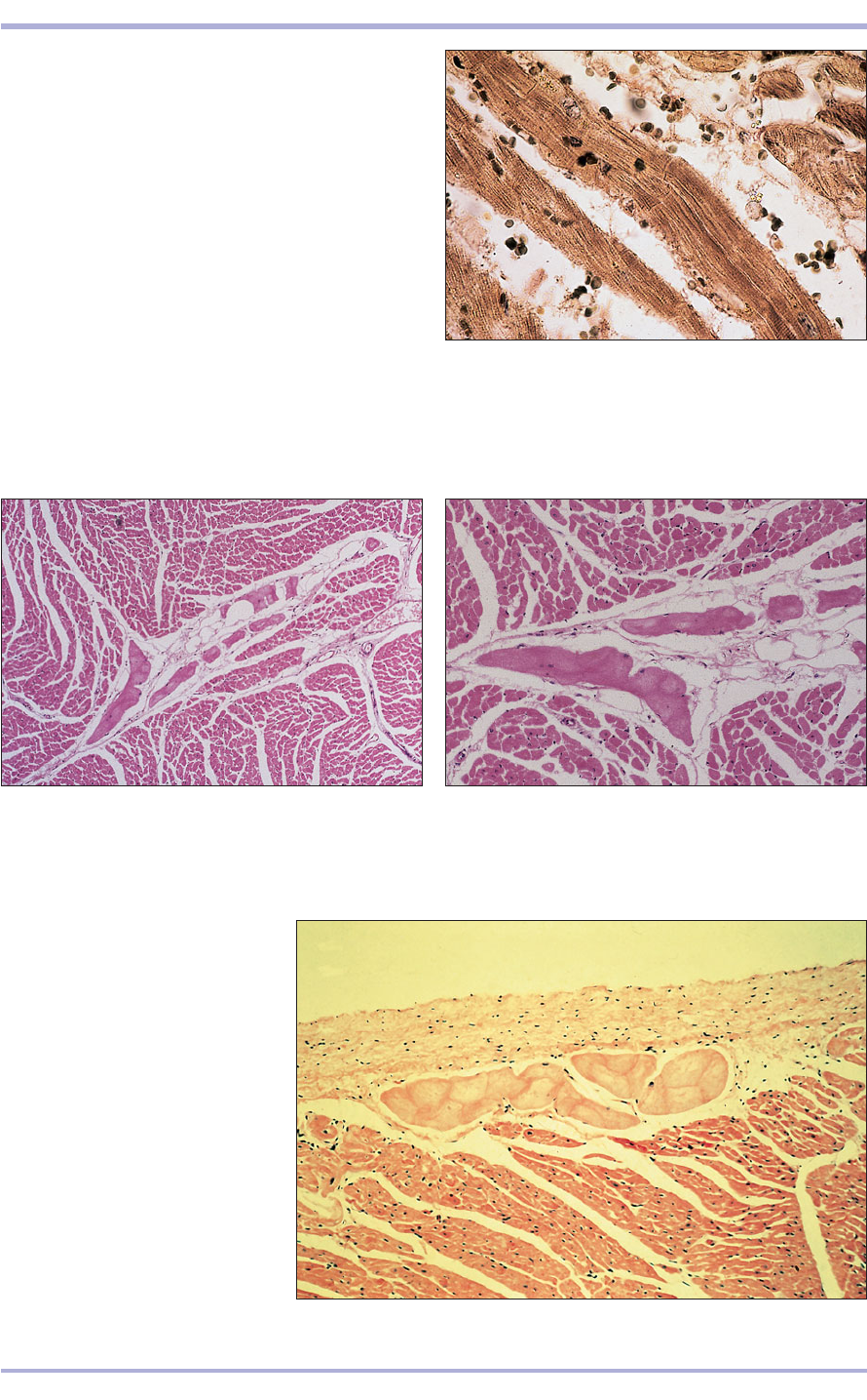
5.13 Cardiac muscle (horse). (1) Cardiac muscle.
(2) Purkinje’s fibres. H & E. ×62.5.
69
haemolymphocytes through the coelom of earth-
worms are formed from myocardium.
As all fish and amphibians and most reptiles (all
except the crocodilians) possess a three-chambered
heart with paired atria and a single ventricle, there
are some structural differences between the hearts
of these animals. For example, a ridge of
myocardium helps direct the flow of blood through
the ventricle so as to reduce the mixing of oxy-
genated and deoxygenated blood in non-crocodil-
ian reptiles. Blood flow through twin aortic arches
and atrioventricular valves is controlled by typical
heart valve leaflets composed of myxoid connective
tissue that are covered by a thin endothelial lining
in all vertebrates.
5.12 Cardiac muscle (horse). (1) Cardiac muscle fibre.
(2) Vascular connective tissue of the endomysium.
(3) Intercalated disc. Heidenhain’s iron haematoxylin.
×200.
5.12
5.14. Cardiac muscle (horse). (1) Cardiac muscle.
(2) Purkinje’s fibres. (3) Loose vascular connective tissue.
H & E. ×125.
5.14
5.13
5.15 Cardiac muscle (horse).
(1) Endocardium. (2) Impulse-
conducting fibres (Purkinje’s fibres).
(3) Muscle fibre. H & E. ×125.
5.15
1
1
1
1
2
2
3
2
3
2
1
3
2
Muscle

5.175.16
Clinical correlates
Myopathies, primary disorders of muscle struc-
ture, may be congenital, metabolic or inflamma-
tory. Viral, bacterial, fungal, parasitic, protozoal
and metazoal agents can be implicated in infec-
tive myopathies.
Where there is frank inflammation the disease
would be termed a myositis. In eosinophilic
myositis, a condition that affects the masticatory
muscles of dogs, in particular in the German
Shepherd breed (5.16), the affected muscle is dif-
fusely infiltrated by numerous eosinophils
accompanied by lesser numbers of lymphocytes
and other inflammatory cells. There is muscle
degeneration and atrophy. Production of abnor-
mal antibodies which attack these muscles is
believed to initiate the process, which then
becomes dominated by a cellular response.
Progressive destruction of these muscles leads to
fixation of the jaws.
Other non-neoplastic conditions that affect
the muscle may be loosely divided into neu-
ropathies which result from disturbance of
innervation and myasthenic conditions of the
motor end plate. Muscle atrophy (reduction in
cross-sectional area of the muscle fibres) will
tend to occur.
Under certain circumstances, all types of mus-
cle can be vulnerable to pathological deposition
of mineral salts. This may be induced by condi-
tions characterized by persistent hypercalcaemia
(see 6.28) or at sites of previous injury.
Primary neoplasms of muscle are quite rare
with malignant tumours (rhabdomyosarcoma,
5.17) outnumbering benign ones (rhabdomyoma).
Muscle can also be affected by neoplasms of asso-
ciated connective tissue origin.
70
Comparative Veterinary Histology with Clinical Correlates
5.17 Rhabdomyosarcoma in a 10-year-old male
cat. Large, rather pleomorphic, elongate to strap-
like cells invade and replace skeletal muscle. These
tumours, although uncommon, are highly malignant.
H & E. ×100.
5.16 Eosinophilic myositis. Masticatory muscles from a
dog. Phosphotungstic acid haematoxylin. ×125.
