Aughey E., Frye F.L. Comparative veterinary histology with clinical correlates
Подождите немного. Документ загружается.

Connective tissue can be classified as follows:
• Embryonal connective tissues.
• Mesenchyme and mucoid connective tissues.
• Connective tissue proper, including loose
(areolar) and dense (regular and irregular)
tissues, as well as the special types, reticular,
elastic and adipose.
• Cartilage and bone.
• Blood cells and blood-forming tissues.
Embryonal, mesenchymal
and mucoid connective
tissues
Connective tissue is derived from mesenchyme, the
loose embryonic packing tissue of mesodermal ori-
gin. Mesenchymal cells have long slender processes
and are embedded in an amorphous gelatinous sub-
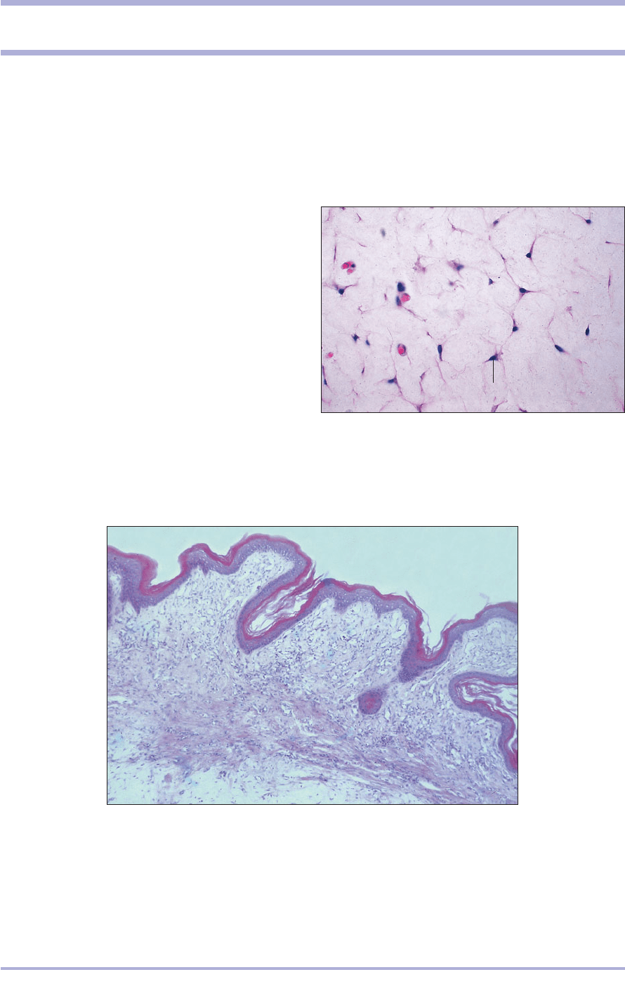
stance, the extracellular matrix (3.1). Mucoid con-
nective tissue is found in the embryo, and also
occurs in limited regions in adult animals, the comb
and wattle of the chicken, and around a healing
wound. There are few cells, which are usually stel-
late undifferentiated fibroblasts; the ground sub-
stance is abundant and gelatinous with very few
fibres. This type of tissue stains poorly with haema-
toxylin and eosin (H & E) (3.2), but stains well with
mucin dyes.
31
3. CONNECTIVE TISSUE
3.1 Umbilical cord (foal). (1) Nucleus of the stellate
mesenchymal cell. (2) Long cell processes. (3) Extracellular
matrix. (4) Blood vessels. H & E. ×125.
3.1
3.2 Mucoid connective tissue. Comb (chicken). (1) Stratified squamous
epithelium. (2) Lamina propria. (3) Mucoid connective tissue. H & E. ×50.
3.2
1
1
2
3
2
3
4
Connective tissue proper
Connective tissue proper fills the interstices of tissues
and organs, and forms a continuous structure that
carries blood vessels and nerves throughout the body.
The relative proportions of the basic components –
fibres, cells and extracellular matrix – determine the
functional characteristics of the tissue, giving it ten-
sile strength in ligaments and tendons and mechan-
ical stability in cartilage and bone, and acting as a
fluid-transport medium: blood.
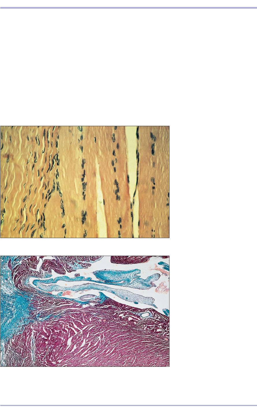
Collagen fibres are thick (2–10 +m in diameter)
and unbranched; when fresh they are white when
unstained and wavy in section. They stain pink
with eosin and green with Masson’s trichrome,
which distinguishes them from muscle fibres (3.3
and 3.4). Elastic fibres are relatively thin (about
1 +m in diameter), less wavy than collagen fibres,
unbranched and yellow in unstained material.
Because they stain poorly with H & E, selective
staining is used (3.5–3.7). Reticular fibres, narrow
bundles of collagen fibrils, are fine and delicate,
branching extensively to form a supporting
network. These also do not stain with standard
methods, so a selective process such as silver
impregnation is used (3.8).
32
Comparative Veterinary Histology with Clinical Correlates
3.3 Tendon/muscle insertion (dog).
(1) Collagen fibres; note wavy
appearance. (2) Fibrocytes.
(3) Striated muscle fibres. H & E.
×200.
3.3
3.4 Heart (kitten). The collagen
fibres and valve cusps are stained
green and the heart muscle is stained
red. Masson’s trichrome. ×50.
3.4
1
2
3

33
3.6 Elastic artery (horse). The elastic
fibres are selectively stained.
Weigert’s elastin. ×62.5.
3.6
3.5 Ligamentum nuchae (ox). The
elastic fibres are stained red and
the collagen fibres are stained
blue/green. Gomori’s trichrome. ×250.
3.5
3.8 Lymph node (dog). The reticular fibres are silver
plated by this method and appear as black strands. The
delicate network of these fibres supports the lymphatic
tissue. Gordon and Sweet. ×125.
3.8
3.7 Elastic artery (horse). The elastic fibres are selectively
stained. Weigert’s elastin. ×500.
3.7
Connective Tissue
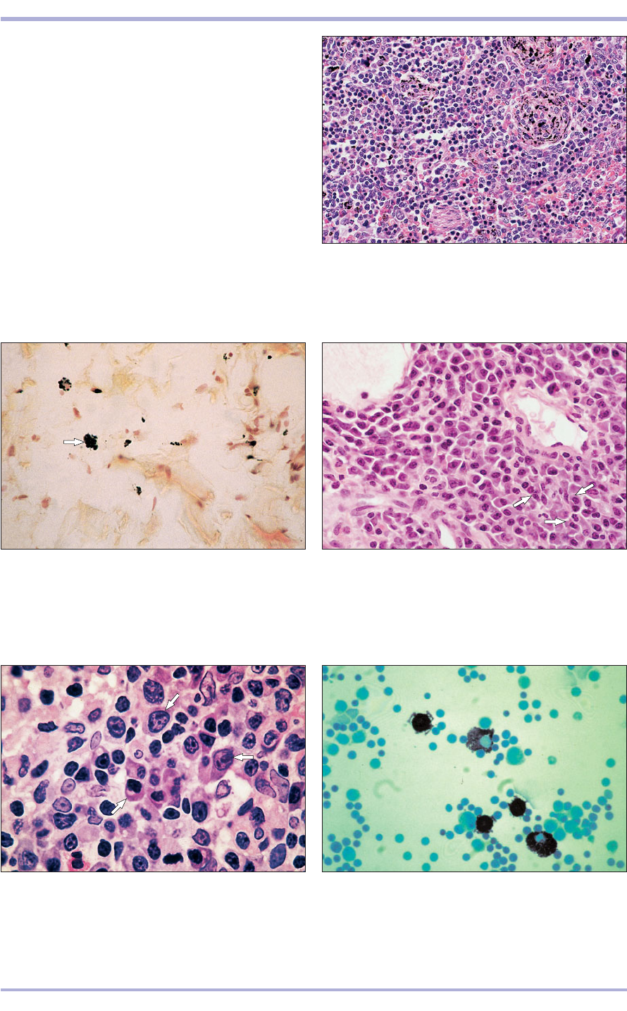
Macrophages, also referred to as histiocytes, are
part of the mononuclear phagocyte system. They
are large, free, mobile phagocytic cells with a round
nucleus. They are part of a large population of
scavengers capable of ingesting cell debris, taking
an active part in the protection of the body by
eliminating some micro-organisms. Identification
may be achieved by using the ability of the cell to
engulf particulate matter, such as carbon particles
injected in vitro (3.11 and 3.12).
Plasma cells have a basophilic cytoplasm and the
nucleus is eccentric with densely clumped chromatin
distributed beneath the nuclear membrane to give
34
Cell types
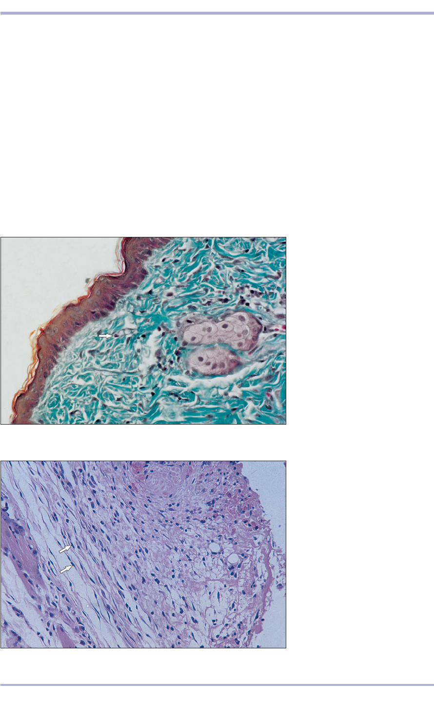
Fibroblasts/fibrocytes are the commonest cell type,
synthesizing collagen, elastin and reticular fibres,
and the extracellular matrix. The fibroblast is the
active form, is elongated and spindle-shaped, and
has abundant cytoplasm and an oval- or cigar-
shaped nucleus. It is found in sites of active repair
or growth. The fibrocyte is the less active stage of
the cell, acting in a maintenance capacity. The cyto-
plasm is reduced in volume and is less reactive to
stains. The nucleus is flattened and the chromatin
is condensed (3.9 and 3.10).
3.10 Fibroblasts in a healing wound
(dog). Nucleus (arrowed) of the
fibroblast. (1) Cytoplasm of the
fibroblast. (2) Collagen fibres. H & E.
×125.
3.10
3.9 Dense irregular connective tissue
(dog). Nucleus of the fibrocyte
(arrowed). (1) Collagen fibres cut in
transverse section. (2) Collagen fibres
cut in longitudinal section. Masson’s
trichrome. ×125.
3.9
1
1
2
2
Comparative Veterinary Histology with Clinical Correlates
35
3.12 Loose connective tissue (dog). A tissue macrophage
is arrowed. Carbon injected, with H & E counterstain.
×200.
3.12
3.11 Spleen (dog). The macrophages have phagocytosed
the carbon particles. Carbon injected, with H & E
counterstain. ×50.
3.11
3.14 Plasma cell (dog). Lymph node. Plasma cells are
arrowed. H & E. ×500.
3.14
3.13 Plasma cell (dog). Lymph node. Plasma cells are
arrowed. H & E. ×200.
3.13
a characteristic clock face appearance. Plasma cells
are involved in the body’s immune response and
represent the cellular source of circulating
immunoglobulin (antibody). They are common
both to loose connective tissue and to the lymphatic
system (3.13 and 3.14).
Mast cells are round or ovoid and the nucleus is
often obscured by the cytoplasmic granules. They are
metachromatic (stain purple with a blue dye such as
toluidine blue or Giemsa) and contain heparin and
histamine and other mediators of inflammation.
Mast cell degranulation causes a local irritant effect
caused by the release of histamine (3.15).
3.15 Mast cell (sheep). The granules stain purple with
the blue dye, toluidine blue. Metachromasia. Toluidine
Blue. ×500.
3.15
Connective Tissue
36
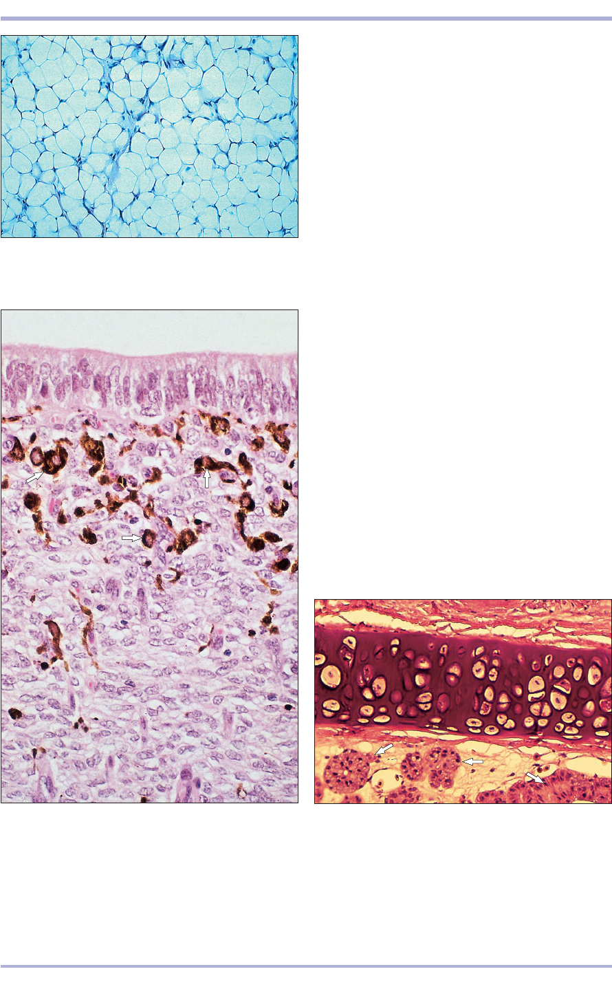
3.18 Nasal septum of green iguana. A supporting sheet of
hyaline cartilage is seen in the centre. Immediately beneath
are saline-secreting nasal salt glands (arrowed). H & E. ×100.
3.18
Fat cells (adipocytes) arise from pericapillary
mesenchymal cells. They accumulate fat in the
cytoplasm as lipid droplets that coalesce until the
cell is filled with one large droplet. The cytoplasm
forms a peripheral rim and the nucleus is displaced
to lie immediately beneath the plasma membrane.
Fat cells may occur singly or in groups in loose
connective tissue to become adipose tissue. Fat
may be white, in which each cell has a single large
droplet, or brown, in which small individual
droplets are scattered throughout the cytoplasm.
Processing dissolves the fat and leaves a network
of lacy, empty spaces in H & E sections. The cells
are usually described as appearing like a signet ring
with the nucleus constituting the signet (3.16).
Pigment cells are derived from neural crest
ectoderm. However, cells carrying pigment granules
(melanin, erythrin, xanthin) are often found in
connective tissue and are called chromatophores
(literally, pigment carriers) and can be macrophages
(melanophages, erythrophages, xanthophages;
3.17<3.20).
Neutrophils (polymorphonuclear leucocytes in
mammals; azurophils, see p. 58, in some lower
vertebrates), eosinophils, lymphocytes and monocytes
are blood cells commonly found in loose connective
tissue. They are migratory and move freely between
3.16 Adipocytes. Foot pad (dog). The fat has been lost
during processing and the lacy network is formed by
cytoplasm and cell membranes. H & E. ×50.
3.16
3.17 Melanocytes. Uterus (ewe). The melanocytes are
arrowed. H & E. ×200.
3.17
Comparative Veterinary Histology with Clinical Correlates

37
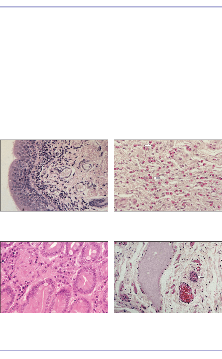
3.19 Serosal surface of chameleon small intestine. Note heavily pigmented
coelomic surface and zone between longitudinal and circular muscular layers.
H & E. ×50.
3.19
3.20 The hepatic parenchyma of many amphibians and reptiles is
characterized by abundant aggregates of melanin pigment contained in
melanophages (arrowed). Illustrated is a section of liver from an African
clawed frog (Xenopus laevis). The hepatocytes are arranged in ray-like cords
extending outward from a thin-walled central vein, next to which are small
tributaries of the hepatic artery and bile duct. H & E. ×100.
3.20
Connective Tissue
38
vessels and nerves. It contains many scattered cells
of various types, blood and lymphatic vessels, and
a loose network of fine collagenous, reticular and
elastic fibres. It is widespread throughout the body,
surrounding vessels and nerves, and is found in
serous membranes, the lamina propria of mucous
membranes, subcutaneous tissue and the superficial
layer of the dermis (3.25). Amorphous ground sub-
stance is particularly abundant in loose connective
tissue. It is composed of a group of carbohydrates,
the glycosaminoglycans, which may be complexed
with a protein to form proteoglycans. These sub-
stances stain poorly with H & E.
Dense connective tissue
Composed principally of thick collagenous fibres,
dense connective tissue contains few cells. Fibrous
elements predominate and the commonest cell is the
3.24 Pericyte. Loose connective tissue (cat). The pericyte is
arrowed in the wall of the arteriole. (1) Vein. (2) Lymphatic
vessel. (3) Fibrocyte. (4) Extracellular matrix. H & E. ×100.
3.24
3.23 Lymphocytes. Duodenum (ox). Lymphocytes are
present in the lamina propria of the duodenum. H & E.
×125.
3.23
3.22 Eosinophils. Colon (horse). The eosinophils are
arrowed. H & E. ×125.
3.22
3.21 Polymorphonuclear leucocytes. Bronchus (ox).
The polymorphonuclear leucocytes have invaded the
connective tissue of the lamina propria in response to
infection. H & E. ×125.
3.21
the blood vessels and the surrounding tissue in
response to local conditions (3.21–3.23).
Endothelial cells and pericytes form a special cell
population in connective tissue, retaining the capacity
to divide and to synthesize collagen and the
extracellular ground substance. The endothelium is
often fenestrated in the capillary bed and it controls
the tissue fluid content locally. Pericytes are pale
staining, connective tissue cells lying adjacent to the
capillary endothelium. They are comparatively
undifferentiated and can give rise both to fibroblasts
and to smooth muscle cells in areas of tissue repair,
as well as assist in the revascularization and repair
of damaged blood vessels (3.24).
Loose areolar connective tissue
Loose areolar connective tissue is found as packing
material throughout the body and carries the blood
2
3
1
4
3
Comparative Veterinary Histology with Clinical Correlates
39
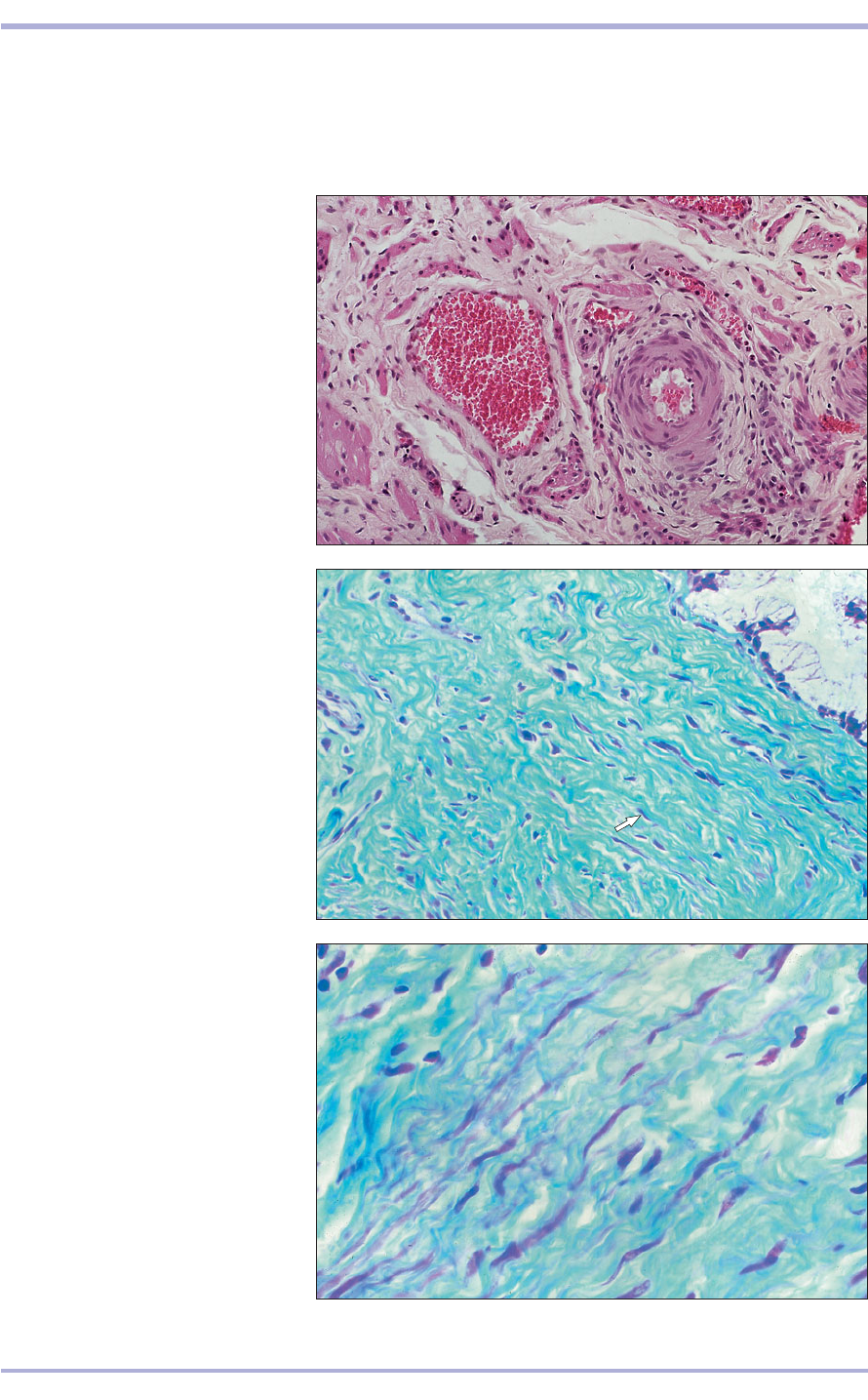
3.25 Loose connective tissue (cat).
Blood vessels: (1) artery; (2) vein.
(3) Nerves. (4) Extracellular matrix.
(5) Fibrocytes. (6) Collagen fibres.
(7) Smooth muscle of the uterine wall.
H & E. ×100.
3.25
3.26 Dense connective tissue (dog).
Fibrocytes (arrowed). (1) Collagen
fibres. (2) Blood vessels. Masson’s
trichrome. ×100.
3.26
1
2
2
2
1
4
5
6
7
3
3.27 Dense connective tissue. Vagina
(sheep). The collagen fibres are stained
green. Masson’s trichrome. ×160.
3.27
fibrocyte. In dense regular connective tissue the
fibres may be arranged in rows to provide tensile
strength in tendons and ligaments, and as sheets in
aponeuroses (see 3.3 and 3.4). In dense irregular
connective tissue the fibres are arranged in different
planes to allow stretching without tearing of the sur-
face membrane, as in the dermis and the vagina
(3.26 and 3.27).
Connective Tissue
40
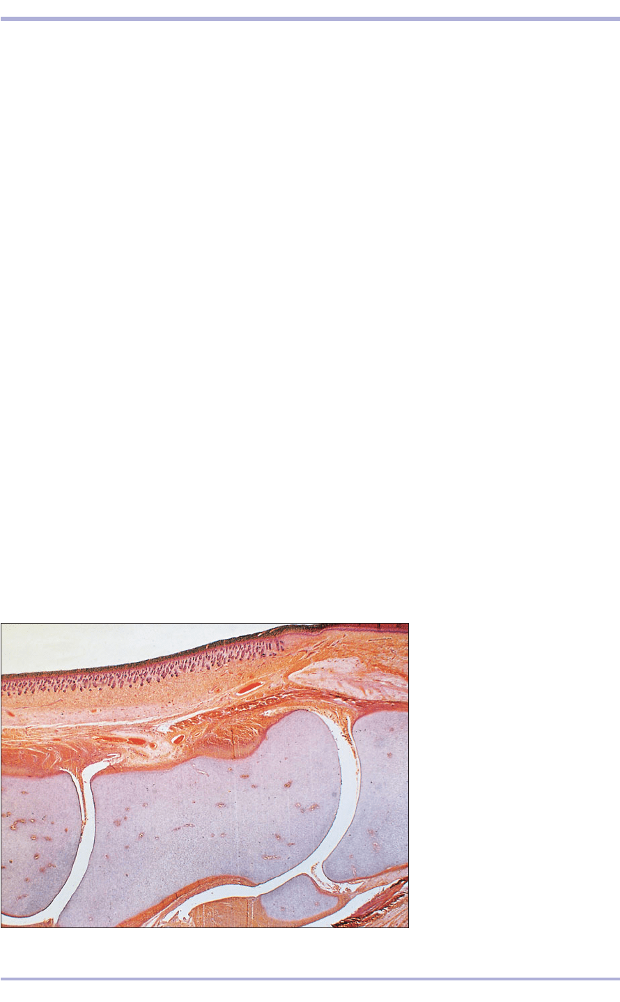
3.28 Developing hoof (foal). (1) Skin.
(2) Hyaline cartilage models of the
digits. (3) Joint cavity. H & E. ×25.
3.28
Special types of connective tissue
Reticular tissue is composed of numerous reticular
fibres and stellate reticular cells, forming a sup-
portive network for structures such as the spleen,
lymph node, kidney and bone marrow. Elastic tis-
sue, characterized by numerous regularly or irreg-
ularly arranged elastic fibres, is exemplified by the
ligamentum nuchae and the elastic fascia of the
ruminant abdomen. Adipose tissue consists of
groups of adipocytes (see above).
Cartilage and bone
Cartilage
Cartilage is a specialized form of connective tissue
combining a degree of rigidity with flexibility and
strength. There are three types of cartilage: hyaline,
elastic and fibrocartilage; differing only in the dis-
tribution of the main components: the cells, fibres
and matrix.
Hyaline cartilage
This type of cartilage is bluish/white in the fresh state
and is the most prevalent form. In the embryo, the pre-
cursors of the long bones begin as cartilage models
(3.28). As the neonate grows, the cartilaginous tem-
plate undergoes progressive mineralization. In post-
natal life, cartilage is present in the rings of the trachea
and in plates in the larynx and nose. With ageing and
under certain conditions of hypervitaminosis-D
3
and
hypercalcaemia, cartilage may become pathologically
mineralized. Cartilage also caps the ends of bones in
articulating joints (3.29).
At predetermined sites in the embryo, mesenchy-
mal cells round off and differentiate into chondro-
blasts (cartilage-forming cells) and secrete a matrix
consisting of proteoglycans and collagen fibrils. The
space occupied by each cell is a lacuna and once the
matrix is laid down, the cells are called chondrocytes
(cartilage cells; 3.30). Chondrocytes are capable of
dividing and several cells may come to occupy a
lacuna; then they are known as an isogenous group
or cell nest (3.31). Compared with the bulk of the
matrix, which stains poorly with H & E, the matrix
in the immediate vicinity of the cells stains intensely
with metachromatic dyes because of the presence of
glycosaminoglycans. Mesenchymal tissue surrounds
the developing cartilage and forms a fibrous cover-
ing, the perichondrium. The inner layer of the
perichondrium is capable of generating new chon-
droblasts. Cartilage is thus able to grow from the
pericardium by appositional growth, and by inter-
stitial growth from within by chondrocyte division
and deposition of new matrix. It is avascular – the
cells are nourished by diffusion.
1
2
2
2
3
3
Comparative Veterinary Histology with Clinical Correlates
