Aughey E., Frye F.L. Comparative veterinary histology with clinical correlates
Подождите немного. Документ загружается.

6.25
6.28
6.27
6.26
81
Cardiovascular System
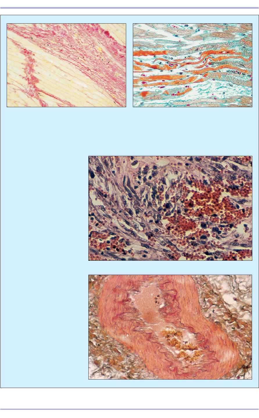
6.27 Haemangiosarcoma in a
horse. The tumour cells are spindle-
shaped with large, often
hyperchromatic, nuclei and are
arranged into loosely interlacing
bundles that form irregular, blood-
filled channels and spaces. H & E.
×125.
6.25, 6.26 Myocardial degeneration and fibrosis in a 12-year-old dog with cardiomyopathy. In 6.25, the
collagenous tissue which replaces muscle bundle is stained red. Sirius Red. ×45. In 6.26, degenerative muscle
fibres are seen stained strongly orange in the centre. The striations of the muscle fibres are also demonstrated
with this stain. Masson’s trichrome. ×180.
6.28 Early arteriosclerotic
mineralization in a large pulmonary
artery (iguana). The mineral
deposition is highlighted in red.
Alcian blue/PAS. ×125.
The respiratory system has two functions: conduc-
tion and respiration. Conduction is carried out via a
continuous system of tubes carrying air from the nos-
trils into the nasal cavity through the nasopharynx
and larynx to the trachea and bronchi. The air is
warmed, moistened and filtered in these passages
before reaching the organs responsible for respira-
tion: the lung parenchyma, the respiratory bronchi-
oles, alveolar sacs and alveoli. It is between the alve-
oli and the capillaries that gas exchange takes place.
The paranormal sinuses are cavities found in
skull bones. The mucoendosteum lining these cav-
ities is continuous with the mucous membrane of
the nasal cavity.
7. RESPIRATORY SYSTEM
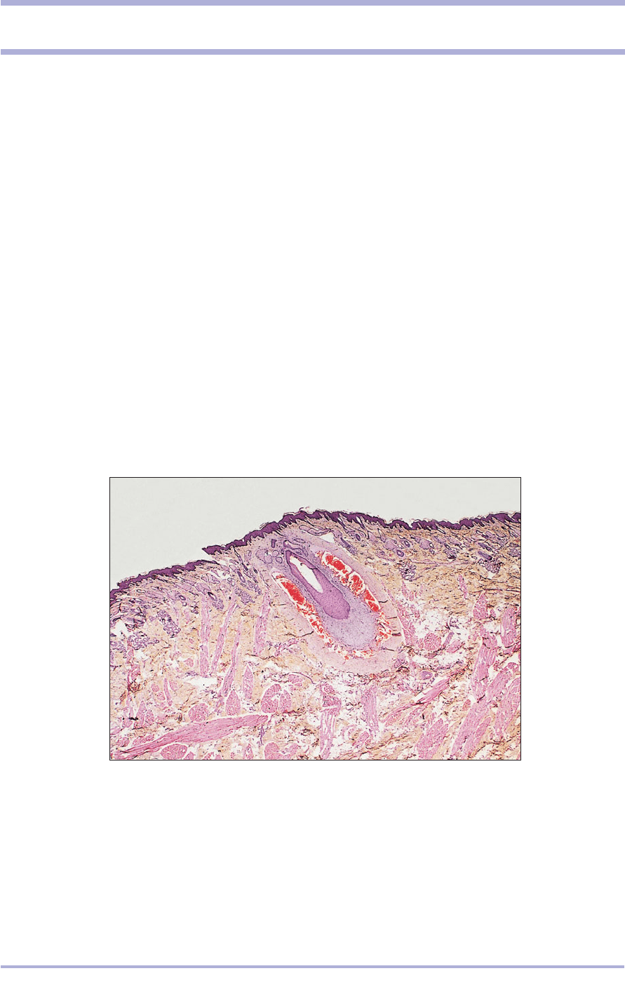
7.1 Skin. Nostril (horse). (1) Sinus hair. (2) Lamina propria.
(3) Sebaceous glands. (4) Sweat glands. H & E. ×20.
7.1
1
2
4
4
3
Conduction of air
The skin around the nostrils has long tactile hairs
and numerous sebaceous and sweat glands (7.1).
Respiratory epithelium lines all but the finer
divisions of the respiratory tract and consists of
pseudostratified columnar ciliated cells and mucus-
secreting goblet cells. The lamina propria is con-
tinuous with the perichondrium or periosteum
where appropriate, is very vascular, contains both
collagen and elastic fibres, and warms the inspired
air. Seromucous glands secrete into the lumen
through the epithelium (7.2–7.4).
82
83
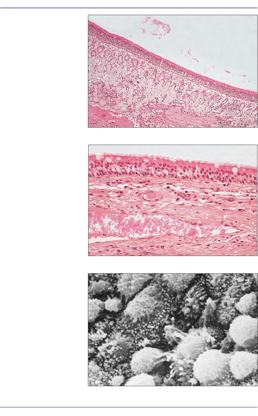
7.2 Respiratory epithelium (horse).
(1) Pseudostratified columnar ciliated
epithelium with goblet cells.
(2) Lamina propria. (3) Seromucous
glands. (4) Smooth muscle. H & E.
×62.5.
7.2
2
3
4
1
7.3 Respiratory epithelium (horse).
(1) Pseudostratified columnar ciliated
epithelium with goblet cells.
(2) Lamina propria. (3) Blood vessels.
H & E. ×160.
7.3
7.4 Respiratory epithelium (horse).
The cilia project from the surface of
the epithelial cell as fine strands;
active mucus-secreting cells lie
between the ciliated cells (arrowed).
Scanning electron micrograph. ×1500.
7.4
2
3
1
3
Respiratory System
Cilia beat towards the pharynx, an area com-
mon both to the digestive and to the respiratory
system. The auditory tube connects the pharynx
to the middle ear and is common to the digestive,
respiratory and auditory systems (7.5). The gut-
tural pouch of equids is a diverticulum of the audi-
tory tube. The digestive surface is covered by a
stratified squamous epithelium that is continuous
with the oral cavity, and the respiratory surface by
respiratory epithelium continuous with the nasal
cavities (7.6 and 7.7). The epiglottis is a flap-like
structure projecting into the pharynx. A plate of
elastic cartilage provides internal support. The
upper digestive surface mucous membrane is cov-
ered by a non-keratinizing stratified squamous
epithelium (7.8) and the lower respiratory surface
by respiratory epithelium (7.9). Taste buds may be
present on the laryngeal aspect.
84
Comparative Veterinary Histology with Clinical Correlates
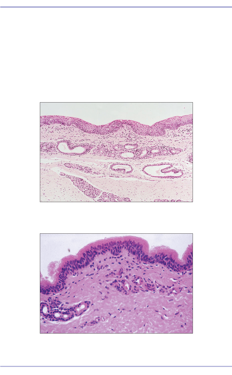
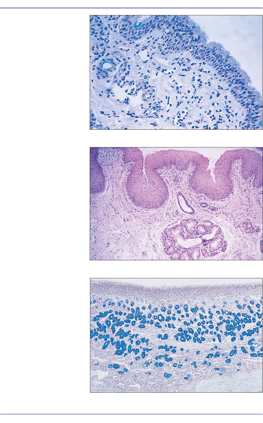
7.5 Auditory tube (horse). (1) Respiratory epithelium. (2) Lamina propria.
(3) Seromucous glands. H & E. ×100.
7.5
7.6 Guttural pouch (horse). (1) Respiratory epithelium. (2) Lamina propria.
(3) Seromucous glands. H & E. ×250.
7.6
3
3
1
2
2
3
1
3
85
7.7 Guttural pouch (horse). (1)
Respiratory epithelium; the goblet
cells are individually stained blue.
(2) Lamina propria. (3) Seromucous
glands. Alcian blue/PAS. ×200.
7.7
2
1
3
7.8 Epiglottis (horse). (1) Stratified
squamous epithelium. (2) Lamina
propria. (3) Seromucous glands.
H & E. ×200.
7.8
2
3
1
7.9 Epiglottis (horse). (1) Stratified
columnar epithelium with mucus-
secreting cells stained blue.
(2) Lamina propria. (3) Seromucous
glands. Alcian blue/PAS. ×125.
7.9
2
3
1
Respiratory System
The larynx is lined by respiratory epithelium. The
lamina propria is continuous with the perichon-
drium of the laryngeal cartilages. The vocal cords
are covered by stratified squamous epithelium (7.10
and 7.11). The trachea extends from the larynx to
the bifurcation of the extrapulmonary bronchi.
These tubes have the same structure: a lining of res-
piratory epithelium rising on a lamina propria of
loose connective tissue with elastic fibres and mixed
seromucous glands opening into the lumen.
Incomplete rings of hyaline cartilage keep the lumen
patent and smooth muscle fibres bridge the gap at
the dorsal aspect of the trachea (7.12). In the
bronchus the hyaline cartilage has a plate-like
arrangement. The smooth muscle forms a spiral and
appears as discontinuous blocks in transverse sec-
tions (7.13 and 7.14). A fibrous adventitial coat cov-
ers the trachea and the extrapulmonary bronchi.
86
Comparative Veterinary Histology with Clinical Correlates
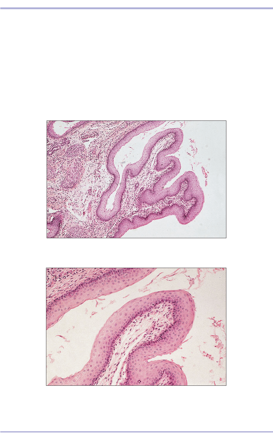
7.10 Larynx (vocal cord; dog). (1) Stratified squamous epithelium. (2) Lamina
propria. (3) Simple tubular glands. H & E. ×62.5.
7.10
7.11 Larynx (vocal cord; dog). (1) Stratified squamous epithelium
covering the vocal cord. (2) Connective tissue core of the lamina propria.
(3) Parasympathetic ganglion. H & E. ×160.
7.11
2
1
3
1
2
3
3
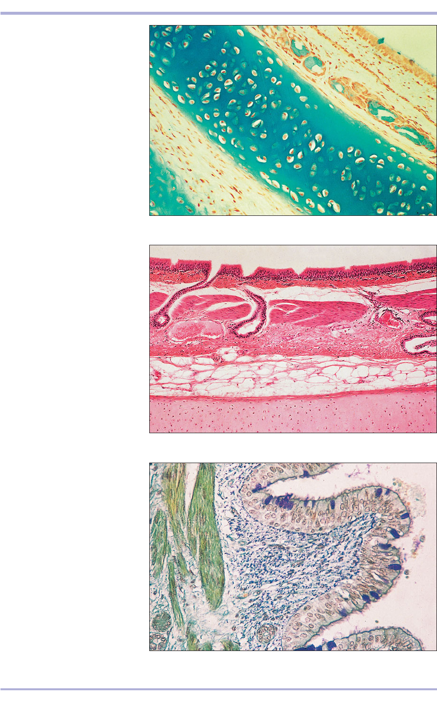
7.12 Trachea (sheep). (1) Respiratory
epithelium. (2) Lamina propria.
(3) Seromucous glands. (4) Hyaline
cartilage. Gomori’s trichrome. ×25.
7.12
1
3
4
2
7.14
7.13
87
Respiratory System
2
1
2
3
4
4
5
1
3
6
7.13 Bronchus (sheep).
(1) Respiratory epithelium.
(2) Lamina propria. (3) Smooth
muscle. (4) Simple tubular glands
open into the lumen through the
epithelium. (5) Perichondrium.
(6) Hyaline cartilage. H & E. ×25.
7.14 Bronchus (ox). (1) Respiratory
epithelium; the goblet cells are
stained specifically. (2) Lamina
propria. (3) Smooth muscle.
(4) Simple tubular glands.
Gomori/aldehyde fuchsin. ×200.
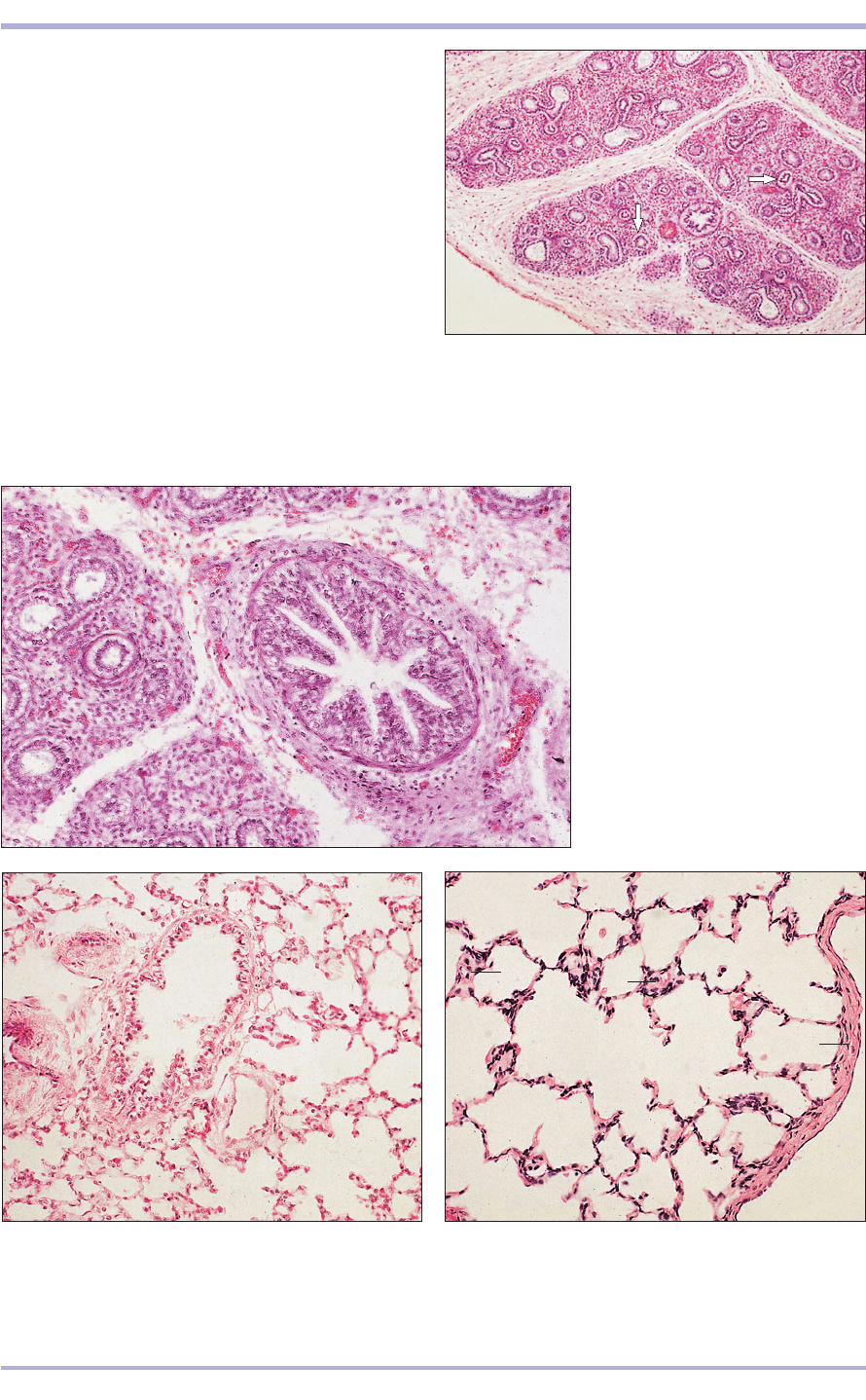
7.18 Adult lung (sheep). (1) Elastic connective tissue
with an outer layer of mesothelium of the visceral pleura.
(2) Alveoli. (3) Blood vessels. H & E. ×62.5.
7.18
88
7.15 Bovine fetal (160-day) lung. Blood vessels
(arrowed). (1) Pleural mesothelium. (2) Mesenchyme.
(3) Duct system. H & E. ×5.
7.15
7.16 Bovine fetal (160-day) lung.
(1) Large duct lined by a simple
columnar epithelium. (2) Smooth
muscle. (3) Vascular mesenchyme.
(4) Small ducts. H & E. ×25.
7.16
1
2
3
3
3
1
3
4
2
3
3
2
2
1
7.17 Fetal lung at term (cow). (1) Respiratory bronchiole
lined by simple columnar epithelium. (2) Alveoli. H & E.
×62.5.
7.17
1
2
2
Respiration
The bronchi, both extrapulmonary and intrapul-
monary, bring air to the lungs and branch out
within the lungs into the bronchioles, which culmi-
nate in clusters of minute sacs: the alveoli. In the
fetal lung the duct system is developed whereas the
respiratory part develops slowly (7.15–7.17).
Expansion begins with the first respiratory move-
ments after birth. Thereafter, the lung expands in
tandem with the growth of the animal.
Each lung is covered by elastic connective tissue
with an outer layer of mesothelium, the visceral
pleura (7.18). Connective tissue septa divide the
lung into lobes and lobules and the intrapulmonary
bronchi have the same structure as the extrapul-
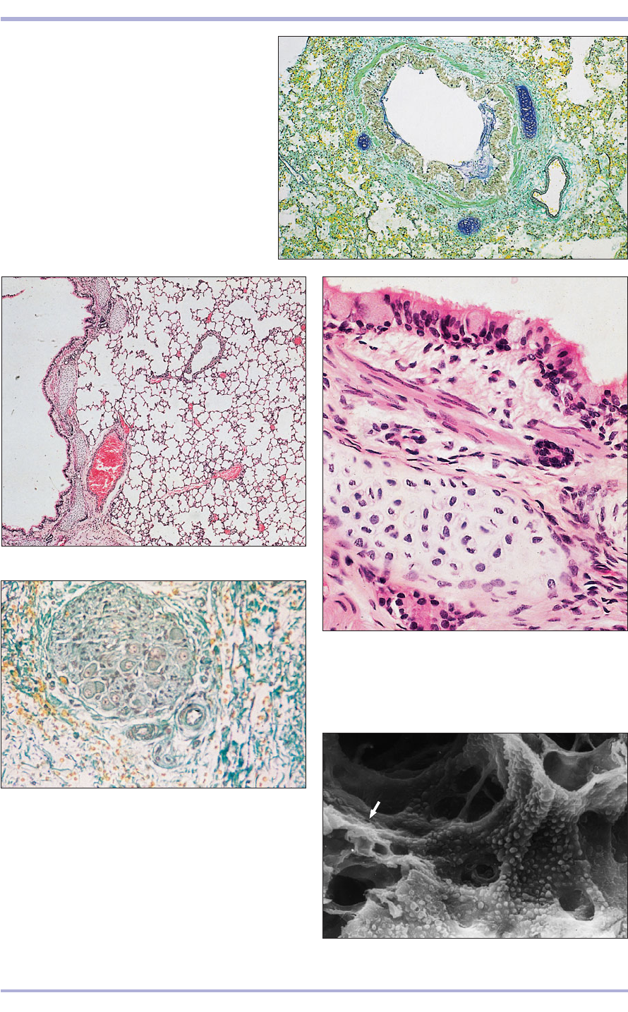
monary bronchi (7.19–7.23). The epithelium of
Comparative Veterinary Histology with Clinical Correlates
7.19
5
6
6
4
1
2
3
7.19 Intrapulmonary bronchus. Lung (cow).
(1) Lumen lined by respiratory epithelium.
(2) Lamina propria. (3) Smooth muscle.
(4) Hyaline cartilage. (5) Blood vessel.
(6) Alveoli. Gomori/aldehyde fuchsin. ×52.
7.20 Intrapulmonary bronchus. Lung (sheep).
(1) Lumen lined by respiratory epithelium.
(2) Lamina propria. (3) Smooth muscle.
(4) Hyaline cartilage. (5) Simple tubular
glands. (6) Blood vessel. (7) Bronchiole.
(8) Alveolar duct. (9) Alveoli. H & E. ×62.5.
7.20
1
1
3
7
8
9
4
2
5
6
89
Respiratory System
7.21 Intrapulmonary bronchus. Lung (sheep).
(1) Respiratory epithelium. (2) Lamina propria.
(3) Smooth muscle. (4) Hyaline cartilage. H & E. ×200.
7.21
7.22 Intrapulmonary bronchus. Lung (cow). A
parasympathetic ganglion is present surrounded
by vascular connective tissue of the lamina
propria.Gomori/aldehyde fuchsin. ×160.
7.22
1
2
3
4
7.23 Intrapulmonary bronchus (dog). (1) The
intrapulmonary bronchus is lined by respiratory
epithelium (arrowed). (2) Alveoli. Scanning electron
micrograph. ×500.
7.23
1
2
2
90
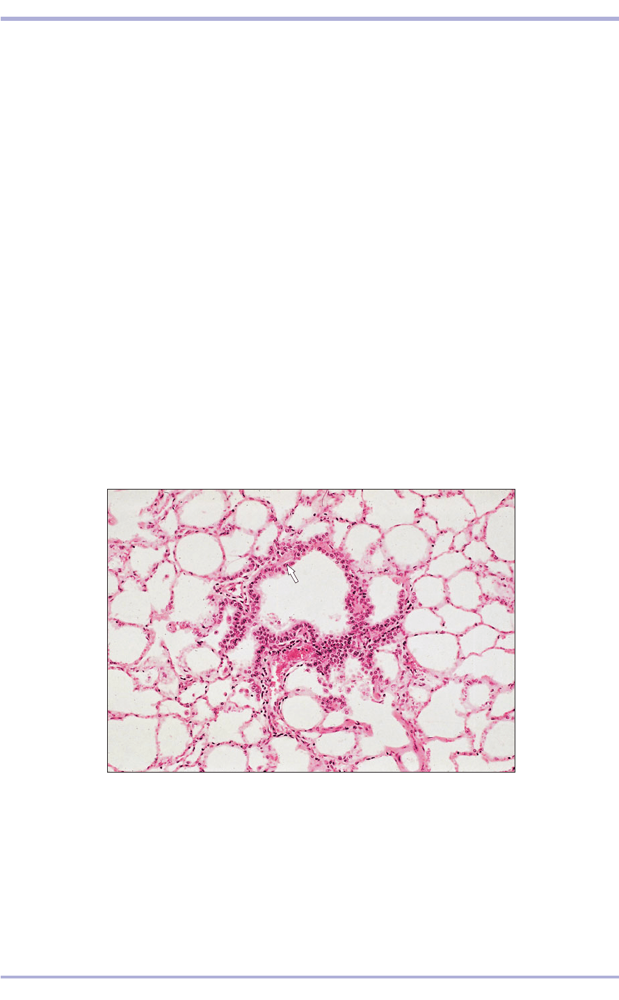
7.24 Bronchiole. Lung (sheep). (1) The bronchiole is lined by cuboidal/low
columnar epithelium with ciliated and non-ciliated cells (arrowed). (2) Smooth
muscle. (3) Alveolar duct. (4) Alveoli. The free cells in the lumen are
macrophages. H & E. ×125.
7.24
1
2
2
2
3
4
4
the bronchioles is columnar or even cuboidal and
ciliated. In the smaller bronchioles the epithelium
is thinner, the lamina propria is elastic, the smooth
muscle forms a complete ring and there is no
adventitia (7.24 and 7.25).
Non-ciliated bronchiolar cells (Clara cells) are
tall, dome-shaped and protrude into the bronchio-
lar lumen. They replace the mucus-secreting gob-
let cells at this level. Both ciliated cells and Clara
cells are present in the terminal and respiratory
bronchioles (in the dog and cat they are lined by the
latter exclusively). Clara cells divide to form other
Clara or ciliated cells and have an important role in
the repair of damaged epithelium. Their secretion
also keeps the small airways patent.
Respiratory bronchioles are lined with a low
columnar or cuboidal epithelium with ciliated and
bronchiolar cells, an elastic lamina propria and a
smooth muscle layer. This opens into the alveolar
duct lined by squamous epithelium, interrupted by
atria and alveoli along its length (7.26). Alveoli are
the functional exchange part of the lung. The septa
are very thin, with both elastic and collagen fibres,
and contain one of the most extensive capillary net-
works in the body. Cells of the immune system,
derived from blood monocytes, are also present
and migrate through the alveolar epithelium into
the air space, where they phagocytose particulate
matter and micro-organisms to become dust cells
(alveolar phagocytes). The respiratory membrane
where gas exchange takes place consists of capil-
lary endothelial cells, alveolar epithelial cells and a
fused basement membrane (7.27). The squamous
alveolar cell, the lining cell responsible for gas
exchange, is a type I pneumocyte. The great alve-
olar cell secretes surfactant to reduce surface ten-
sion. It is a type II pneumocyte, is cuboidal and
projects into the lumen.
Comparative Veterinary Histology with Clinical Correlates
