Shani G. Radiation Dosimetry: Instrumentation and Methods
Подождите немного. Документ загружается.

400 Radiation Dosimetry: Instrumentation and Methods
19. Rosenfeld, A. B. et al., IEEE Trans. Nucl. Sci., 46, 1766,
1999.
20.
Van Dam, G. et al., Radiother. Oncol., 19, 345, 1990.
21.
Edwards, C. R. et al., Phys. Med. Biol., 42, 2383, 1997.
22.
Buehler, M. G. et al., IEEE Trans. Nucl. Sci., 40, 1442,
1993.
23.
Brucker, G. J. et al., IEEE Trans. Nucl. Sci., 42, 33, 1995.
24.
Schwank, J. R. et al., IEEE Trans. Nucl. Sci., 43, 2671,
1996.
25.
Buehler, M. G. et al., IEEE Trans. Nucl. Sci., 43, 2679,
1996.
26.
Grusell, E. and Rikner, G., Phys. Med. Biol., 38, 785,
1993.
27.
Piermattei, A. et al., Med. Phys., 22, 835, 1995.
28.
Nuclear Associates, Diagnostic Imaging and Radiation
Therapy Catalog,
1999.
29.
Heukelom, S. et al., Phys. Med. Biol., 36, 47, 1991.
30.
Khivrich, V. I. et al., IEEE Trans. Nucl. Sci., 43, 2687,
1996.
31.
Bellem, R. D. et al., IEEE Trans. Nucl. Sci., 41, 2139,
1994.
32.
Falco, T. et al., Med. Phys., 25, 814, 1998.
33.
ICRU 37, Stopping powers for electrons and positrons,
1984.
34.
ICRU 22, Measurement of low-level radioactivity,
1972.
Ch-08.fm Page 400 Friday, November 10, 2000 12:03 PM

401
9
Gel Dosimetry
CONTENTS
I. Introduction and Basic Concepts NMR Relaxation ..............................................................................................401
II. Application of Ferrous Ions..................................................................................................................................409
III. Application of BANG Polymer Gel ......................................................................................................................416
References .......................................................................................................................................................................422
I. INTRODUCTION AND BASIC CONCEPTS
In 1984, Gore et al. [1] proposed that the radiation-induced
changes in the well-established Fricke solution could be
probed with nuclear magnetic resonance (NMR) relax-
ation measurements rather than using the conventional
spectrophotometry. Gore et al. realized that the NMR spin-
lattice and spin-spin relaxation rates (1
T
1
and 1
T
2
,
respectively) of the dosimeter are related to the amount
of Fe
3
present in the Fricke solution and, since these
relaxation parameters govern the contrast of MR images,
it was possible to observe the radiation-induced changes
in Fricke phantoms by MRI. This initial work indicated
that there was a possibility for a 3-D dosimetry based on
MRI and ferrous sulphate (Fricke) dosimeters. Following
the initial proposal, a number of groups reported on MR-
based radiation dosimetry. Most imaging applications
used ferrous sulphate solutions incorporated into gel
matrices (Fricke-gels) to stabilize the spatial integrity of
the dosimetry. Various gel matrices were investigated for
Fricke-gel MR-based dosimetry, including gelatin, agar-
ose, and sephadex. Each system had its advantages and
limitations. A few papers have suggested that dose distri-
butions can be determined directly from MR images based
on signal intensity and calibration curves. However, the
dosimeters require high doses, typically 10–40 Gray (Gy),
for radiation-induced changes to be readily observed by
NMR. The Fe ions diffuse even in the gel matrices and
the spatial information eventually is destroyed. The time
between the start of irradiation to the end of the dose
measurement should be no more than about two hours.
It was well-known that irradiating polymers or mac-
romolecules could alter their molecular dynamics and
structure. This made polymer systems good candidates for
MRI-based dosimetry. For many polymers, the changes
occurred only at very high doses (
10
4
Gy). However, by
choosing a suitable polymer or polymer-solvent system,
the dose range could be extended to a range more exten-
sive than that offered by Fricke dosimeters (e.g., 50 cGy
to 100 Gy with aqueous polyacrylamide).
While the relaxation times had been shown to be very
sensitive probes of the viscosity and molecular weight of
polymer systems, the changes in the relaxation times of
irradiated systems were not expected a priori to be related
to dose in a simple manner.
Polymer-gel dosimeters present many advantages over
Fricke-gel systems (principally since there is significantly
less diffusion of polymers within the gel matrix so that,
theoretically, the radiation-induced changes maintain their
spatial integrity indefinitely). There are still a number of
features (cost, duration of polymerization reactions after
irradiation, monomer toxicity) which seem less attractive.
Radiation-induced polymerization of polymer-gel
dosimeters is clearly visible. Experience with optical
changes in gel dosimeters has initiated a new realm in gel
dosimetry, with dose measurement using optical tech-
niques and image reconstruction in two and three dimen-
sions. [2] Most of the reactions involving the free radicals
initially produced by the radiation are very rapid and are
essentially complete within a microsecond. However, sub-
sequent reactions of non-radical products, or of large free
radicals on polymer chains, may be quite slow. Figure 9.1
is an example of the agarose/ferrous system. [3]
NMR R
ELAXATION
Nuclear magnetic resonance (NMR) methods have been
very useful in the study of the structure, composition, and
molecular dynamics of various materials. Of particular
interest are NMR relaxation or relaxometry studies of
radiation chemical dosimeters such as the ferrous sulfate–
doped or Fricke gels and polymer gels. The radiation-
induced changes in the solutes of these aqueous dosimeters
affect the relaxation properties of the water protons
(hydrogen nuclei) constituting most of the magnetization
signal studied using proton NMR or magnetic resonance
Ch-09.fm Page 401 Friday, November 10, 2000 12:04 PM
402
Radiation Dosimetry: Instrumentation and Methods
imaging (MRI) methods. Models show that the NMR
dose response of the gel dosimeters is governed by two
mechanisms: the chemical response of the gel to radia-
tion and the response of NMR parameters to radiation
products. NMR dose response models allow for the
absorbed radiation dose to be determined from funda-
mental physical variables rather than a calibration of the
dosimeter’s response. [4]
If the material is irradiated with a time-dependent
magnetic field or radio frequency (rf) energy, such as rf
pulse sequences, the equilibrium magnetization may be
perturbed such that the magnetization becomes time-
dependent and possesses components transverse to the
external magnetic field. Once the perturbing rf energy is
removed, the magnetization begins to relax to its equilib-
rium value and direction. The longitudinal and transverse
magnetizations relax at the characteristic longitudinal and
transverse rates
R
1
and
R
2
, which are simply the inverse
of the relaxation times
T
1
and
T
2
.
The macroscopic equilibrium magnetization
M
(bold
type represents a vector quantity) of a material placed in
an external magnetic field
H
(
0,0,
H
0
)), oriented by con-
vention along the
z
-axis, results from the superposition of
the magnetic moments of individual nuclei in the material.
Only nuclei with a non-zero spin possess a nuclear mag-
netic moment,
I
, where
is the gyromagnetic ratio
specific to a particular nucleus, 2
, and
I
(
(
I
x
,
I
y
,
I
z
)) is the
quantum mechanical spin operator. In MRI gel dosimetry
the nuclei of interest are mainly the hydrogen nuclei or
protons on the water and macromolecules in the gels. The
following discussions is limited to these nuclei of spin 1
2.
The overall magnitude and direction of
M
(
(0,0,
M
0
))
depend on the behavior of individual nuclear magnetic
moments or spins interacting with the magnetic field. The
spins may be classically described as precessing about the
z
-axis at the Lamor frequency of
0
H
0
and aligned
with or against
H
. Both orientations represent a different
spin state with a different energy level. Spins aligned along
the field are in the lower energy state of
E
1
2
H
0
,
and those aligned against the field are in the higher energy
state of
E
1
2
H
0
. This description of spin behavior
concurs with that determined quantum-mechanically by
solving the equation of motion of
,
(9.1)
where
H
0
(
H
H
0
I
z
) is the Zeeman Hamiltonian
representing the interaction between the spin and the mag-
netic field. Taking the expectation value of the solution
for
(
t
) also yields an oscillatory time dependence for
the
x
,
y
(
t
) component and a constant
z
component aligned
with or against
H
.
For a material composed of an ensemble of spins, the
components of the net macroscopic magnetization are the
average of the expectation values of the corresponding
components, , multiplied by the number of spins,
N:
(9.2)
where
i
designates the axis (
i
x
,
y
,
z
). At equilibrium
the phase of the oscillating
x
,
y
component for individual
spins varies randomly over all spins; i.e., there is no phase
coherence between the
x,y
of the spins. Thus, the average
is zero, and no net transverse magnetization
M
x,y
is observed. The average depends on the number
of spins directed along the magnetic field and the number
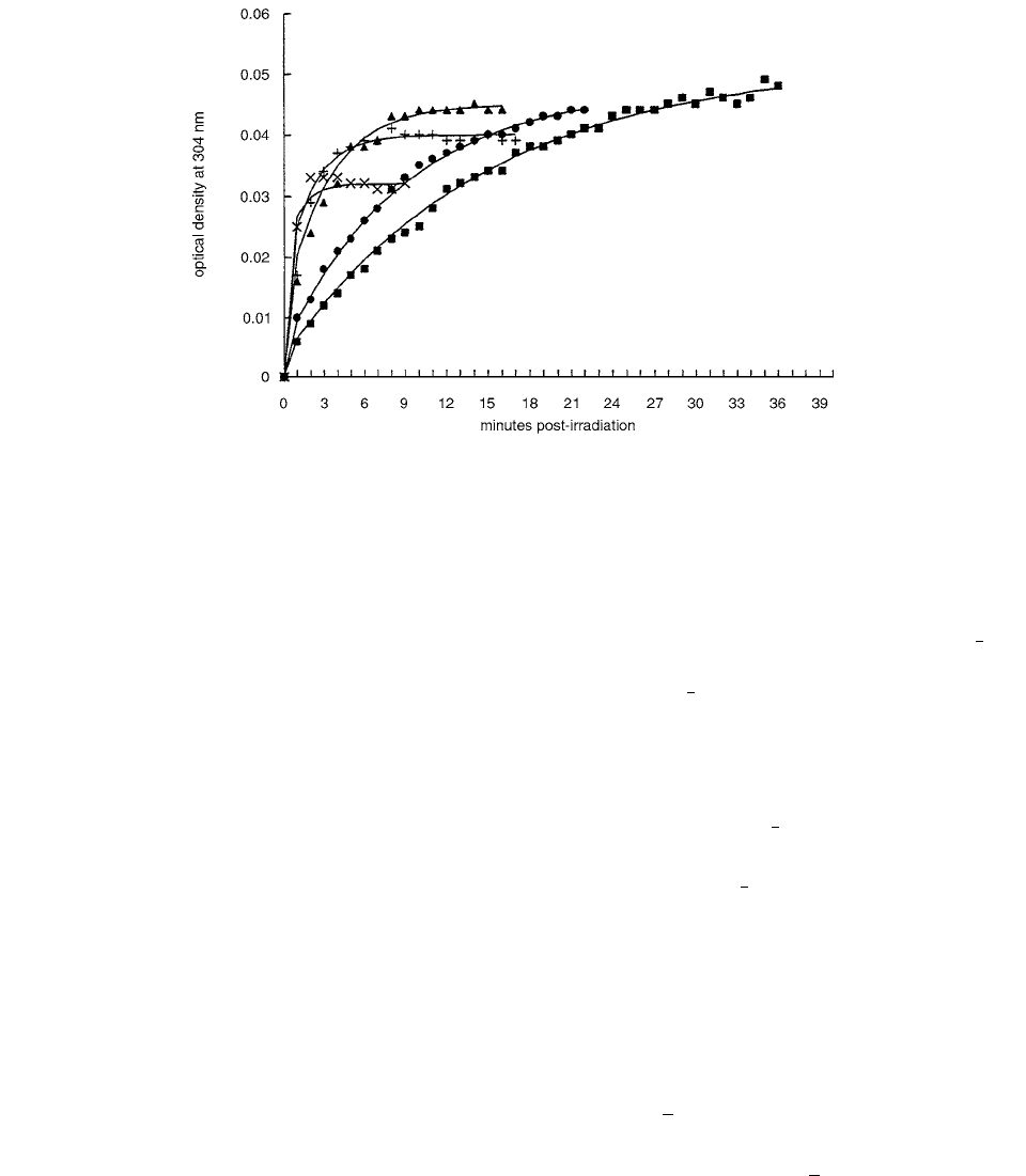
FIGURE 9.1
Optical density of agarose/ferrous gels after 1.87 Gy of gamma irradiation. 0.1 mM Fe
2
(
), 0.2 mM Fe
2
(
), 0.3
mM Fe
2
(
), 0.5 mM Fe
2
(
), 0.9 mM Fe
2
(
). (From Reference [3]. With permission.
h
h
t()d
td
--------------
i
h
---
t(),H
0
[]
h
i
〈〉
M
i
N
i
〈〉
xy,
〈〉
z
〈〉
Ch-09.fm Page 402 Friday, November 10, 2000 12:04 PM

Gel Dosimetry
403
directed against. Since more spins are aligned along the
direction of the magnetic field (
E
0
0) than
against it (
E
0
0),
M
z
is greater than zero.
The overall equilibrium magnetization of the ensemble of
spins is thus given by
M
(0, 0,
M
0
).
A system of nuclear spins placed in an external homo-
geneous static magnetic field will possess an equilibrium
magnetization
M
(0, 0,
M
0
). If the material is irradiated
with a time-dependent magnetic field or radio frequency
(rf) energy oscillating at the Larmor frequency,
0
, res-
onant absorption of the rf energy occurs and perturbs the
equilibrium magnetization such that
M
becomes time-
dependent, and and . For a given field
H
0
, different types of nuclei, each characterized by a dif-
ferent
y
, will have different Larmor frequencies and, there-
fore, each type can be selectively irradiated; i.e., only
protons will absorb rf energy of 25 MHz in a 0.6-T field.
Once perturbed, the magnetization tends to return to or
relax to its equilibrium value
M
(0, 0,
M
0
).
The perturbation and relaxation dynamics of the mac-
roscopic magnetization can be described by the phenom-
enological Bloch equations:
(9.3)
where
T
1
and
T
2
are the relaxation time constants charac-
terizing the relaxation of the respective magnetization
components. The first terms on the right-hand side of the
two expressions in Equation 9.3 describe the motion of
the macroscopic magnetization in the presence of an
applied field,
H
.
The field applied during MR imaging or NMR
experiments typically has the form
H
(
H
1
cos(
0
t
),
H
1
sin(
0
t
),
H
0
), where the
z
term is the static field and
the
x
and
y
terms are the perturbing transverse fields of
the rf energy pulses of magnitude
H
1
. During the appli-
cation of short pulses, usually only microseconds long,
little magnetization relaxation occurs and the magnetiza-
tion dynamics may be approximated by ignoring the sec-
ond right-hand terms in Equation 9.3. In this case the
magnetization in a frame of reference rotating about the
z
-axis at
0
(
H
0
) becomes:
(9.4)
where the prime designates the rotating reference frame,
and
H
1
is arbitrarily taken to point along the x-axis. Thus,
applying a transverse rf field will cause M to rotate away
from the
z-axis at a frequency
1
H
1
. The angle of
the rotation of M is determined by
H
1
p
, where
p
is the duration of the rf pulse. This angle is used to
describe the rf energy pulses in the pulse sequences that
manipulate the magnetization during MR imaging or
NMR experiments. At a more fundamental level, M
z
is
affected by the coupling between the rf energy and the
spins, since the coupling creates non-zero transition prob-
abilities between the eigenstates. As a result, the absorp-
tion of rf energy can affect the populations of the eigen-
states and hence the average . The M
xy
is also
affected by the coupling, as the coupling also creates
phase coherence in the time dependence of the average
and , such that the average magnetic moments
gain magnitude.
Once the perturbing rf energy is removed, the Bloch
equations reduce to:[4]
(9.5)
such that only the relaxation terms remain. The longitudi-
nal magnetization grows exponentially to its equilib-
rium value
M
0
with a characteristic time T
1
, the longitudinal
relaxation time, whereas the transverse magnetization
decays exponentially to zero with a characteristic
time T
2
, the transverse relaxation time.
There are four water proton groups in a Fricke gel
dosimeter: the bulk water protons, the water protons hydrat-
ing the Fe
2
and Fe
3
ions, and the water protons hydrating
the gel. The exchange of the water protons between all four
groups determines the overall observed relaxation. Rapid
exchange has been observed for the overall R
1
and R
2
of
water in Fricke gels containing gelatin, but not for the R
2
of water in gels containing agarose. [4]
Within the limit of fast exchange, the longitudinal mag-
netization recovery is characterized by a single exponential
and hence by a single overall water spin-lattice relaxation
rate R
1
. This rate is simply the sum of the inherent rates,
each weighted by the fraction of water protons p
i
in their
respective groups, as follows:
(9.6)
1
2
---
h
1
2
---
h
M
z
M
0
M
xy
0
M
z
d
dt
----------
MH()
z
M
0
M
z
T
1
----------------------
and
M
x, y
d
dt
-------------
MH()
x, y
M
x, y
T
2
----------
M
y
M
0
H
1
t()and M
z
M
0
H
1
t()cossin
z
〈〉
x
〈〉
y
〈
〉
M
z
d
dt
------------
M
0
M
z
T
1
------------------------
and
M
xy,
d
dt
----------------
M
xy,
T
2
-------------
M
z
M
xy,
R
1
p
3
R
1
3
p
2
R
1
2
p
gel
R
1
gel
1 p
3
p
2
p
gel
()R
1
water
k
3
R
1
3
R
1
water
()Fe
3
[]k
2
R
1
2
R
1
water
()
Fe
2
[] k
gel
R
1
gel
R
1
water
()gel[]R
1
water
r
1
3
Fe
3
[] r
1
2
Fe
2
[]r
1
gel
gel[]R
1
water
Ch-09.fm Page 403 Friday, November 10, 2000 12:04 PM
404 Radiation Dosimetry: Instrumentation and Methods
where the r
1
i
are the relativities of the respective proton
groups and specify the ability of a solute to enhance spin-
lattice relaxation of water protons. Equation 9.6 indicates
that under a fast exchange regime, R
1
varies linearly with
the concentration of ions. The spin-lattice relaxation rate
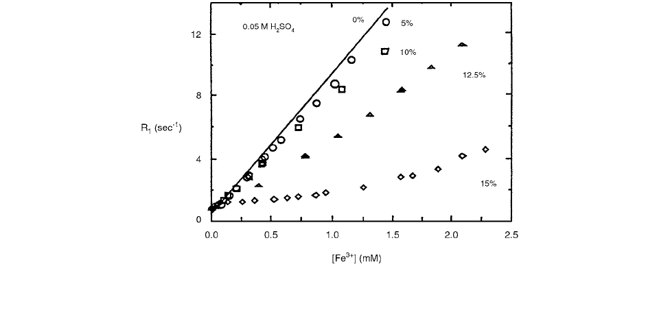
dependence on ferric concentration is shown in Figure 9.2.
Equation 9.6 may be expressed as follows by noting
that [Fe
2
] [Fe
3
] [Fe
2
]
0
:
(9.7)
where
The final expression for the R
1
dose response is
(9.8)
where N
A
is Avogadro’s number,
is the density in kgliter,
e is the number of Joules per electron volt, and G(Fe
3
) is
the chemical yield for Fe
3
in number of ions per 100
electron volts, given by
(9.9)
The single spin-spin relaxation rate
R
2
observed for
water and monomer protons suggests that the protons on
the bulk water, monomer, and water hydrating the poly-
acrylamide gelatine and monomer are all under fast
exchange. Under the fast-exchange regime, the water and
monomer
R
2
is the sum of the inherent water R
2
weighted
by the fraction of water proton in the respective group:
(9.10)
where the superscripts are: b bulk water, p polyacry-
lamide, and g gelatin. k
p
[p] is the fraction of
water-proton-hydrating polymer per weight fraction of
polymer in the dosimeter. Equation 9.10 illustrates how
an increase in R
2
with dose results from an increase in
polymer concentration. Using the fast exchange model for
the water in a PAG gel dosimeter, the following dose
response model results: [4]
(9.11)
where is the polymer relaxivity,
[p]D is the
polymer yield in units of percent weight fraction of poly-
mer formed per Gy, and R
2
(0Gy) is the spin-spin relaxation
rate of an un-irradiated PAG gel dosimeter.
One of the most important qualities possessed by a
dosimeter gel is that it forms both phantom and detector.
The advantages to be expected with gel dosimetry com-
pared with conventional dosimeters may thus be summa-
rized in the following properties: [5]
• independence on radiation direction, radiation
quality, and dose rate for conventional clinical
beams
• absorbed dose integration in the dosimeter (of
utmost importance for dynamic treatment and
multiple beams)
FIGURE 9.2 The spin-lattice relaxation rate dependence on the ferric-ion concentration for the different gel concentrations (% by
weight) labeled on the graph and 0.05-M sulfuric acid. (From Reference [4]. With permission.)
R
1
r
eff
3
r
2
()Fe
3
[] Fe
2
[]R
1
0Gy()
R
1
0Gy() r
2
Fe
2
[]
0
r
gel
gel[]
0
R
1
water
()
R
1
r
eff
3
r
2
()GFe
3
()
10
eN
A
----------
D R
1
0Gy()
G Fe
3
()
Fe
3
[]eN
A
10
D
---------------------------
R
2
p
p
R
2
p
p
g
R
2
g
1 p
p
p
g
()R
2
b
k
p
R
2
p
R
2
b
()p[] p
g
R
2
g
1 p
g
()R
2
b
{}
p
p
R
2
r
p
G
p
{}DR
2
0Gy()
r
p
G
p
Ch-09.fm Page 404 Friday, November 10, 2000 12:04 PM
Gel Dosimetry 405
• evaluation of a complete volume
• potential for true three-dimensional dosimetry
utilizing a high spatial resolution
• possibility to measure in a phantom that is
equivalent to anatomical soft tissue
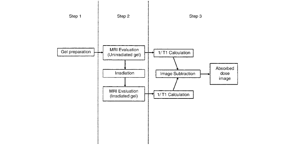
The contributions to uncertainty of the different steps
included in FeGel relative absorbed-dose measurements
(Figure 9.3) can be evaluated according to the guidelines
given by the International Organization for Standardization
recommendations. The uncertainty is divided into two cat-
egories, according to the way in which its numerical value
was estimated: [5]
• type A uncertainty determined by statistical
methods
• type B uncertainty determined by other means,
e.g., estimated or obtained from a calibration
certificate, etc.
In step 1 (Figure 9.3), the gel is produced, and if a
linear and spatially uniform 1T
1
dose response is assumed,
the uncertainty introduced by this step is negligible. The
next steps include MRI acquisition (step 2, Figure 9.3) and
background subtraction (step 3, Figure 9.3) to obtain the
final absorbed dose image.
The local water proton NMR relaxation rates (R
1
1T
1
and R
2
1T
2
) in the gel increase in proportion to
the absorbed radiation dose. This is believed to be caused
by the increase of the population of those water molecules
which are bound to the surfaces of the rigid polymer
particles. [6] These bound water molecules exchange their
protons with the polymer and with the bulk water mole-
cules. The relaxation rates that can be measured from MRI
images are averaged over a voxel and represent the bulk
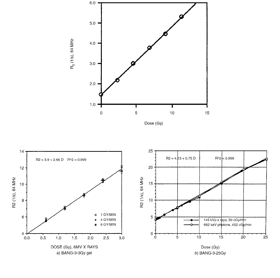
water relaxation. Figure 9.4 shows R2-dose calibration
curve.
Figure 9.5 may indicate that the polymer gel dosimeter
is independent of photon energy and dose rate; it is true
only within certain limits for dose rate. Different gel for-
mulations, and especially different monomers, have great
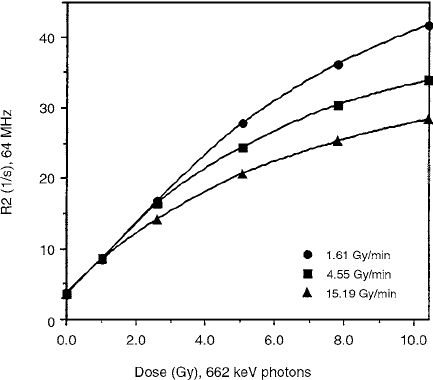
effect on dose rate dependence. The “supersensitive”
BANG-3 formulation shows independence of dose rate up
to a certain dose, above which the curves which represent
higher dose rates begin to saturate (Figure 9.6).
High LET radiations saturate the polymer gels at
lower doses than lower LET radiations. However, the
extent of this effect depends on the choice of the mono-
mers and the response modifiers. BANG-1 formulation
(bis acrylamide gelatin water) did not show mea-
surable LET dependence, whereas the BANG-3 series
(gelatin methacrylic acid water) showed a significant
effect. [6]
A formulation of a tissue-equivalent polymer gel
dosimeter for the measurement of three-dimensional dose
distributions of ionizing radiation has been developed by
Maryanski et al. [7] It is composed of aqueous gelatin
infused with acrylamide and N,N-methylene-bisacryla-
mide monomers and made hypoxic by nitrogen saturation.
Irradiation of the gel, referred to as BANG, causes localized
polymerization of the monomers, which, in turn, reduces
the transverse NMR relaxation times of water protons.
FIGURE 9.3 A schematic description of the procedure used for the FeGel system, including the background subtraction. (From
Reference [5]. With permission.)
Ch-09.fm Page 405 Friday, November 10, 2000 12:04 PM
406 Radiation Dosimetry: Instrumentation and Methods
The dose dependence of the NMR transverse relaxation
rate, R
2
, is reproducible (less than 2% variation) and is
linear up to about 8 Gy, with a slope of 0.25 s
1
Gy
1
at
1.5 T. Magnetic resonance imaging may be used to obtain
accurate three-dimensional dose distributions with high
spatial resolution. Since the radiation-induced polymers
do not diffuse through the gelatin matrix, the dose dis-
tributions recorded by BANG gels are stable for long
periods of time and may be used to measure low-activity
radioactive sources. Since the light-scattering properties
of the polymerized regions are different from those of
the clear, non-irradiated regions, the dose distributions
are visible, and their optical densities are dependent on
dose. [7]
The formation of cross-linked polymers in the irra-
diated regions of the gel increases the NMR relaxation
rates of neighboring water protons. Therefore, the radiation-
induced polymerization in polymer gels plays a role
similar to that of the radiation-induced oxidation of
ferrous ions in Fricke gels, with four important advan-
tages. First, in polymer gels the spatial distribution of
NMR relaxation rates, which reflects the distribution of
dose, is stable and does not change with time. Second,
Fricke gels have an intrinsically high electrical conduc-
tivity. Consequently, the RF field is strongly attenuated.
The polymer gel dosimeters do not contain ionic species
and show insignificant RF attenuation. Third, polymer-
ized regions can be seen visually. Last, polymer gels
FIGURE 9.4 R
2
(dose) calibration. (From Reference [6]. With permission.)
FIGURE 9.5 a), b) Dose response for BANG-3-type gels irradiated with photons at low to medium dose rates: a) 3Gy, b) 25Gy.
(From Reference [6]. With permission.)
Ch-09.fm Page 406 Friday, November 10, 2000 12:04 PM
Gel Dosimetry 407
are considerably more sensitive to radiation than the
Fricke gels.
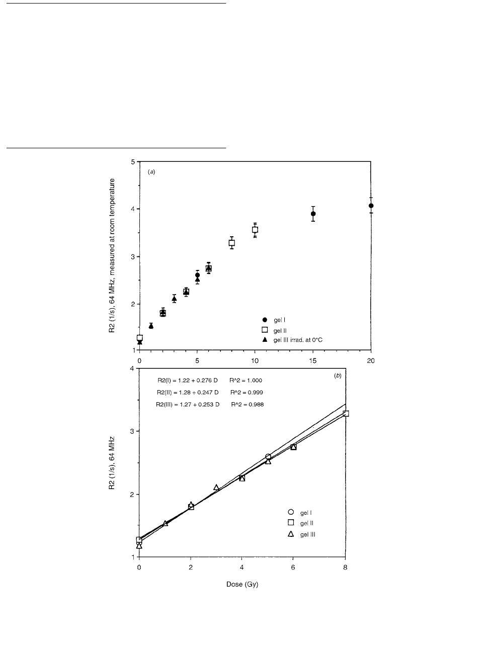
Three BANG gels were prepared by Maryanski et al.
Gels I and II were contained in tissue culture flat flasks
made of polystyrene, and gel III was prepared in a rectan-
gular Lucite box. Each gel was irradiated by 4 4-cm
2
fields using 6-MV x-rays produced by a Varian 2100C
linear accelerator at a dose rate of 4 Gy min
1
. The fol-
lowing doses were delivered at d
max
: 5, 10, 15, 20 Gy to
gel I; 2, 4, 6, 8, 10 Gy to gel II; 1, 2, 3, 4, 5, 6 Gy to gel
III. Gels I and II were irradiated at an ambient room tem-
perature. Gel III was irradiated at 0°C, immediately after
being removed from an ice-and-water bath. All gels were
stored in a refrigerator at 4°C immediately after irradiation.
Figure 9.7 shows values of transverse relaxation rates
(R
2
) for gels I, II, and III as a function of dose. The pooled
data (Figure 9.7a) show that the dose response was highly
reproducible over a wide range of doses. For doses below
8 Gy, the dose response is well-fitted by a straight line;
Figure 9.7b shows the individual straight-line fits for each
gel. For example, a relaxation rate of 2.0 s
-1
corresponds
to doses of 2.83, 2.92, and 2.90 Gy in the three different
gels, a variation of less than 2% from the mean value.
The signal in a spin-echo MR image produced by a
dose D is given by
(9.12)
where S is the signal at echo time TE.
Radiation dose distributions in three dimensions from
tomographic optical density scanning of polymer gels were
described by Gore et al. [8] The dosimetric data stored
within the gels were measured using optical tomographic
densitometry. The dose-response mechanism relied on the
production of light-scattering micro-particles, which result
from the polymerization of acrylic comonomers dispersed
in the gel. The attenuation of a collimated light beam
caused by scattering in the irradiated optically turbid
medium was directly related to the radiation dose over the
range 0–10 Gy. An optical scanner has been developed
which incorporates a He-Ne laser, photodiode detectors,
and a rotating gel platform.
The intensity of a monochromatic light beam passing
through the medium is attenuated exponentially under nar-
row-beam conditions. If
(x, y) denotes the optical atten-
uation coefficient per unit length in a section of the object,
then the intensity exiting the sample at position x is I(x)
when the incident intensity is I
0
:
(9.13)
A schematic diagram that illustrates the operation of the
prototype device is shown in Figure 9.8.
After acquisition, the projection data are transferred
to an image reconstruction program.
Progress in the development of polymer gel dosimetry
using MRI was reported by Maryanski et al. [9] The dose
distribution image produced in the tissue-equivalent gel by
radiation-induced polymerization and encoded in the spatial
distribution of the NMR transverse relaxation rates (R
2
) of
the water protons in the gel is permanent. Maps of R
2
are
constructed from magnetic resonance imaging data and serve
as a template for dose maps, which can be used to verify
complex dose distributions from external sources or brachy-
therapy applicators. The integrating, three-dimensional, tis-
sue-equivalent characteristics of polymer gels make it possi-
ble to obtain dose distributions not readily measured by
conventional methods. An improved gel formulation
(BANG-2) has a linear dose response that is independent of
energy and dose rate for the situations studied to date.
The so-called BANANA (acronym based on Bis,
Acrylamide, Nitrous Oxide, And Agarose) and BANG
(acronym based on Bis, Acrylamide, Nitrogen, and Gelatin)
formulations of polymer gels differ in their gel matrices,
which are agarose and gelatin, respectively; 1% by
weight agarose in BANANA gel was replaced by 5% by
weight gelatin in BANG gel. This resulted in a lower
background R
2
( 1T
2
), transverse NMR relaxation rate
of the water protons) for the nonirradiated gel, and a
more transparent medium in which the irradiated region
is clearly visible.
An improved polymer-gel formulation was developed,
containing 3% N,N-methylene-bisacrylamide (referred to
as bis), 3% acrylic acid, 1% sodium hydroxide, 5% gela-
tin, and 88% water, where all percentages are by weight
(see Table 9.1). This gel differs from BANG mainly in the
substitution of acrylic acid for acrylamide. Henceforth, it
FIGURE 9.6 Dose response saturation and dose rate depen-
dence at higher dose rates in BANG-3 gel formulation. (From
Reference [6]. With permission.)
STE() S 0()e
R
2
D()TE
Ix() I
0
e
x,y()yd
Ch-09.fm Page 407 Friday, November 10, 2000 12:04 PM
408 Radiation Dosimetry: Instrumentation and Methods
will be referred to by the acronym BANG-2. Dissolved
oxygen, which inhibits free-radical polymerization reac-
tions, is removed from the mixture by passing an inert
gas such as nitrogen through it when it is above the gelling
temperature and prior to sealing the vessel.
In all preparations gelatin type A (acid-derived), approx-
imately 300 Bloom (a gel strength indicator), was used. The
water was from an ion-exchange purifier, and the nitrogen
gas contained less than 100 ppm oxygen.
To establish the tissue equivalence of the gel, its physical
density was determined by weighing the gel and measuring
its volume at room temperature, one day after gelation.
TABLE 9.1
Chemical Composition of the BANG-2 Gel
Component Formula
Weight Fraction
in the Gel
Gelatin (C
17
H
32
N
5
O
6
)
x
0.05
Acrylic acid CH
2
CHCOOH 0.03
Bis (CH
2
CHCONH)
2
CH
2
0.03
Sodium hydroxide NaOH 0.01
Water H
2
O 0.88
From Reference [9]. With permission.
FIGURE 9.7 (a) The dose dependence of the water-proton NMR transverse relaxation rate R
2
, combining the data from three
separately prepared BANG gels, over the range 0–20 Gy. The error bars indicate 5% relative error. (
b) The dose dependence of the
water-proton NMR transverse relaxation rate
R
2
, measured in three separately prepared BANG gels, over the range 0–8 Gy. The
separate linear fits were
R
2
(I) 1.22 0.276D, 1.000; R
2
(II) 1.28 0.247D, 0.999; R
2
(III) 1.27 0.253D,
0.988. (From Reference [7]. With permission.)
r
2
r
2
r
2
Ch-09.fm Page 408 Friday, November 10, 2000 12:04 PM

Gel Dosimetry 409
These measurements were repeated several times. The
measured density was 1.03 0.005 g/cm
3
. The atomic
composition, electron density, and average atomic number
of the BANG-2 gel of human muscle tissue, and of water
are compared in Table 9.2.
All gels used were held in glass bottles, flasks, or beakers,
so as to obviate the problem of oxygen contamination that
occurs in containers made from oxygen-permeable plastics.
II. APPLICATION OF FERROUS IONS
Ferrous sulphate gel analyzed by relaxation time measure-
ments with NMR-imaging 3D determinations of absorbed
dose was discussed by Gambarini et al. [10] The dose-
response curve slope is about 0.2 s
1
Gy
1
, and the G-
factor turns out to be ~185 ions per 100 eV of absorbed
energy.
The spin-echo images were obtained employing a mul-
tiple spin-echo sequence. Since the spin-spin relaxation
rate 1T
2
proved to have a dose sensitivity higher than the
spin-lattice relaxation rate 1T
1
, only the value of 1T
2
was
generally evaluated. The transverse relaxation rate was
determined by a multiple spin-echo sequence with 16 ech-
oes; the echo times were T
E
(28 20n) ms where n
0,…,15, and the repetition time was T
R
2.5 s. The T
2
values were calculated utilizing a one-exponential fit with
a nonlinear, least-squares, three-parameter algorithm. The
matrix size was 256 256 mm
2
, and the voxel size was
10 1.2 1.2 mm
3
. The results of the fit, as can be
seen from Figure 9.9, support the validity of the one-
exponential analysis and confirm the mono-exponential
trend of the magnetization recovery process in agar, or
other gels, doped with paramagnetic ions.
The set-up for the gel preparation completely elimi-
nates steam loss and makes reproducibility of the opera-
tions possible. This equipment consists of a cylindrical
Pyrex container whose cover is supplied with an opening
for the thermometer, a water-cooled coil for continuous
TABLE 9.2
Comparison of Elemental Composition (Weight Fractions are Denoted as W
k
), Electron Densities, and
Average Atomic Numbers for BANG-2 Gel, Human Muscle Tissue, and Water.
Material w
C
w
H
w
N
w
o
w
Na
(kg m
–3
)
e
( 10
29
m
–3
)
e
/
( 10
26
kg
–1
)
BANG-2 0.0564 0.1051 0.0135 0.8173 0.0058 1030 3.42 3.32 7.14
Muscle 0.1230 0.1020 0.0350 0.7289 0.0008 1040 3.44 3.31 6.93
Tissue
Water 0.00 0.1111 0.00 0.8889 0.00 1000 3.34 3.34 7.22
a
Calculated as .
Source: From Reference [9]. With permission.
FIGURE 9.8 A schematic diagram of the prototype scanner. The mirrors translate left to right to obtain projections of the gel optical
attenuation. Between each translation, the gel is rotated by a second stepping motor. (From Reference [8]. With permission.)
Z
a
Z
k
w
k
Z
k
Ch-09.fm Page 409 Friday, November 10, 2000 12:04 PM