Shani G. Radiation Dosimetry: Instrumentation and Methods
Подождите немного. Документ загружается.

Ch-04.fm Page 300 Friday, November 10, 2000 12:01 PM

301
5
Film Dosimetry
CONTENTS
I. Introduction............................................................................................................................................................301
II. Examples of Photographic Film Dosimetry..........................................................................................................301
III. Radiochromic Film Dosimetry ..............................................................................................................................310
References .......................................................................................................................................................................329
I. INTRODUCTION
Film dosimetry is attractive due to its high spatial resolu-
tion, wide accessibility, and the flexibility to place the film
in humanoid phantoms. Also, the short measuring time and
the fact that the film dosimetry is intrinsically two-dimen-
sional and integrating in time are appreciated. Film is
potentially the ideal detector for determining dose distribu-
tion for dynamic beams and for studying the combination
of stationary beams treated sequentially. Film dosimetry is
widely used to obtain the relative dose distribution of elec-
tron and photon beams in water, in plastics, and in inho-
mogeneous phantom. Film dosimetry in phantoms is
advantageous because of high spatial resolution, short treat-
ment unit immobilization time, and 2D information.
Modern film processing units improve the reproduc-
ibility and reliability of film dosimetry and make it an
attractive method for many applications. Fast film digitiz-
ers connected to a computer, equipped with proper eval-
uation software, allow rapid and accurate analysis of large
films in a short time.
The most common setup in relative dose measurements
with films is to sandwich a film within a phantom of water-
equivalent material with the film plane-parallel to the cen-
tral axis of the radiation field. With the parallel geometry,
two particular precautions must be taken: there must be
heavy pressure on the phantom to avoid any air gap on
either side of the film, and there must be perfect alignment
of the film edge with the surface of the phantom. In addi-
tion, the artifacts that result from a thin air layer trapped
between the packaging material, the paper spacer, and the
film are responsible for the inaccurate dose measurement
in the build-up region of the electron depth dose.
Conventional silver halide film has a highly nonlinear
photon energy response, especially at low energies. It has
radiation interaction properties markedly different from
those of tissue. Along with variations introduced by the
necessary post irradiation processing, this type of film is
extremely difficult to use for accurate analytical dosimetry.
Most radiochromic systems are chemical radiation sen-
sors consisting of solid or liquid solutions of colorless
leuco dyes; these become colored without the need for deve-
lopment when exposed to ionizing radiation. Various radi-
ochromic forms, such as thin films, thick films and gels,
liquid solutions, and liquid-core waveguides, have been in
routine use for dosimetry of ionizing radiation over a wide
range of absorbed doses (10
2
to 10
6
Gy). Radiochromic
film is used for general dosimetry of ionizing radiation in
high-gradient areas of electron and photon beams in a wide
energy range. The film allowing approximately tissue-
equivalent dosimetry has been applied to mapping of dose
distribution in brachytherapy.
Radiochromic film (RCF) is of great interest as a planar
dosimeter for radiation oncology applications. It consists
of a thin, radiosensitive, 7–23-
m thick, colorless leuco
dye bonded to a 100-
m-thick mylar base. RCF turns deep
blue in color upon irradiation. RCF is approximately tis-
sue-equivalent. GafChromic MD-55 is usable at doses
from less than 1 Gy up to 12 Gy when measured at the
wavelength of maximum sensitivity (676 nm) and up to
about 500 Gy when measured at a wavelength of low
sensitivity. The absorption spectra of GafChromic (GC)
film contain two peaks with wavelengths in the range
610–680 nm and, thus, the dose-response curve as mea-
sured by an optical densitometer or spectrophotometer will
be highly dependent on the light source spectrum and sen-
sor material.
II. EXAMPLES OF PHOTOGRAPHIC
FILM DOSIMETRY
The use of film as a dosimeter is still limited, due to the
various difficulties associated with films such as energy
dependence, film orientation, and sensitometric nonlinearity.
On the other hand, film is probably one of the best detectors
for studying spatial distribution of dose or energy imparted.
The dosimetric resolution is limited only by the grain size
Ch-05.fm Page 301 Friday, November 10, 2000 12:01 PM
302
Radiation Dosimetry: Instrumentation and Methods
and the size of the aperture of an optical densitometer.
Commercially available laser-scanning densitometers
afford film dosimetry with a resolution of a few microns.
Unlike other detectors such as ionization chambers, diode
detectors, thermoluminescent detectors (TLDs), scintilla-
tion detectors, and diamond detectors where dose informa-
tion must be recorded before the readout is cleared for
another irradiation, radiographic films allow repetitive
readouts and provide a permanent record of the dosimet-
ric measurements. Films may be customized in various
sizes and shapes to fit any dosimetric application. Due to
the relatively small thickness, a film can be treated very
close to a Bragg-Gray cavity. The physical flexibility of
a film is also suitable for the curved and cylindrical sur-
face dose mapping when other detectors are impractical.
In relative dose measurements, the optical density may
be taken as proportional to the dose without any correc-
tion, since the collisional stopping power ratio of emul-
sion to water varies slowly with electron energy.
High-energy photon and electron dosimetry was car-
ried out by Cheng and Das using CEA film. [1] The
packaging of the CEA films is distinctly different from
that of the other films, in that each film is vacuum- sealed
in a shiny polyester-made, waterproof packet about 130
.
thick, permitting film dosimetry to be carried out even in
water phantom. Aside from the difference in packaging,
the CEA films have a clear polyester film base as opposed
to a bluish-dye film base found in the Kodak Readypack
XV and XTL films. It is not clear if the difference in film
base may have any effect on image quality. The CEA
films come in two types: TLF (localization) and TVS
(verification), similar to the XTL and XV films of the
Kodak Readypack.
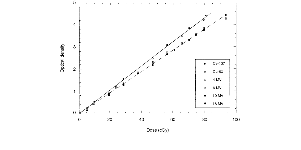
Figure 5.1 shows the variation of net optical density with
dose for the TVS film for a range of photon energies from
137
Cs to 18 MV. The base optical density of the CEA films
is on the order of 0.06, compared to about 0.2 for the Kodak
films. As shown in Figure 5.1, the linear portion of the char-
acteristic curve covers a range of optical density up to 4.3.
This wide range of linearity offers a convenient means in
percent depth dose and isodose measurements. It is inter-
esting to note that the film is faster to rays than to
bremsstrahlung
x-rays from a linear accelerator but is inde-
pendent of energy for each type of radiation. This feature
is particularly attractive for high-energy x-rays, as one
single sensitometric curve can be used for a wide range of
x-ray energies. A straight line is fitted to each of the
characteristic curves for rays and x-rays with a regression
coefficient of 0.999. For gamma rays, the line of regres-
sion is [1]
(OD)
, rays
0.054 07
dose, (5.1)
while for x-rays, the line of regression is
(OD)
X, rays
0.047 65
dose (5.2)
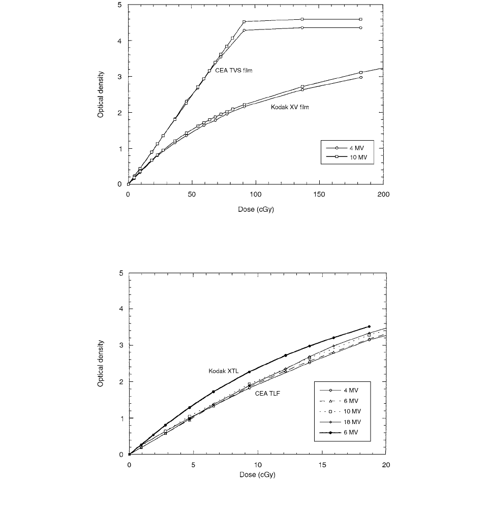
Figure 5.2 compares the characteristic curves for the
CEA TVS film and the Kodak XV film for two x-ray
energies, 4 and 10 MV. For the CEA TVS film, the linear
portion extends over the dose range up to 90 cGy, followed
by an abrupt saturation above 90 cGy. The CEA TVS
curves have a linear relationship, with a coefficient of
regression very close to 1. For the Kodak XV film, on the
other hand, the optical density varies curvilinearly with
FIGURE 5.1
Characteristic curves of the TVS films for photon beams. (From Reference [1]. With permission.)
Ch-05.fm Page 302 Friday, November 10, 2000 12:01 PM
Film Dosimetry
303
dose up to 200 cGy. The characteristic curve of the Kodak
XV films exhibits a longer tail and a smaller gamma
compared to that of the CEA TVS film.
The size of the silver halide crystals is generally smaller
in the CEA TVS film than in the Kodak XV film. Indeed,
the silver halide crystals in the CEA TVS film are of fairly
uniform size and shape, resulting in a linear characteristic
curve. On the other hand, the silver halide crystals of the
Kodak XV film are of different sizes and shapes, with the
largest more than 10 times bigger than the smallest, result-
ing in a nonlinear characteristic curve. Furthermore, the
CEA TVS film has a higher silver concentration (about 42
g
m
2
) compared to regular x-ray films (about 7 g
m
2
).
The characteristic curves for the TLF film over the
photon energy range 4-18 MV are shown in Figure 5.3.
The film saturates at about 30 cGy, which is considerably
higher than the Kodak Readypack XTL film. Unlike the
CEA TVS film, the characteristic curves for the CEA TLF
films are slightly curvilinear over the range of dose up to
30 cGy. However, if the data is considered only in the
range of 0–15 cGy, the curves are all straight lines with
a coefficient of regression near unity. The CEA TLF film
FIGURE 5.2
Comparison of the characteristic curves of CEA TVS film and Kodak XV film for different photon energies. (From
Reference [1]. With permission.)
FIGURE 5.3
Characteristic curves of the CEA TLF film for photon energies 4–18 MV. For comparison, characteristic curve of
Kodak XTL film is also shown for a 6-MV beam. (From Reference [1]. With permission.)
Ch-05.fm Page 303 Friday, November 10, 2000 12:01 PM
304
Radiation Dosimetry: Instrumentation and Methods
is also energy-independent within the experimental
uncertainty.
A method of film dosimetry for high-energy photon
beams was proposed by Burch et al. [2] which reduced the
required film calibration exposures to a set of films obtained
for a small radiation field size and shallow depth (6 cm
6 cm at 5-cm depth). It involves modification of a compres-
sion-type polystyrene film phantom to include thin lead
foils parallel to the vertical film plane at approximately 1 cm
from both sides of the film emulsion. The foils act as high
atomic number filters which remove low-energy Compton
scatter photons that otherwise would cause the film sensi-
tivity to change with field size and depth.
High-energy photon beams used in radiation oncology
are considered to interact primarily by the Compton scat-
tering process with tissue. However, when film is placed
in a tissue-equivalent material, photoelectric interactions
associated with the silver atoms in the emulsion cause the
film to over-respond relative to the tissue-equivalent mate-
rial. For the low-energy photons, film dose may be as
much as 25 times the tissue dose at the same physical
location.
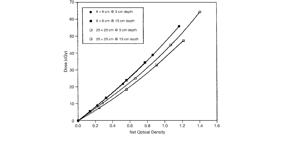
The results for the 6-cm
6-cm and 25-cm
25-cm
fields shown in Figure 5.4 emphasize the importance of
the film sensitivity as a function of field size and depth.
As the field size was increased, the dose required to
produce a given density on the film was reduced. For
sizes smaller than 10 cm
10 cm, the sensitivity change
is small (not shown); and for sizes less than 6 cm
6 cm,
no change in sensitivity occurs. For small fields with
less scatter, the change in sensitivity with depth is not
apparent.
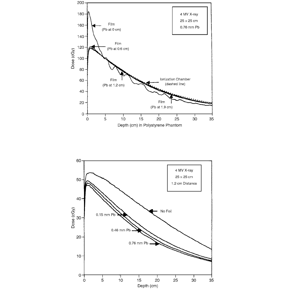
The effects of foil-to-film separation distance and foil
thickness were investigated in order to obtain a single
optimum distance-thickness combination, and the results
are presented in Figures 5.5 and 5.6. In Figures 5.5 and
5.6, the dose was calculated using data from the 6-cm
6-cm calibration films. Film/foil separation distances of
0, 0.6, 1.2, and 1.9 cm and lead foil thicknesses of 0, 0.15,
0.30, 0.46, and 0.76 mm were included in the investiga-
tion. At 0-cm film/foil separation distance, the curve
shows the effect of electrons coming from the lead due to
interactions within the foil. This intensification effect
exaggerates the shape of the depth-dose curve and the non-
uniform film/foil contact is apparent in the data (Figure 5.5).
At 0.6 cm the scattered electrons are absorbed in the inter-
vening polystyrene. Further increase in film/foil separation
distance produced only minor changes in the calculated
dose curve. Figure 5.6 shows the effect of changing foil
thickness. A single thickness of 0.15 mm causes a signif-
icant decrease in the calculated dose, with only subtle
changes as additional layers are added. The best match to
ion chamber percentage depth-dose data was observed for
lead foil thickness of 0.46 mm with a 1.2 cm film/foil
separation distance.
The use of radiographic film for the dosimetry of large
(i.e.,
10 cm
10 cm) photon radiotherapy beams is
complicated, due to a dependence of film emulsion sen-
sitivity on depth within the phantom. This dependence is
caused by a relative increase in depth in the population
of lower-energy, scattered photons in the spectrum and
the subsequent photoelectric absorption of these photons
by the film emulsion. This effect becomes most pro-
nounced with increases in the photon population in the
FIGURE 5.4
Film sensitivity dependence upon field size and upon depth. (From Reference [2]. With permission.)
Ch-05.fm Page 304 Friday, November 10, 2000 12:01 PM
Film Dosimetry
305
energy region below approximately 400 keV, where the
mass attenuation coefficient of emulsion increases rapidly,
approximately as 1
. As the low-energy spectral
component increases, the tissue equivalence of film emul-
sion diverges from that of tissue-equivalent materials, due
to an approximate dependence of the photoelectric
interaction cross section. [3]
In order to examine possible dependencies of emul-
sion exposed by radiosurgical beams, a series of sensito-
metric curves was established for a range of depths and
field sizes. For each exposure a 10-in
12-in sheet of
Kodak X-Omat V film from a single batch (#194 05 2)
was sealed within a light-tight Solid Water (Gammex
RMI, Inc.) cassette. The cassette consists of two 2-cm-
thick slabs of solid water sealed around three edges by
nylon screws and a rubber O-ring. [3]
To minimize possible variations due to film-processing
conditions, a Kodak X-Omat RP processor was used by
Robar and Clark for which the throughput is very high
and quality assurance is performed daily. Developer tem-
perature fluctuated by less than
0.5°F between pro-
cessing sessions. Optical density was measured using a
FIGURE 5.5
Lead foils placed adjacent to the film show an exaggerated response at shallow depth and a wavy appearance due to
undulation in their surface. Each curve is normalized to 5-cm depth. (From Reference [2]. With permission.)
FIGURE 5.6
The effect of foil thickness on the shape of the depth-dose curve. (From Reference [2]. With permission.)
hv()
3
Z
3
Ch-05.fm Page 305 Friday, November 10, 2000 12:01 PM

306
Radiation Dosimetry: Instrumentation and Methods
scanning densitometer. Base-plus-fog optical density was
subtracted from scanned optical densities for each film.
In order to relate optical densities to absolute doses,
the dose to water corresponding to each of these depths
and for each MU setting was calculated from
(5.3)
where
D
(
d
,
A
) is the dose at depth
d
for field size
A
at the
phantom surface,
MU
is the number of monitor units
given,
PDD
(
d
,
A
) is the percent depth dose and
S
t
(
A
) is
the total scatter factor. Both
S
t
(
A
) and
PDD
(
d
,
A
) were
measured in a water phantom using a
p
-type silicon elec-
tron diode .
Figure 5.7a shows the sensitometric curves obtained
for the large (20-cm
20-cm) photon beam at depths of
1.0 cm,10.0 cm, and 20.0 cm. For each depth a curve was
fitted to the measured sensitometric data using the single-
target/single hit equation
(5.4)
where
OD
and
D
are the measured optical density and
given dose, respectively. The saturation density of the film,
OD
sat
, was estimated by delivering a large dose (500 cGy)
to a film in phantom, and it was held constant in the fitting
algorithm. The fitting parameter
represents emulsion
sensitivity and was allowed to vary. For this large field,
the curves diverge markedly, indicating that using a sen-
sitometric curve that is not depth-specific would introduce
significant error in converting optical density to dose. In
order to minimize this error to below approximately 10%,
for example, it is necessary to confine the dose range to
less than 80 cGy. In contrast, Figure 5.7b shows sensito-
metric curves corresponding to the same depths for the
radiosurgical (2.5-cm-diameter) field. For this small field,
the curves agree to within the reproducibility of film devel-
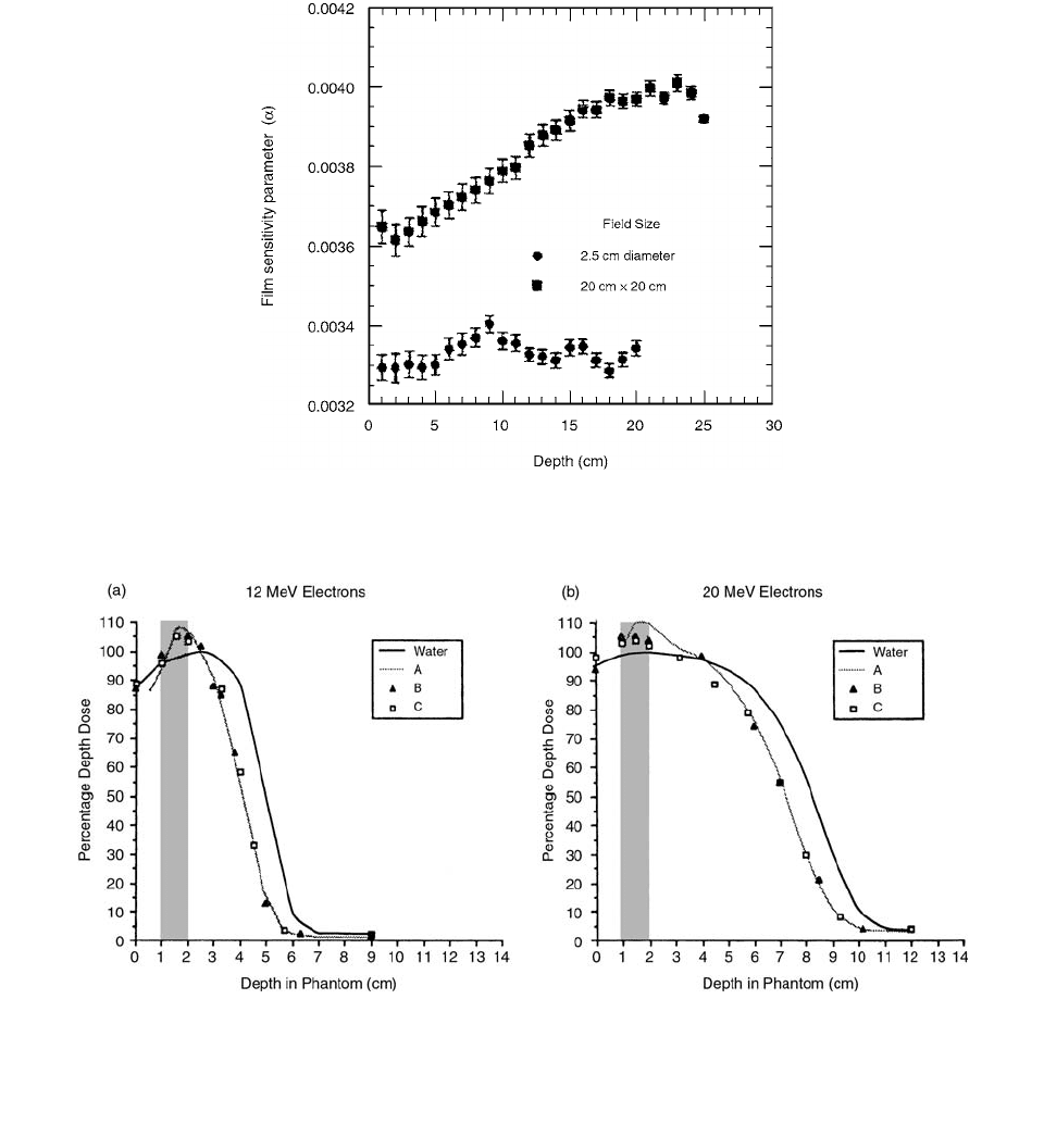
opment and scanning. By determining the value of
for
each curve, film sensitivity was obtained as a function of
depth, as illustrated for the 20-cm
20-cm and 2.5-cm-
diameter fields (Figure 5.8). As expected from the dispar-
ity in the sensitometric curves in Figure 5.7a, the film
sensitivity for the large (20-cm
20-cm) field increases
systematically with depth. While small fluctuation of the
values of
is apparent for the radiosurgical field, no
systematic variation of sensitivity with depth is apparent.
Film dosimetry is most problematic for lower-energy
photon beams (e.g.,
60
Co and 4-MV) due to their lower
primary-to-scatter ratio. Figure 5.7 shows the importance
of corrections in converting scanned optical density to dose
even for the 6-MV beam.
Film is often used for dose measurements in hetero-
geneous phantoms. In those situations perturbations are
produced which are related to the density and atomic
number of the phantom material and the physical size and
orientation of the dosimeter. Significant differences were
observed by El-Khatib et al. between the dose measure-
ments within the inhomogeneity. [4] These differences
were influenced by the type and orientation of the dosim-
eter in addition to the properties of the heterogeneity.
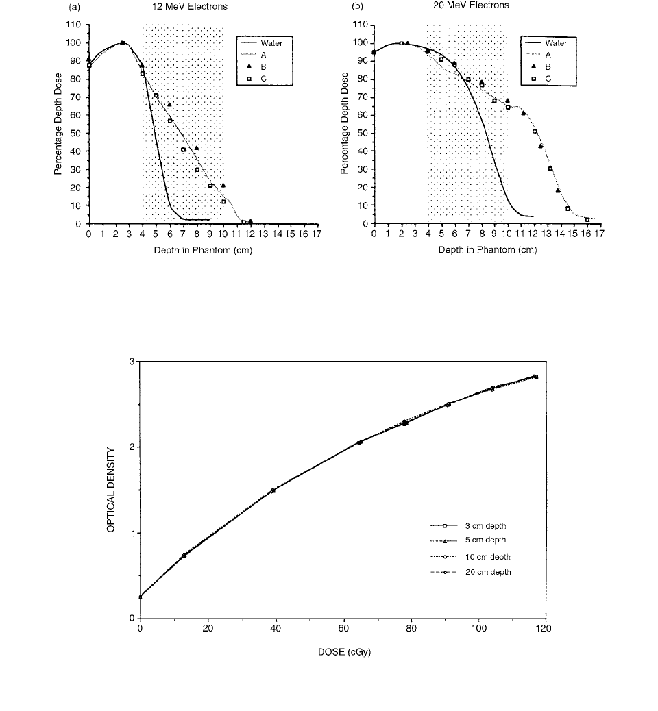
To investigate how the differences in dose measure-
ments are related to the dosimeter configuration, the EGS4
Monte Carlo system, together with the PRESTA algorithm,
was used to model the experimental conditions. The per-
centage depth doses measured in the phantom containing
1-cm bone are shown in Figure 5.9 and are compared to
Dd, A()
MUPDD d, A()S
t
A()
100
---------------------------------------------------
OD OD
sat
110
D
()
FIGURE 5.7
The sensitometric curves established experimen-
tally for (a) the 20-cm
20-cm radiotherapy field and (b) the
2.5-cm-diameter radiosurgical field, shown for depth in solid
water phantom of 1.0 cm, 10.0 cm, and 20.0 cm. (From Refer-
ence [3]. With permission.)
Ch-05.fm Page 306 Friday, November 10, 2000 12:01 PM
Film Dosimetry
307
the depth doses measured in the homogeneous water phan-
tom. All film measurements were done with the film in the
ready pack with the inner paper removed. The doses were
normalized to the maximum dose in the homogeneous
polystyrene phantom, measured with whatever dosimeter
was used to measure the doses at depth. The doses within
the bone represent dose-to-unit density material within the
bone rather than dose to the bone itself, since a correction
by ratio of collisional stopping powers was not done. At
several cm beyond the bone, the PDD measured with all
dosimeter configurations are identical. The opposite effect
for the two film orientations is observed for the phantom
containing the cork (Figure 5.10). The PDD is unaffected
in the initial 4 cm of polystyrene and there is greater
penetration of the beam in cork. Within the cork the dif-
ference measured with the film in different orientations is
as much as 6%, and the TLD measurements are lower than
the measurements with film. These greater discrepancies
are attributed to the fact that the inhomogeneity is much
larger and lies in a region of high dose gradient.
A method for the creation of a complete 3D dose
distribution from measured data was discussed by
FIGURE 5.8
The curve-fitting parameter
, representing emulsion sensitivity, as a function of depth for the 20-cm
20-cm
radiotherapy field and the 2.5-cm-diameter radiosurgical field. (From Reference [3]. With permission.)
FIGURE 5.9
The percentage depth doses measured in a polystyrene phantom containing 1-cm hard bone at 1-cm depth using
(A) film in parallel orientation to the beam, (B) film in perpendicular orientation to the beam, and (C) TLD are shown and compared
to the PDD measured in a homogeneous water phantom for electron beams of nominal energy (a) 12 MeV and (b) 20 MeV. (From
Reference [4]. With permission.)
Ch-05.fm Page 307 Friday, November 10, 2000 12:01 PM
308
Radiation Dosimetry: Instrumentation and Methods
Stern et al. [5] It involved the measurement with film of
the dose distribution in a series of “beam’s-eye-view”
(BEV) planes (planes perpendicular to the beam central
axis at different depths). Film measurements were made
using Kodak Readypack XV-2 film (Eastman Kodak Co.,
Rochester, NY) sandwiched between sheets of water-
equivalent solid. Exposed films were digitized using a
laser digitizer with spot size set to 0.42 mm and pixel
size 0.45 mm.
Film image data files were next converted into the
planning system’s standard grayscale image file format.
When necessary, the number of pixels was reduced using
nearest-neighbor sampling. The images were entered into
the planning system and aligned with the planning system
representation of the beam by translating and rotating the
coordinate system of the displayed film image. Optical
density values were converted to dose values using the
appropriate measured film sensitometric curve.
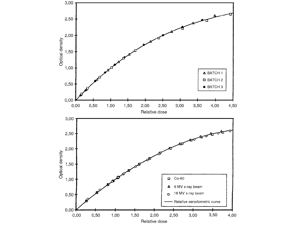
The sensitometric curves obtained at four different
depths ranging from
d
max
to 20 cm are plotted vs. absolute
dose in Figure 5.11. The largest variation in dose for a
given optical density determined from this set of curves is
FIGURE 5.10
The percentage depth doses measured in a polystyrene phantom containing 6-cm cork at 4-cm depth using (A) film in
parallel orientation to the beam, (B) film in perpendicular orientation to the beam, and (C) TLD are shown and compared to the PDD
measured in a homogeneous water phantom for electron beams of nominal energy (a) 12 MeV and (b) 20 MeV. (From Reference [4].
With permission.)
FIGURE 5.11
Film sensitometric curves derived from four sets of BEV films placed at depths of 3, 5, 10, and 20 cm, respectively.
(From Reference [5]. With permission.)
Ch-05.fm Page 308 Friday, November 10, 2000 12:01 PM
Film Dosimetry
309
2.3%, at an optical density of approximately 2.3. Variation
is less than 1% over most of the measured dose range.
This demonstrates that a single sensitometric curve mea-
sured at one depth can be accurately used for optical
density-to-dose conversion of BEV films at all depths for
the experimental situation used.
A feasibility study for mailed film dosimetry has been
performed by Novotny et al. [6] The fading of the latent
image before film processing is only 3% per month and
the normalized sensitometric curve is not modified after
a period of 51 days between irradiation and processing.
All film experiments have been performed with Kodak
Readypack paper envelope films (25.4 cm
30.5 cm).
All films were processed with an automatic processing
unit, either an Agfa Gevamatic 1100 (processing time 90 s
and temperature 35
1°C) or a modern AGFA Curix 260
(processing time 90 s and temperature 35
1°C). No
dependence on the photon energy has been observed in
the shape of the normalized sensitometric curve for
60
Co,
6-MV, and 18-MV beams (Figure 5.12).
No increase is observed in the fog value for un-
irradiated films after mailing or storing for more than
75 days; the average reading is 0.16
0.01 for the two
series of films, either processed immediately after irradi-
ation or processed after mailing.
Fading is expressed as the ratio of the optical density
of a faded film and the optical density of the reference film,
as a function of the time
t
between film irradiation and film
processing. The interval
t
is equal to 0 for the reference
film. The dependence of fading on time
t
, for films stored
in the laboratory, is presented in Figure 5.13. A straight
line is fitted through the measured data using the least-
squares method. A decrease of about 3% in optical density
is observed per month of delay between film irradiation
and film processing (corresponding to a given dose of 0.45
Gy and optical density of about 1.3). A smaller decrease
of about 1.8% per month in optical density is observed for
films irradiated with dose of 1.5 Gy (corresponding to
optical density 2.7). These decreases correspond to esti-
mated variations on dose of 3.8% and 3.4%, respectively.
FIGURE 5.12
Relative sensitometric curve for three photon beam qualities (
60
Co, 6-MV x-rays, 18-MV x-rays). (From Reference [6].
With permission.)
Ch-05.fm Page 309 Friday, November 10, 2000 12:01 PM