Shani G. Radiation Dosimetry: Instrumentation and Methods
Подождите немного. Документ загружается.

320 Radiation Dosimetry: Instrumentation and Methods
was established by Chiu-Tsao et al. [13] The net optical
density was found to be a power function of dose with
exponents of 0.858 and 0.997 for the Macbeth and LKB
densitometers, respectively. Film sensitivity with the LKB
densitometer was about double that with the Macbeth
densitometer.
The dose-response curves of the high-sensitivity
GafChromic films were plotted for
125
I,
137
Cs, and
60
Co
and shown in Figures 5.31a, b, and c, respectively, for
doses up to 200 Gy. The curves for the same radionuclide
obtained with the LKB and Macbeth densitometers were
compared to the figure. The net OD increases with
increasing dose. The values with the LKB were about
twice those with the Macbeth, depending on the dose.
This means that the film sensitivity measured with red
light from the He-Ne laser is higher than that obtained
with the broadbeam spectrum. The data obtained with the
Macbeth densitometer were fitted to a power function of
dose, with exponents of about 0.858, 0.839, and 0.849
for
125
I,
137
Cs, and
60
Co, respectively. As for the LKB
densitometer, the curves were linear for
137
Cs and
60
Co.
The curve for
125
I, however, was better-fitted to a power
function of dose with a power of 0.997, i.e., essentially
linear.
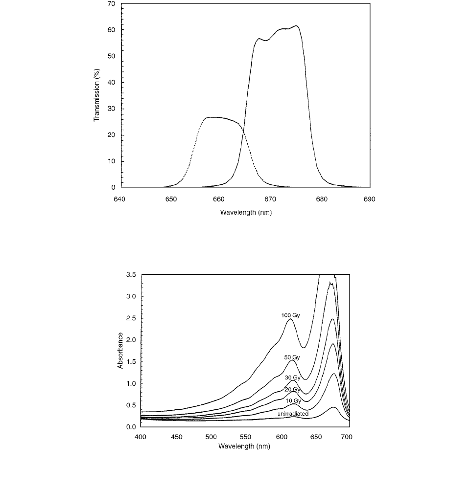
FIGURE 5.28 Transmission spectra for the 660 nm filter (dashed line) and 621 nm filter (solid line) measured using the Du 640B
spectrometer. (From Reference [12]. With permission.)
FIGURE 5.29 Absorbed spectra of GafChromic film (MD-55-2) for the dose levels 0, 10, 20, 30, 50, and 100 Gy. The spectra were
measured on a Beckman DU 640B spectrometer. (From Reference [12]. With permission.)
Ch-05.fm Page 320 Friday, November 10, 2000 12:01 PM
Film Dosimetry 321
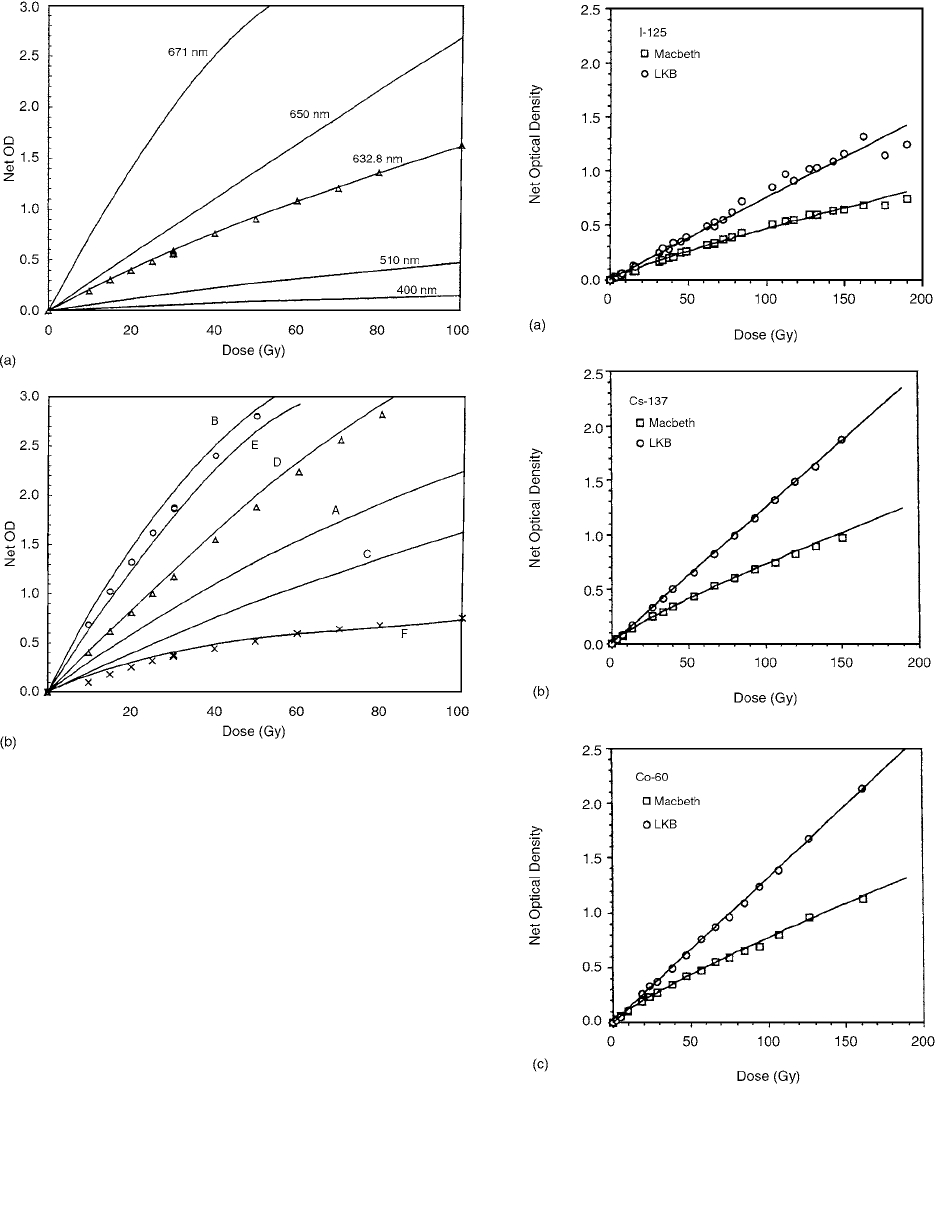
FIGURE 5.30 (a) Predicted dose response curves when mea-
sured at fixed wavelengths 400 nm, 510 nm, 632.8 nm, 650 nm,
and 671 nm for GafChromic film (MD-55-2), determined from
its absorption spectra using a Beckman DU 640B spectropho-
tometer at 31.9 days post-irradiation (solid curves). Measured
data (
) using the He-Ne laser is also shown for comparison
with the predicted dose response curve at fixed wavelength of
632.8 nm. Three samples were given the same total dose of 30
Gy and used to check for measurement reproducibility. (b) Pre-
dicted dose response curves (solid curves) for a tunable light
source to the minor peak (A), major peak (B), and for valley
between peaks (C), and for complex light sources: an LED light
source coupled to a 660-nm band-pass filter (D), an LED light
source coupled to a 671-nm filter (E), and a broadband light
source (F). Measured dose response curves are also shown for
comparison, namely a 660-nm filtered densitometer (
), 671-nm
filtered densitometer (O) and a broadband densitometer (x). Three
samples were given the same total dose of 30 Gy and were used
to check for measurement reproducibility. (From Reference [12].
With permission.)
FIGURE 5.31 The dose-response curves of the high-sensitivity
GafChromic film for three radionuclides,
125
I,
137
Cs, and
60
Co, in
a, b, and c, respectively. The square and circle symbols are for
the reading using Macbeth and LKB densitometers, respectively.
The solid curves represent the corresponding function fit. (From
Reference [13]. With permission.)
Ch-05.fm Page 321 Friday, November 10, 2000 12:01 PM
322 Radiation Dosimetry: Instrumentation and Methods
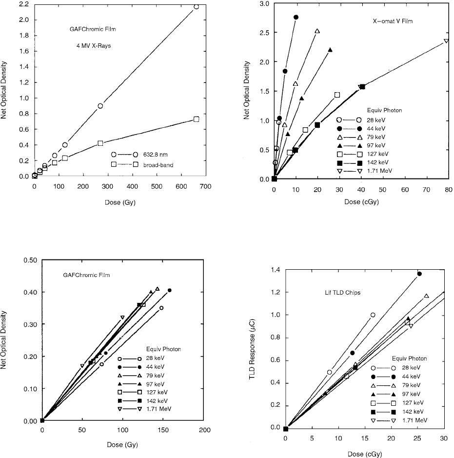
The dose response of the radiochromic film for 4-MV
x-rays is shown in Figure 5.32. [14] The x-rays were
produced by a Clinac 4 linear accelerator, and the film
was irradiated at a depth of 1.25 cm in an acrylic phan-
tom set up at an SSD of 41 cm. The dose rate was about
900 cGy/min. The lower curve shows the results from
optical density measurements using broadband absorp-
tion, and the upper curve shows the results of measure-
ments using 632.8-nm light.
A family of dose-response curves for radiochromic film
for different photon energies is shown in Figure 5.33. These
curves are in the low-dose range for this film, although the
doses are about 250 times the doses used for the silver
halide film curves (Figure 5.34) and about 600 times the
doses used for the LiF chip curves (Figure 5.35). The curves
show linearity of dose response, and the similarity of slopes
shows that the dependence on photon energy is slight. The
optical densities are for 632.8-nm light.
An “improved film” was developed with higher sensi-
tivity and better uniformity than the model MD-55 films by
Meigooni et al. [15] The improved film is a multilayered
structure composed of a nominal 30-
m thickness of the
FIGURE 5.32 Dose response curves for radiochromic film.
(From Reference [14]. With permission.)
FIGURE 5.33 Family of dose response curves for radiochromic
film. (From Reference [14]. With permission.)
FIGURE 5.34 Family of dose-response curves for silver halide
film. Each curve is for one x-ray beam (one equivalent photon
energy). (From Reference [14]. With permission.)
FIGURE 5.35 Family of dose-response curves for LiF TLD
chips. Each curve is for one type of x-ray beam (one equivalent
photon energy). (From Reference [14]. With permission.)
Ch-05.fm Page 322 Friday, November 10, 2000 12:01 PM
Film Dosimetry 323
sensitive material, sandwiched between two pieces of 75-
m-
thick polyester base material and two pieces of 13
m lam-
inated material, yielding a total thickness of 206
m. In
contrast, MD-55 film is composed of a layer (23-
m-thick)
of sensitive material on one side of a 100-micron polyester
base. These films are colorless and transparent before being
exposed to ionizing radiation. As they are irradiated, their
color changes to blue, due to the polymerization of radio-
chromic dye. The darkness of the blue color depends on the
absorbed dose and can be measured with a laser densitom-
eter with a wavelength ranging from 610–675 nm.
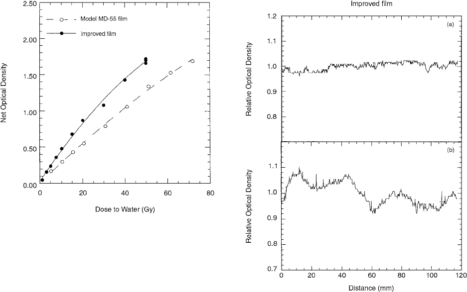
Figure 5.36 shows the measured sensitometric curves
of the improved radiochromic film and MD-55 film as a
function of absorbed dose. This figure indicates that the
improved film requires about 40% less dose than MD-55
film to yield an optical density of 0.5. This difference is
reduced to about 30% for an optical density of about 1.5.
The combination of all sources of uncertainties in these
measurements is estimated to be less than 5%. This
uncertainty includes the reproducibility of the laser scan-
ning system, uncertainty of determination of absorbed
dose given to the film, and standard deviation of the optical
density measured along a path of about 5 mm in a uni-
formly exposed film.
The uniformity of the improved radiochromic film and
MD-55 film along two orthogonal directions was deter-
mined by measuring the optical density of a film irradiated
with a uniform radiation field. Figure 5.37a shows very
good uniformity (within 4%) along one direction of the
improved radio-chromic film. A similar result was
obtained at a distance of 5 cm from the beam axis, along
the same direction of the film. However, along the per-
pendicular direction (Figure 5.37b), a non-uniformity of
up to 15% was observed. This non-uniformity is not con-
sistent from one sample of film to another.
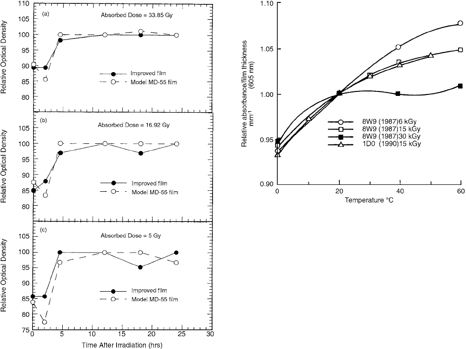
Figure 5.38 shows the variation of relative optical
density of the improved radiochromic film and MD-55
film as a function of time after irradiation for absorbed
dose as shown in the figures.
Evaluation of the influence of irradiation temperature
differences on the response of three widely used radiochro-
mic film dosimeters was made by McLaughlin et al. [16]
Freestanding nylon base film (mean thickness 48
m)
FWT-60-00, thin-coated (~7
m) sensor on 100-
m poly-
ester base GafChromic DM-1260 dosimetry media, and
freestanding polyvinylbutyral base film (mean thickness
23
m) Risø B3 were investigated.
The irradiations were made with gamma-rays from two
60
Co sources, one at an absorbed dose rate of 12.2 kGy/h
and the other at 30.0 kGy/h. During irradiation the films
(three films for each irradiation) were held between two
electron equilibrium layers of 3-mm-thick polystyrene and
sealed at 50% relative humidity in a triple layer pouch of
FIGURE 5.36 Sensitometric curves of the improved radiochro-
mic film (solid symbol) compared with that of MD-55 film (open
symbol). These results were obtained by irradiating the films to
doses ranging from 1–72 Gy of gamma rays from a
60
Co tele-
therapy unit. (From Reference [15]. With permission.)
FIGURE 5.37 Relative optical density of a uniformly irradiated
improved radiochromic film (a) measured in one direction along
the film and (b) measured along the orthogonal direction. (From
Reference [15]. With permission.)
Ch-05.fm Page 323 Friday, November 10, 2000 12:01 PM
324 Radiation Dosimetry: Instrumentation and Methods
30-
m-thick polyethylene–aluminum foil laminate. Each
packet was conditioned for 30 min at each irradiation
temperature before irradiation, then irradiated, and then
stored at room temperature immediately after irradiation.
Figures 5.39, 5.40, and 5.41 show, respectively, the
temperature dependence of the gamma-ray responses of
the three most recent batches of radiochromic films, FWT-
60-00 (batches 8W9-1989 and 1D0-1990), GafChromic
1260 (batch 09031501-1989), and Risø B3 (batch 85-128-
1989). In all three cases, it is seen that a different trend
occurs at different absorbed doses, particularly at the higher
temperatures.
A technique of readout was developed by Kellermann
et al. [17] to measure the optical density distributions of
the film in purely directed light. This technique imple-
ments radiochromic film dosimetry near the film’s
absorption maximum by using a single-mode top-surface
emitting laser diode (675.2 nm). The effective sensitivity
of the film, compared with a helium-neon laser densito-
meter (632.8 nm), is increased approximately threefold.
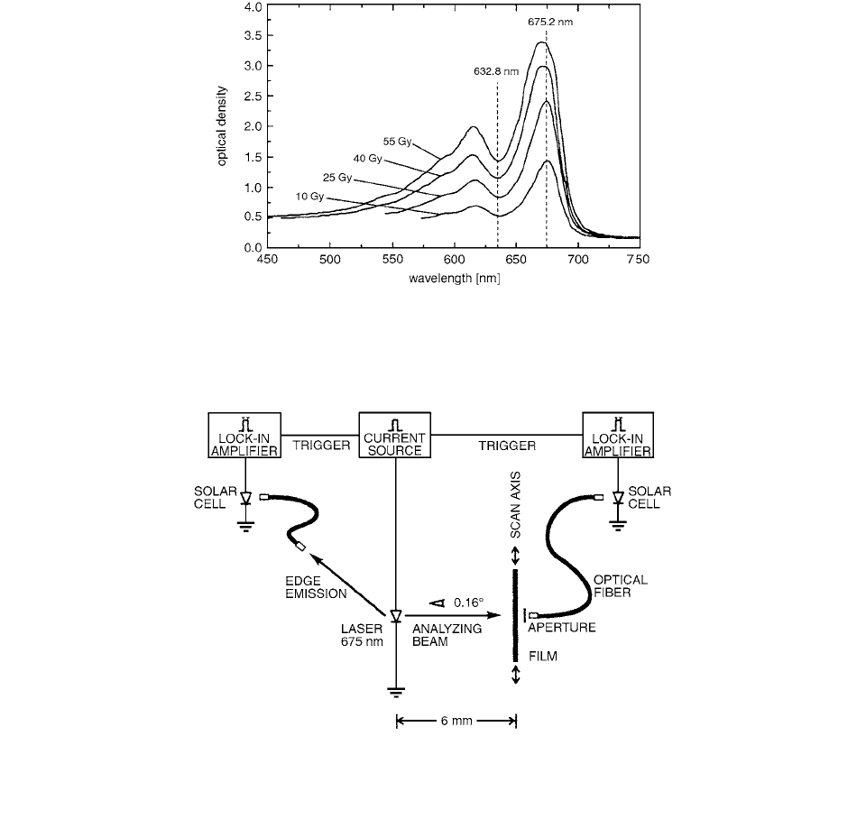
Figure 5.42 shows the optical density of the radiochro-
mic film depending on the wavelength of the analyzing
beam after irradiation with four different doses. [18] In
the helium-neon laser densitometer, the film is scanned
with an analyzing beam and the transmission is measured
by a photomultiplier. The emission wavelength (632.8 nm)
of the analyzing beam lies in the valley of the film’s
absorption spectrum. Since the absorption spectrum of the
radiochromic film contains two peaks centered at approx-
imately 615 nm and 675 nm, it has been suggested [17]
that significant improvement in response might be
achieved by tailoring the wavelength of the incident den-
sitometer light to one of these peaks. Densitometers with
a light-emitting diode (LED) (660 nm, spectral line width
30 nm) and with an LED coupled to a band-pass filter
(670 nm, bandwidth 11 nm, GatChromic densitometer)
have been presented, but their spectral line width is too
broad to match the absorption maximum exactly, and the
improvement in response is reduced.
The radiochromic film’s sensitive layer consists of
microcrystals of radiation-sensitive monomer uniformly
dispersed in a gelatin binder. When the microcrystals are
exposed to ionizing radiation, polymerization occurs and
the polymers alter the crystal color to various shades
of blue. The optical density increases continuously with
the absorbed dose. The optical density depends neither
FIGURE 5.38 Variation of the optical density with time after
irradiation for the improved radiochromic film (solid symbol)
compared to MD-55 film (open symbol). These results were
obtained by simultaneous irradiation of pieces of each film-type
absorbed doses of 5, 16.92, and 33.85 Gy using a
60
Co telether-
apy unit. (From Reference [15]. With permission.)
FIGURE 5.39 Temperature dependence, relative to that at
20°C, of the gamma-ray response of two batches of radiochromic
film FWT-60-00. Batch 8W9 was irradiated at three absorbed
doses (6, 15, and 30 kGy) and batch 1D0 was irradiated at 15
kGy. The absorbances per unit thickness (
A mm
1
) were mea-
sured at 605-nm wavelength. (From Reference [16]. With per-
mission.)
Ch-05.fm Page 324 Friday, November 10, 2000 12:01 PM
Film Dosimetry 325
on photon energy in the range 0.1 to 20 MeV nor on
the energy of secondary electrons in the range 0.01 to
20 MeV.
GafChromic MD-55™ irradiated in a uniform radia-
tion field showed a non-uniformity in optical density of
up to 15% in one direction and within 4% in the direction
orthogonal to it. This would result in a dose error of
or %.
A laser diode with an emission wavelength of 675.2 nm
was chosen by Kellermann et al. as a light source to tran-
silluminate the radiochromic film. At 675.2 nm, near the
film’s absorption maximum, a dose gradient in the radiation
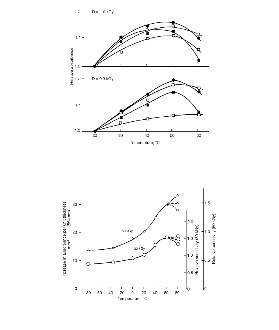
FIGURE 5.40 Temperature dependence, relative to that at 20°C, of the gamma-ray response of radiochromic film GafChromic
Dosimetry Media, DM-1260, batch 09031501, irradiated at two absorbed doses. The wavelengths of absorbance measurements:
•
650 nm; 600 nm; X 510 nm; ◊ 400 nm. (From Reference [16]. With permission.)
FIGURE 5.41 Temperature dependence of the gamma-ray response of radiochromic film Risø B3, batch 85-128, irradiated at two
absorbed doses. The two right ordinates show the response relative to that at 20°C. The absorbances per unit thickness (
A mm
1
)
were measured at 554-nm wavelength. (From Reference [16]. With permission.)
10% 2.5
Ch-05.fm Page 325 Friday, November 10, 2000 12:01 PM
326 Radiation Dosimetry: Instrumentation and Methods
field leads to a change in optical density that is up to three
times larger than the change that is caused by the same
dose gradient in the valley (632.8 nm) of the film’s absorp-
tion spectrum. The laser light propagating in the active
region excites a transverse electrically polarized (TE
0
) sur-
face mode in the waveguide structure on the top of the
laser diode through grating coupling. The surface mode
couples both into the vacuum light cone, resulting in top-
surface emission with narrow beam divergence, and back
to the active region, leading to a gain mechanism and thus
to single-mode emission.
The film is fastened (with double-sided adhesive tape)
to a stage that allows precise movements along the scan-
ning direction (and orthogonal to it) in 50-
m steps along
a distance of 75 mm. The schematic of Figure 5.43 dis-
plays the measurement setup.
After irradiation, the optical density of radiochromic
film increases logarithmically. Therefore, the optical den-
sity measurements with the densitometry system are done
four days after the end of irradiation, by which time the
changes in optical density are small.
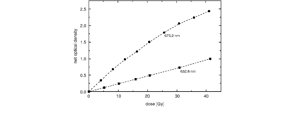
The calibration curve—change in optical density at
675.2 nm vs. radiation dose—is shown in Figure 5.44. The
curve rises rapidly, but the upward gradient becomes smaller
with higher doses. In comparison, the optical density vs.
radiation dose at 632.8 nm is flat but approximately linear.
[19] At 675.2 nm, a dose gradient from 0 Gy to 20 Gy in
the radiation field leads to a change in optical density that is
3.1 times larger than at a wavelength of 632.8 nm. This ratio
decreases to 1.9 with a dose gradient from 20 Gy to 40 Gy.
Film digitizers are, compared with mechanical scanners,
much faster, while their spatial resolution is even higher.
FIGURE 5.42 Optical density of the radiochromic film (Gafchromic MD-55
TM
, Model No. 37-041), depending on the wavelength
of the analyzing beam after irradiation with four different doses. [18] The wavelength of the helium-neon, laser (632.8 nm) lies in
the valley of the film’s absorption spectrum, while the wavelength of the laser diode (675.2 nm) used by Kellermann et al. is near
the film’s absorption maximum. (From Reference [17]. With permission.)
FIGURE 5.43 The schematic measurement setup is sketched. The laser light transmits the film and is sent via an optical fiber to a
solar cell, where it is transformed to an electrical signal. This is measured with a lock-in amplifier. The light of the edge emission
is used as a reference signal. (From Reference [17]. With permission.)
Ch-05.fm Page 326 Friday, November 10, 2000 12:01 PM
Film Dosimetry 327
Most commercially available digitizers are based on either
a scanning He-Ne laser beam or a one-dimensional charge-
coupled device (CCD) combined with a standard fluores-
cent lamp. Mersseman and De Wagter [20] investigated a
CCD-based digitizer.
The principles of the digitizer are as follows. A broad
light beam from the fluorescent lamp (Philips F17T8/
TL841) crosses the light-diffusion plate and travels
through the scrolling film to be digitized. Two fixed ver-
tical and two movable horizontal shutters subsequently
collimate this light beam to a narrow rectangular field that
covers the film lengthwise. The transmitted light is sub-
sequently reflected by a stationary mirror, focused by the
40-mm lens and finally detected by the linear CCD ele-
ment. The CCD detector reads line per line at 300 dots
per inch (dpi) over its full length of 35.6 cm (14 in). In
order to achieve a 150, 75, and 60 dpi resolution, the film
digitizer combines data from nearby pixels. A 12-bit
analog-to-digital converter digitizes the signal. Conver-
sion tables (built-in or loaded from the host computer) are
allowed to further translate the data. The resulting data
matrix is transferred to the host computer (Pentium PC)
using an SCSI interface.
Nuclear Associates’ GafChromic Dosimetry Media
is a radiation-sensitive imaging film designed for use in
medical and industrial quality control measurement
applications. The media is colorless and grainless and
offers a very high spatial resolution (1200 LP/mm), mak-
ing it invaluable for measuring dose distribution around
small brachytherapy sources and stereotactic radiosur-
gery fields. [21] GafChromic Dosimetry Media MD-55
(Model 37-041) comprises clear film sheets that
are suitable for medium-dose measurement applications
covering the 1.5 to 50-Gy dose range, when used with the
radiochromic densitometer. Higher doses (100 Gy and
greater) can be measured using other commercially avail-
able densitometers. MD-55 has a higher sensitivity than
HD-810 and was developed for use in external-beam radio-
therapy and brachytherapy applications. MD-55 is a lam-
inated film composed of two pieces of 2.65-mil polyester
base, each with a nominal 15-micron-thick coating.
HD-810 (Model 37-040) films are low-sensitivity
clear film sheets suitable for high-dose beam pro-
filing and dose-mapping applications in the 10 to 300-Gy
dose range, when used with the radiochromic densitome-
ter. Higher doses (up to 2500 Gy) can be measured using
other commercially available densitometers. This media
has a nominal 7-micron-thick radiation-sensitive layer on
a 3.9 mil polyester base.
GafChromic Dosimetry Media is composed of materials
with low atomic numbers, so the film can be considered
tissue-equivalent for most radiation therapy applications.
Additionally, this material has low sensitivity to ambient
room light, which simplifies handling procedures and
enhances image stability. Its response is dose rate indepen-
dent; it is linear dose response and energy independence
above 100 keV. [21]
TECHNICAL DATA
MD-55: Polyester Base: 67-
m; Sensitive Layer: 15-
m;
Pressure-Sensitive Adhesive Layer: 44.5
m; Polyester
Base: 25
m; Pressure-Sensitive Adhesive Layer: 44.5
m;
Sensitive Layer: 15
m; Polyester Base: 67
m.
HD-810: Sensitive Layer: 7
m; Adhesive Layer: 1.5
m;
Conductive Layer: 0.05
m; Polyester base: 99
m.
FIGURE 5.44 Calibration curve—change in optical density at 675.2 nm vs. radiation. The curve rises rapidly, but the upward gradient
becomes smaller with higher doses. In comparison, optical density vs. radiation dose at 632.8 nm is flat but approximately linear.
(From Reference [17]. With permission.)
5 5
8 10
Ch-05.fm Page 327 Friday, November 10, 2000 12:01 PM
328 Radiation Dosimetry: Instrumentation and Methods
37-041 MD-55 GAFCHROMIC Dosimetry Media;
Package of Five Sheets, Weight: .10 lb (.04 kg).
37-040 HD-810 GAFCHROMIC Dosimetry Media:
Package of Five Sheets, Weight: .50 lb (.24 kg)
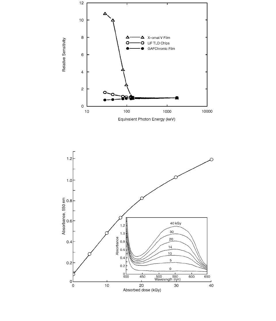
The GAFCHROMIC sensitivity to energy compared
to that of silver based film is shown in Figure 5.45.
Another promising thin film polymer dosimeter for high
doses consists of a film or gel cast from an alcohol solu-
tion of blue tetrazolium or triphenyl-tetrazolium chloride
combined with a polymer matrix material (e.g. polyvinyl
alcohol, polyvinyl pyrrolidone), McLaughlin [22]. Figure
5.46 shows the radiation response properties of one exam-
ple of this film system, in this case with a thickness of
30
m.
FIGURE 5.45 GafChromic sensitivity to energy composed to that of silver based film on LiF TLD. (From Reference [21]. With
permission.)
FIGURE 5.46 Response of radiochromic film of tetrazolium chloride in polyvinyl pyrrolidone (in terms of increase in absorbance
measured at 522 nm wavelength) as a Junction of the gamma ray absorbed dose. The inset shows the increase in the absorption
spectral amplitudes with dose. (From Reference [22]. With permission.)
5 5
8 10
Ch-05.fm Page 328 Friday, November 10, 2000 12:01 PM
Film Dosimetry 329
REFERENCES
1. Cheng, C-W. and Das, I. J., Med. Phys., 23, 1225, 1996.
2.
Burch, S. E. et al., Med. Phys., 24, 775, 1997.
3.
Robar, J. L. and Clark, B. G., Med. Phys., 26, 2144,
1999.
4.
El-Khatib, E. et al., Med. Phys., 19, 317, 1992.
5.
Stern, R. L. et al., Med. Phys., 19, 165, 1992.
6.
Novotny, J. et al., Phys. Med. Biol., 42, 1277, 1997.
7.
Stevens, M. A. et al., Phys. Med. Biol., 41, 2357, 1996.
8.
McLaughlin, W. L. et al., Nucl. Inst. Meth., A302, 165,
1991.
9.
Vatnitsky, S. M. et al., Phys. Med. Biol., 42, 1887,
1997.
10.
Zhu, Y. et al., Med. Phys., 24, 223, 1997.
11.
Klassen, N. V. et al., Med. Phys., 24, 1924, 1997.
12.
Reinstein, L. E. et al., Med. Phys., 24, 1935, 1997.
13.
Chiu-Tsao, S-T. et al., Med. Phys., 21, 651, 1994.
14.
Muench, P. J. et al., Med. Phys., 18, 769, 1991.
15.
Meigooni, M. F. et al., Med. Phys., 23, 1883, 1996.
16.
McLaughlin, W. L. et al., in Proc. High Dose Dosim-
etry for Radiation Protection,
IAEA, 1991, 305.
17.
Kellermann, P. O. et al., Phys. Med. Biol., 43, 2251,
1998.
18.
Ertl, A. et al., Phys. Med. Biol., 43, 1567, 1998.
19.
Meigooni, A. C. et al., Med. Phys., 23, 1803, 1996.
20.
Mersseman, B. and De Wagter, C., Phys. Med. Biol.,
43, 1803, 1998.
21.
Nuclear Associates, Diagnostic Imaging and Radiation
Therapy Catalog,
1999.
22.
McLaughlin, W. L., in Proc. High Dose Dosimetry for
Radiation Protection,
IAEA, 1991, 3.
Ch-05.fm Page 329 Friday, November 10, 2000 12:01 PM