Russ J.C. Image Analysis of Food Microstructure
Подождите немного. Документ загружается.

on using just two of the sections, and the precision can be improved by processing
more pairs of planes, so that the total number of events (feature ends) is increased,
as discussed in Chapter 1. Additional sampling is also needed if the specimen is not
uniform.
CALIBRATION
After the various measurements of feature size, shape, position, and brightness
or color described in this chapter have been made, the resulting data are often used
for feature identification or recognition. There are several different approaches for
performing this step, which will be discussed at the end of the chapter. All of these
procedures require image calibration. Both counting results, which are usually
reported as number per unit area, and feature measurements (except for some shape
descriptors) rely on knowing the image magnification. In principle, this is straight-
forward. Capturing an image of a known scale, such as a stage micrometer, or a
grid of known dimension (many of which are traceable standards to NIST or other
international sources) allows easy determination of the number of pixels per micron,
or whatever units are being used. For macroscopic images, such as those obtained
using a copy stand, rulers along the sides or even a full grid on which objects are
placed are commonly employed.
(c)
FIGURE 5.8 (continued)
2241_C05.fm Page 287 Thursday, April 28, 2005 10:30 AM
Copyright © 2005 CRC Press LLC
The assumption, of course, is that the images of the objects of interest have the
same magnification everywhere (no perspective distortion from a tilted view, or
pincushion distortion from a closeup lens, or different magnification in the X and
Y directions from using a video camera and framegrabber, or area distortions because
of piezo creep in an AFM, for example). If the scale is photographed once and then
the calibration is stored for future use, it is assumed that the magnification does not
change. For a light microscope, that is usually a good assumption because the optics
are made of glass with fixed curvatures and, unless some adjustments are made
in the transfer lens that connects the camera to the scope, the same image mag-
nification will be obtained whenever a chosen objective lens is clicked into place.
Focusing is done by moving the sample toward or away from the lens, not altering
the optics.
For a camera on a copy stand, it is usually possible to focus the camera lens,
which does change the magnification. But on the copy stand the scale is usually
included in the image so there is always a calibration reference at hand. Electron
microscopes use lenses whose magnification is continuously adjustable, by varying
the currents in the lens coils. Even in models that seem to provide switch-selected
fixed magnifications, the actual magnification varies with focus, and also depends
on the stability of the electronics. Keeping the magnification constant over time, as
the calibration standard is removed and the sample inserted, is not easy (and not
often accomplished).
Calibrating the brightness or color information is much more difficult than
calibrating the image magnification. Again, standards are needed. For density mea-
surement, film standards with known optical density are available from camera stores
(Figure 5.9), but fewer choices are available for microscopic work. For color com-
parisons, the Macbeth chart shown in Chapter 2 is a convenient choice, but again
is useful only for macroscopic work. A few microscope accessory and supply
companies do offer a limited choice of color or density standards on slides. For
electron microscopy, only relative comparisons within an image are generally prac-
tical. In images such as those produced by the AFM in which pixel value represents
elevation, calibration using standard artefacts is possible although far from routine.
Some of the other AFM modes (tapping mode, lateral force, etc.) produce signals
whose exact physical basis is only partially understood, and cannot be calibrated in
any conventional sense.
One difficulty with calibrating brightness in terms of density for transmission
images, or concentration for fluorescence images, etc., is that of stability. The light
sources, camera response, and digitization of the data are not generally stable over
long periods of time, nor can they be expected to repeat after being turned off and
on again. As mentioned in Chapter 2, some cameras are not linear in their response
and the relationship between intensity and output signal varies with signal strength.
As an example, a camera may be linear at low light levels and become increasingly
non-linear, approaching a logarithmic response at brighter levels. Some manufac-
turers do this intentionally to gain a greater dynamic range. If automatic gain and
brightness circuitry, or automatic exposure compensation is used, there will be no
way to compare one image to another.
2241_C05.fm Page 288 Thursday, April 28, 2005 10:30 AM
Copyright © 2005 CRC Press LLC
The only solution in such cases is to include the standard in every image. With
a macro camera on a copy stand, or with a flatbed scanner as may be used for reading
films or gels, that is the most reasonable and common way to acquire images. The
calibration steps are then performed on each image using the included calibration
data. Obviously, this is much more difficult for a microscopy application.
The procedure for establishing a calibration curve is straightforward, if tedious.
It is illustrated in Figure 5.9, using an image of a commercial film density wedge
(a) (b)
(c) (d)
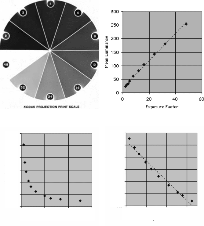
FIGURE 5.9
Intensity calibration: (a) image of density wedge; (b) plot of mean luminance
of each region vs. labeled exposure factor; (c) luminance vs. reciprocal of exposure factor
(optical density); (d) log (luminance) vs. reciprocal of exposure factor.
0
50
100
150
200
250
300
Mean Luminance
0.4 0.4 0.60
1/Exposure Factor
1.3
1.5
1.7
1.9
2.1
2.3
2.5
Log (Luminance)
0.4 0.5 0.6 0.7
1/Exposure Factor
2241_C05.fm Page 289 Thursday, April 28, 2005 10:30 AM
Copyright © 2005 CRC Press LLC
used for darkroom exposure settings. This was scanned with a flatbed scanner, and
in normal use would have been included in the same scanned image as the film or
gel to be quantified. The wedge has ten areas of more-or-less uniform density, with
known (labeled) exposure factor settings (the exposure factor is proportional to the
inverse of the optical density). Measuring the mean pixel brightness value in each
region allows creating a calibration curve as shown in the figure.
Plotting the mean pixel value (luminance) vs. the labeled exposure factor pro-
duces a graph that appears quite linear and could certainly be used for calibration,
but other plots reveal more of the principal of optical density. The luminance drops
exponentially with density as shown in Figure 5.9(c), following Beer’s law. Replot-
ting the data with the log of the luminance as a function of optical density produces
a nearly linear relationship (ideally linear if the wedge, detector, electronics, etc. are
perfect) that spreads the points out more uniformly than the original plot of the raw
data. Once the curve is constructed, it can then be stored and used to convert points
or features in measured images to optical density.
Other types of standards can be used for other applications. Drug doses or
chemical concentration (for fluorescence images), average atomic number (for SEM
backscattered electron images), step height (AFM images), etc. can all be quantified
if suitable standards can be acquired or fabricated. In practically all cases the
calibration curves will be non-linear. That means that the brightness value for each
pixel should be converted to the calibrated units and then the average for the feature
calculated, rather than the mean pixel value being used to look up a calibrated value.
Other statistics such as the standard deviation, minimum and maximum are also
useful as will be shown in some of the examples below.
Color is a more challenging problem. As pointed out in Chapter 2, a tristimulus
camera that captures light in three relatively broad (and overlapping) red, green, and
blue bands cannot be used to actually measure color. Many different input spectra
would be integrated across those bands to give the exact same output. Since human
vision also uses three types of detectors and performs a somewhat similar integration,
it is possible to use tristimulus cameras with proper calibration to match one color
to another in the sense that they would appear the same to a human observer, even
if their color spectra are not the same in detail. That is accomplished by the tristim-
ulus correction shown in Chapter 2, and is the principal reason for using standard
color charts.
In most image analysis situations, the goal is not to measure the exact color but
to use color as a means of segmenting the image or of identifying or classifying the
features present. In Figure 5.10, the colored candy pieces can be thresholded from
the grey background based on saturation. As pointed out in Chapter 3, the background
is more uniform than the features and can be selected relatively easily. After thresh-
olding, a watershed segmentation is needed to separate the touching features. Then
the binary image is used as a mask to select the pixels from the original (combining
the two images while keeping whichever pixel value is brighter erases the back-
ground and leaves the colored features unchanged).
Measuring the red, green, and blue values in the acquired color image is not
very useful, but converting the data to hue, saturation and intensity allows the
identification, classification and counting of the candies. As shown in the plot, the
2241_C05.fm Page 290 Thursday, April 28, 2005 10:30 AM
Copyright © 2005 CRC Press LLC
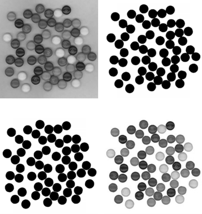
hue values separate most of the features while the intensity or luminance is needed
to distinguish the brown from the orange colors.
There is more information available in this image as well. The ratio of the
maximum to minimum luminance for each feature is a measure of the surface gloss
of the candy piece. That is true in this instance because the sample illumination
consisted of two fluorescent tubes in the flatbed scanner that moved with the detector
bar and thus lit each piece of candy with the same geometry. Another way to measure
surface gloss is to light the surface using a polarizer and record two images with a
(a) (b)
(c) (d)
FIGURE 5.10
Measurement of feature color: (a) original image of candies (see Color Figure
3.67; see color insert folllowing page 150); (b) thresholded; (c) watershed segmentation; (d)
isolated individual features (see Color Figure 4.26a; see color insert following page 150); (e)
plot of hue vs. luminance (brightness) for each feature, showing clusters that count candies
in each class.
2241_C05.fm Page 291 Thursday, April 28, 2005 10:30 AM
Copyright © 2005 CRC Press LLC
polarizing filter set to parallel and perpendicular orientations. The ratio gives a
measure of surface gloss or reflectivity.
For other classes of particles, the standard deviation of the brightness values
within each feature is often a useful measure of the surface roughness.
SIZE MEASUREMENT
Size is a familiar concept, but not something that people are actually very good
at estimating visually. Judgments of relative size are strongly affected by shape,
color, and orientation. Computer measurement can provide accurate numerical values
(provided that good measurement algorithms are used, which is not always the case),
but there is still a question about which of many size measurements to use in any
particular situation. The area covered by a feature can be determined by counting
the number of pixels and applying the appropriate calibration factor to convert to
square micrometers, etc. But should the area include any internal voids or holes?
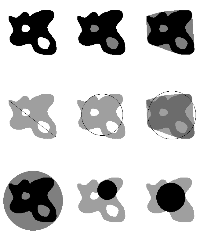
Should it include indentations around the periphery? As shown in Figure 5.11, the
computer can measure the net area, filled area or convex area but it is the user’s
problem to decide which of these is meaningful, and that depends on an understand-
ing of the application.
(e)
FIGURE 5.10 (continued)
Red Yellow Green Cyan Blue Magenta Red
Mean Hue
255
Mean Luminance
0
8 Brown
7 Green
6 Orange
11 Yellow
11 Blue
14 Red
2241_C05.fm Page 292 Thursday, April 28, 2005 10:30 AM
Copyright © 2005 CRC Press LLC
In the majority of cases, people prefer to work with a size parameter that is
linear rather than squared (e.g., mm rather than mm
2
). One of the commonly used
measures is the equivalent circular diameter, which is just the diameter of a circle
whose area would be the same as that of the feature (usually the net area, but in
principle this could be the filled or convex area as well). But there are several other
possibilities. One is the maximum caliper dimension (often called the maximum
Feret’s diameter) of the feature, which is the distance between the two points that
are farthest apart. Another is the diameter of the largest inscribed circle in the feature.
(a) (b) (c)
(d) (e) (f)
(g) (h) (i)
FIGURE 5.11
Measures of size: (a) net area; (b) filled area; (c) convex area; (d) maximum
caliper dimension; (e) equivalent circular diameter (net area); (f) equivalent circular diameter
(convex area); (g) circumscribed circle; (h) inscribed circle; (i) inscribed circle (filled area).
2241_C05.fm Page 293 Thursday, April 28, 2005 10:30 AM
Copyright © 2005 CRC Press LLC
Yet another is the diameter of the smallest circumscribed circle around the feature.
All of these can be measured by computer software, but once again it is up to the
user to determine which is most suitable for a particular application.
For those interested in the details, the convex area is determined by fitting a
taut-string or rubber-band boundary around the feature, as shown in Figure 5.11(c),
to fill in indentations around the periphery. It is constructed as a many-sided polygon
whose sides connect together the extreme points of the feature on a coordinate system
that is rotated in small angular steps (e.g., every 10 degrees). The circumscribed
circle (Figure 5.11g) is fit by analytical geometry using the vertices of the same
polygon used for the convex hull. The maximum caliper dimension (Figure 5.11d)
is also obtained from those vertices. The inscribed circle (Figure 5.11h and 5.11i)
is defined by the maximum point in the Euclidean distance map of the feature, but
as shown in the illustration, is strongly influenced by the presence of internal holes.
There are some other dimensions that can be measured, such as the perimeter
and the minimum caliper dimension. The perimeter can present problems both in
measurement and in interpretation. For one thing, many objects of interest are rough-
bordered and the length of the perimeter will increase as the magnification is
increased. In the particular case in which the increase in length with increasing
magnification produces a straight line plot on log-log axes (a Richardson plot), the
feature is deemed to have a fractal shape and the actual perimeter length is undefined.
A second difficulty with perimeter measurements is the selection of a measuring
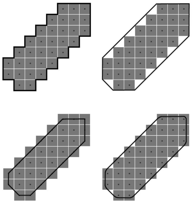
algorithm that is accurate. When counting pixels to measure area, it is convenient
to think of them as being small squares of finite area. But if that same approach is
used for perimeter, the length of the border around an object would become a city-
block distance (as shown in Figure 5.12) that overestimates the actual perimeter (in
fact, it would report a perimeter equal to the sides of a bounding box for any shape).
Many programs improve the procedure somewhat by using a chain-code perim-
eter, either one that touches the exterior of the square pixels or one that runs through
the centers of the pixels as shown in the figure, summing the links in a chain that
are either of length 1 (the side of a square pixel) or length 1.414 (the diagonal of a
square pixel). This is still not an accurate measurement of the actual perimeter,
produces a length value that varies significantly as a feature it rotated in the image,
and also measures the perimeter of a hole as being different from the perimeter of
a feature that exactly fills it.
The most accurate perimeter measurement method fits a locally smooth line
along the pixels while keeping the area inside the line equal to the area based on
the pixel count. This method also rounds corners that would otherwise be perfect
90˚ angles, and is relatively insensitive to feature or boundary orientation, but it
requires more computation than the other methods. The main use of the perimeter
value is usually in some of the shape descriptors discussed below.
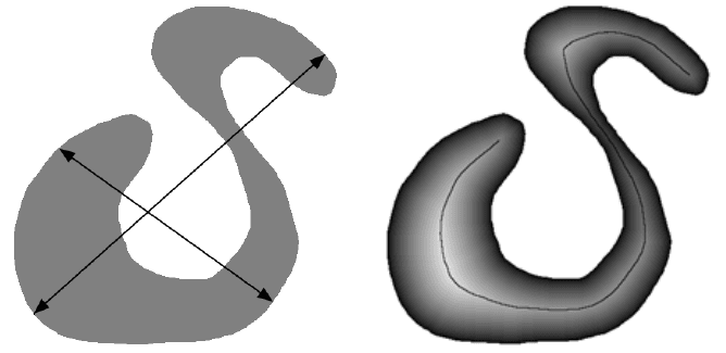
For a feature such as the irregular S-shaped fiber in Figure 5.13, the length
measured as the maximum caliper dimension has little meaning. It is the length
along the midline of the curved feature that describes the object. Likewise, the width
is not the minimum caliper dimension but the mean value of the dimension perpen-
2241_C05.fm Page 294 Thursday, April 28, 2005 10:30 AM
Copyright © 2005 CRC Press LLC
dicular to that midline and averaged along it. The skeleton and Euclidean distance
map provide these values, as described in the preceding chapter. The length of the
skeleton, measured by a smooth curve constructed in the same way as the accurate
perimeter measurement shown above, follows the fiber shape. The skeleton stops
short of the exact feature end points, actually terminating at the center of an inscribed
circle at each end. But the value of the Euclidean distance map at the end pixel is
exactly the radius of that circle, and adding back the EDM values at those end points
to the length of the skeleton provides an accurate measure of fiber length.
(a) (b)
(c) (d)
FIGURE 5.12
Perimeter measurement algorithms (shown by dark line) applied to a feature
composed of pixels (grey squares with centers indicated): (a) city-block method; (b) chain
code surrounding pixels; (c) chain code through pixel centers; (d) area-preserving smooth
(super-resolution) method.
2241_C05.fm Page 295 Thursday, April 28, 2005 10:30 AM
Copyright © 2005 CRC Press LLC
Similarly, the EDM values along the midline of the feature measure the radii of
inscribed circles all along the fiber. The skeleton selects the pixels along the midline,
so averaging the EDM values at all of the pixels on the skeleton provides an accurate
measurement of the fiber width (and other statistics such as the minimum, maximum
and standard deviation of the width can also be determined).
Given this very large number of potential measurements of feature size, which
ones should actually be used? Unfortunately, there is no simple guide. Sometimes
the definition of the problem will specify the appropriate measurement. The grading
of rice as long- or short-grain is defined by the U.S. Department of Agriculture in
terms of the length (the maximum caliper dimension) of the grains, so that is the
proper measurement to use.
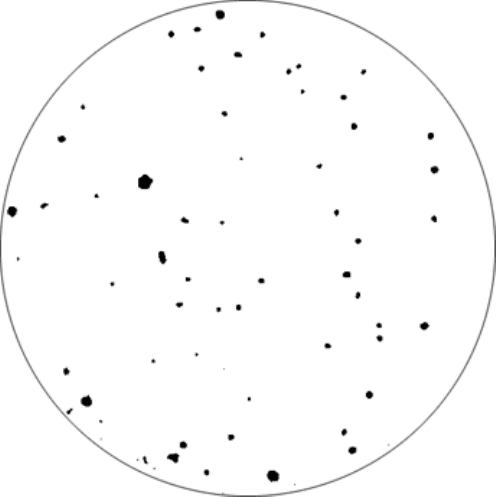
For the example in Figure 5.14, the sample preparation was done by sprinkling
rice onto a textured (black velvet) cloth attached to a vibrating table. The vibrations
separated the grains and the cloth provided a contrasting background, so the resulting
image could be automatically thresholded without any additional processing. Mea-
surement of the lengths of all the grains that were entirely contained in the field of
view produced the histogram shown in the figure, which indicates that this is a long
grain rice (short grained rice would have a more than 5% shorter than 6 mm) with
a more-or-less normal (Gaussian) distribution of lengths.
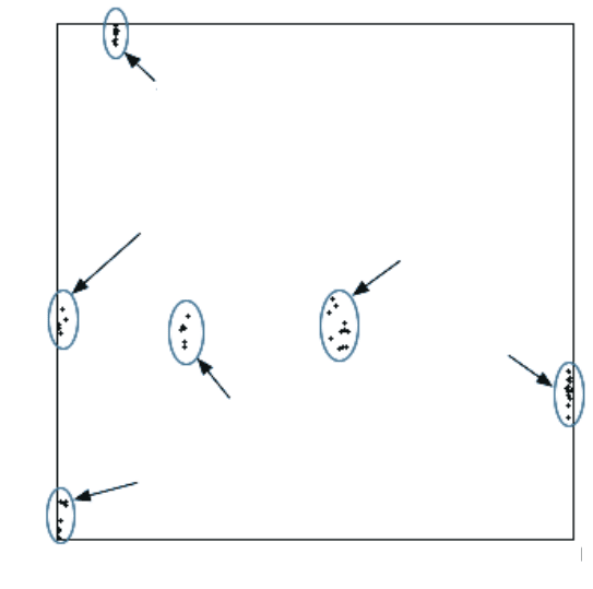
In this particular case, ignoring the features that intersect the edge of the image
does not introduce a serious error (because the range of feature sizes is small, and
it is the percentage of short ones that is of greatest interest). However, we will return
to the problem of edge-touching features below.
(a) (b)
FIGURE 5.13
Measuring fiber length and width: (a) an irregular feature with the (largely
meaningless) maximum and minimum caliper dimensions marked; (b) the feature skeleton
superimposed on the Euclidean distance map. Their combination provides measures for the
length and width of the shape.
2241_C05.fm Page 296 Thursday, April 28, 2005 10:30 AM
Copyright © 2005 CRC Press LLC