Russ J.C. Image Analysis of Food Microstructure
Подождите немного. Документ загружается.

Fourier transforms will be encountered several times in subsequent chapters on
enhancement and measurement. The basis for all of these procedures is Fourier’s
theorem, which states that any signal (such as brightness as a function of position)
can be constructed by adding together a series of sinusoids with different frequencies,
by adjusting the amplitude and phase of each one. Calculating those amplitudes and
phases with a Fast Fourier Transform (FFT) generates a display, the power spectrum,
that shows the amplitude of each sinusoid as a function of frequency and orientation.
Most image processing texts (see J. C. Russ, The Image Processing Handbook, 4th
edition, CRC Press, Boca Raton, FL) include extensive details about the mathematics
and programming procedures for this calculation. For our purposes here the math
is less important than observing and becoming familiar with the results.
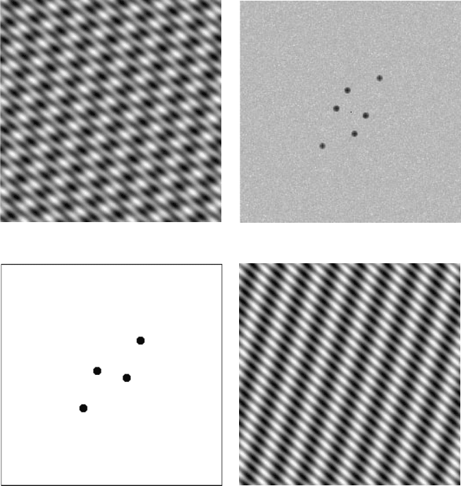
For a simple image consisting of just a few obvious sinusoids, the power spec-
trum consists of just the corresponding number of points (each spike is shown twice
because of the rotational symmetry of the plot). For each one, the radius from the
center is proportional to frequency and the angle from the center identifies the
orientation. In many cases it will be easier to measure spacings and orientations of
structures from the FFT power spectrum, and it is also easier to remove one or
another component of an image by filtering or masking the FFT. In the example
shown in Figure 2.29, using a mask or filter to set the amplitude to zero for one of
the frequencies and then performing an inverse FFT removes the corresponding set
of lines without affecting anything else.
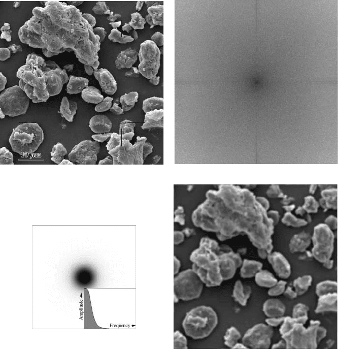
A low pass filter like the Gaussian keeps the amplitude of low frequency sinu-
soids unchanged but reduces and finally erases the amplitude of high frequencies
(large radius in the power spectrum). For large standard deviation Gaussian filters,
it is more efficient to actually execute the operation by performing the FFT, filtering
the data there, and performing an inverse FFT as shown in Figure 2.30, even though
it may be easier for those not familiar with this technique to understand the operation
based on the kernel of neighborhood weights. Mathematically, it can be shown that
these two ways of carrying out the procedure are identical.
There is another class of neighborhood filters that do not have equivalents in
frequency space. These are ranking filters that operate by listing the values of the
pixels in the neighborhood in brightness order. From this list it is possible to replace
the central pixel with the darkest, lightest or median value, for example. The median
filter, which uses the value from the middle of the list, is also an effective noise
reducer. Unlike the averaging or Gaussian smoothing filters, the median filter does
not blur or shift edges, and is thus generally preferred for purposes of reducing
noise. Figures 2.26d and 2.27d include a comparison of median filtering with neigh-
borhood smoothing.
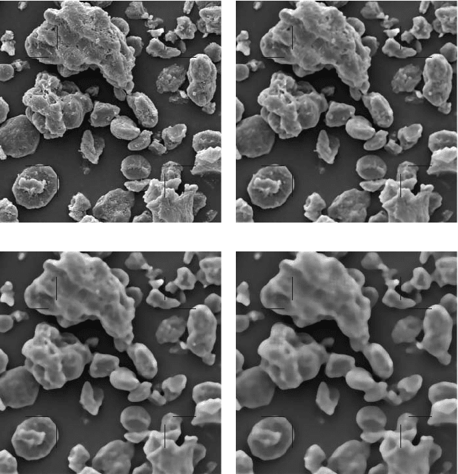
Increasing the size of the neighborhood used in the median is used to define the
size of details — including spaces between features — that are defined as noise,
because anything smaller than the radius of the neighborhood cannot contribute the
median value and is eliminated. Figure 2.31 illustrates the effect of neighborhood
size on the median filter. Since many cameras provide images with more pixels than
the actual resolution of the device provides, it is often important to eliminate noise
2241_C02.fm Page 104 Thursday, April 28, 2005 10:23 AM
Copyright © 2005 CRC Press LLC
that covers several pixels. Retention of fine detail requires a small neighborhood
size, but because it does not shift edges, a small median can be repeated several
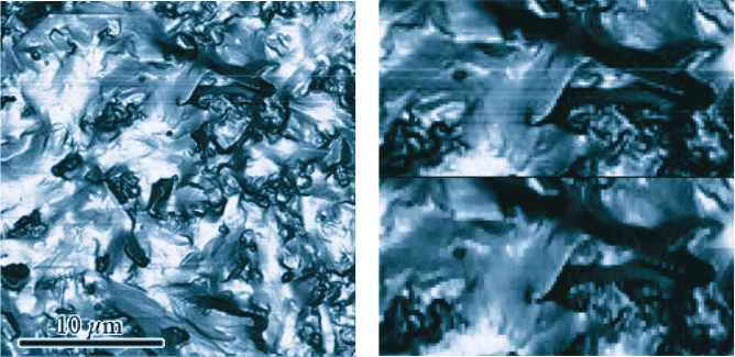
times to reduce noise. If the noise is not isotropic, such as scan line noise from
video cameras or AFMs, or scratches on film, the use of a neighborhood that is not
circular but instead is shaped to operate along a line perpendicular to the noise, the
median can also be an effective noise removal tool (see Figure 2.32).
One problem with the median filter is that while it does not shift or blur edges,
it does tend to round corners and to erase fine lines (which, if they are narrower
(a) (b)
(c) (c)
FIGURE 2.29 Illustration of Fourier transform, and the relationship between frequency pat-
terns in the pixel domain and spikes in the power spectrum: (a) image produced by superpo-
sition of three sets of lines; (b) FFT power spectrum of the image in (a), showing three spikes
(each one plotted twice with rotational symmetry); (c) mask used to select just two of the
frequencies; (d) inverse FFT using the mask in (c), showing just two sets of lines.
2241_C02.fm Page 105 Thursday, April 28, 2005 10:23 AM
Copyright © 2005 CRC Press LLC
than the radius of the neighborhood, are considered to be noise). This can be
corrected by using the hybrid median. Instead of a single ranking operation on all
of the pixels in the neighborhood, the hybrid median performs multiple rankings on
subsets of the neighborhood. For the case of the 3 × 3 neighborhood, the ranking
is performed first on the 5 pixels that form a + pattern, then the five that form an x,
and finally on the original central pixel and the median results from the first two
rankings. The final result is then saved as the new pixel value. This method can be
extended to larger neighborhoods and more subsets in additional orientations. As
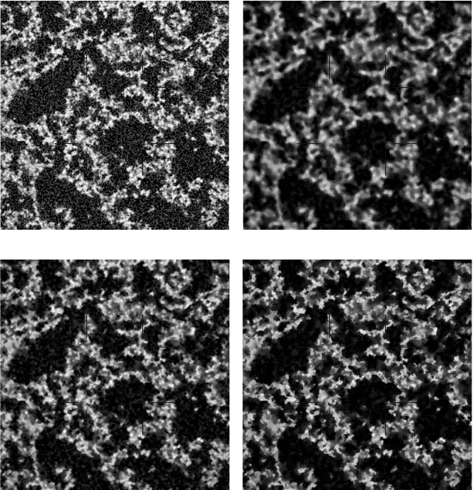
shown in the example in Figure 2.33c, fine lines and sharp corners are preserved.
(a) (b)
(c) (d)
FIGURE 2.30 Low pass (smoothing) filter in frequency space: (a) original (SEM image of
spray dried soy protein isolate particles); (b) FFT power spectrum; (c) filter that keeps low
frequencies and attenuates high frequencies; (d) inverse FFT produces smoothed result.
2241_C02.fm Page 106 Thursday, April 28, 2005 10:23 AM
Copyright © 2005 CRC Press LLC
Another approach to modifying the neighborhood is the conditional median
(Figure 2.33d). In addition to the radius of a circular neighborhood, the user specifies
a threshold value. Pixels whose difference from the central pixel exceed the threshold
are not included in the ranking operation. This technique also works to preserve fine
lines and irregular shapes.
All of these descriptions of a median filter depend on being able to rank the
pixels in the neighborhood in order of brightness. Their application to a grey scale
image is straightforward, but what about color images? In many cases with digital
cameras, the noise content of each channel is different. The blue channel in particular
generally has a higher noise level than the others because silicon detectors are
(a) (b)
(c) (d)
FIGURE 2.31 Effect of the neighborhood size used for the median filter: (a) original image;
(b) 5 pixel diameter; (c) 9 pixel diameter; (d) 13 pixel diameter.
2241_C02.fm Page 107 Thursday, April 28, 2005 10:23 AM
Copyright © 2005 CRC Press LLC
relatively insensitive to short wavelengths and more amplification is required. The
blue channel also typically has less resolution than the green channel (half as many
blue filters are used in the Bayer pattern). Separate filtering of the RGB channels
using different amounts of noise reduction (e.g., different neighborhood sizes) may
be used in these cases.
The problem with independent channel filtering is that it can alter the proportions
of the different color signals, resulting in the introduction of different colors that
are visually distracting. It is usually better to filter only the intensity channel, leaving
the color information unchanged. Another approach uses the full color information
in a median filter. One approach is to use a brightness value for each pixel, usually
just the sum of the red, green and blue values, to select the neighbor whose color
values replace those of the central pixel. This does not work as well as performing
a true color median, although it is computationally simpler.
Finding the median pixel using the color values requires plotting each pixel in
the neighborhood as a point in color space, using one of the previously described
systems of coordinates. It is then possible to calculate the sum of distances from
each point to all of the others. The median pixel is the one whose point has the
smallest sum of distances, in other words is closest to all of the other points. It is
also possible and equivalent to define this point in terms of the angles of vectors to
the other points. In either case, the color values from the median point are then
reassigned to the central pixel in the neighborhood. The color median can be used
in conjunction with any of the neighborhood modification schemes (hybrid median,
conditional median, etc.).
Random speckle noise is not the only type of noise defect present in images.
One other, scratches, has already been mentioned. Most affordable digital cameras
suffer from a defect in which a few detectors are inactive (dead) or their output is
(a) (b)
FIGURE 2.32 Removal of scan line noise in AFM images: (a) original image (surface of
chocolate); (b) application of a median filter using a 5 pixel vertical neighborhood to remove
the scan line noise (top) leaving other details intact (bottom).
2241_C02.fm Page 108 Thursday, April 28, 2005 10:23 AM
Copyright © 2005 CRC Press LLC
always maximum (locked). This produces white or black values for those pixels, or
at least minimum or maximum values in one color channel. A similar problem can
arise in some types of microscopy such as AFM or interference microscopes, where
no signal is obtained at some points and the pixels are set to black. Dust on film
can produce a similar result. Generally, this type of defect is shot noise.
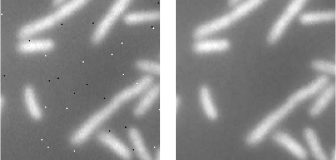
A smoothing filter based on the average or Gaussian is very ineffective with
shot noise, because the errant extreme value is simply spread out into the surrounding
pixels. Median filters, on the other hand, eliminate it easily and completely, replacing
the defective pixel with the most plausible value taken from the surrounding neigh-
borhood, as shown in Figure 2.34.
(a) (b)
(c) (d)
FIGURE 2.33 Comparison of standard and hybrid median: (a) original (noisy CSLM image
of acid casein gel, courtesy of M. Faergemand, Department of Dairy and Food Science, Royal
Veterinary and Agricultural University, Denmark); (b) conventional median; (c) hybrid
median; (d) conditional median.
2241_C02.fm Page 109 Thursday, April 28, 2005 10:23 AM
Copyright © 2005 CRC Press LLC
Periodic noise in images shows up as a superimposed pattern, usually of lines.
It can be produced by electronic interference or vibration, and is also present in
printed images such as the ones in this book because they are printed with a halftone
pattern that reproduces the image as a set of regularly spaced dots of varying size.
Television viewers will recognize moiré patterns that arise when someone wears
clothing whose pattern is similar in size to the spacing of video scan lines as a type
of periodic noise that introduces strange and shifting colors into the scene, and the
same phenomenon can happen with digital cameras and scanners.
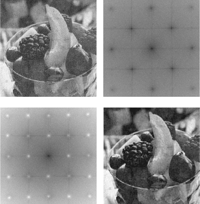
The removal of periodic noise is practically always accomplished by using
frequency space. Since the noise consists of just a few, usually relatively high
frequencies, the FFT represents the noise pattern as just a few points in the power
spectrum with large amplitudes. These spikes can be found either manually or
automatically, using some of the techniques described in the next chapter. However
they are located, reduction of the amplitude to zero for those frequencies will
eliminate the pattern without removing any other information from the image, as
shown in Figure 2.35.
Because of the way that single chip color cameras use filters to sample the colors
in the image, and the way that offset printing uses different halftone grids set at
different angles to reproduce color images, the periodic noise in color images is
typically very different in each color channel. This requires processing each channel
separately to remove the noise, and then recombining the results. It is important to
select the right color channels for this purpose. For instance, RGB channels corre-
spond to how most digital cameras record color, while CMYK channels correspond
to how color images are printed.
(a) (b)
FIGURE 2.34 Removal of shot noise: (a) original image of bacteria corrupted with random
black and white pixels; (b) application of a hybrid median filter.
2241_C02.fm Page 110 Thursday, April 28, 2005 10:23 AM
Copyright © 2005 CRC Press LLC
NONUNIFORM ILLUMINATION
One assumption that underlies nearly all steps in image processing and mea-
surement, as well as strategies for sampling material, is that the same feature will
have the same appearance wherever it happens to be positioned in an image. Non-
uniform illumination violates this assumption, and may arise for a number of dif-
ferent causes. Some of them can be corrected in hardware if detected before the
images are acquired, but some cannot and we are often faced with the need to deal
with previously recorded images that have existing problems.
(a) (b)
(c) (d)
FIGURE 2.35 Removal of periodic noise: (a) image from a newspaper showing halftone
printing pattern; (b) Fourier transform power spectrum with spikes corresponding to the high
frequency pattern; (c) filtering of the Fourier transform to remove the spikes; (d) result of
applying an inverse Fourier transform.
2241_C02.fm Page 111 Thursday, April 28, 2005 10:23 AM
Copyright © 2005 CRC Press LLC
Variations that are not visually apparent (because the human eye compensates
automatically for gradual changes in brightness) may be detected only when the
image is captured in the computer. Balancing lighting across the entire recorded
scene is difficult. Careful position of lights on a copy stand, use of ring lighting for
macro photography, or adjustment of the condenser lens in a microscope, are pro-
cedures that help to achieve uniform lighting of the sample. Capturing an image of
a uniform grey card or blank slide and measuring the brightness variation is an
important tool for such adjustments.
Some other problems are not normally correctable. Optics can cause vignetting
(darkening of the periphery of the image) because of light absorption in the glass.
Cameras may have fixed pattern noise that causes local brightness variations. Cor-
recting variations in the brightness of illumination may leave variations in the angle
or color of the illumination. And, of course, the sample itself may have local
variations in density, thickness, surface flatness, and so forth, which can cause
changes in brightness.
Many of the variations other than those which are a function of the sample itself
can be corrected by capturing an image that shows just the variation. Removing the
sample and recording an image of just the background, a grey card, or a blank slide
or specimen stub with the same illumination provides a measure of the variation.
This background image can then be subtracted from or divided into the image of
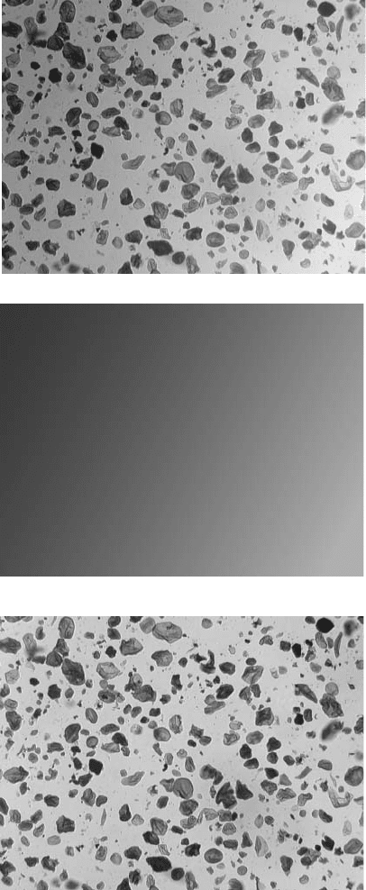
the sample to level the brightness. The example in Figure 2.36 shows particles of
cornstarch imaged in the light microscope with imperfect centering of the light
source. Measuring the particle size distribution depends upon leveling the contrast
so that particles can be thresholded everywhere in the image. Capturing a background
image with the same illumination conditions and subtracting it from the original
makes this possible.
The choice of subtraction or division for the background depends on whether
the imaging device is linear or logarithmic. Scanners are inherently linear, so that
the measured pixel value is directly proportional to the light intensity. The output
from most scanning microscopes is also linear, unless nonlinear gamma adjustments
are made in the amplified signal. The detectors used in digital cameras are linear,
but in many cases the output is converted to logarithmic to mimic the behavior of
film. Photographic film responds logarithmically to light intensity, with equal incre-
ments of density corresponding to equal ratios of brightness. For linear recordings,
the background is divided into the image, while for logarithmic images it is sub-
tracted (since division of numbers corresponds to the subtraction of their logarithms).
In practice, the best advice when the response of the detector is unknown, is to try
both methods and use the one that produces the best result. In the examples that follow,
some backgrounds are subtracted and some are divided to produce a level result.
In situations where a satisfactory background image cannot be (or was not)
stored along with the image of the specimen, there are several ways to construct
one. In some cases one color channel may contain little detail but may still serve as
a measure of the variation in illumination. Another technique that is sometimes used
is to apply an extreme low pass filter (e.g., a Gaussian smooth with a large standard
deviation) to the image to remove the features, leaving just the background variation.
This method is based on the assumptions that the features are small compared to
2241_C02.fm Page 112 Thursday, April 28, 2005 10:23 AM
Copyright © 2005 CRC Press LLC
(a)
(b)
(c)
FIGURE 2.36 Leveling contrast with a recorded background image: (a) original image of
cornstarch particles with nonuniform illumination; (b) image of blank slide captured with
same illumination; (c) subtraction of background from original.
2241_C02.fm Page 113 Thursday, April 28, 2005 10:23 AM
Copyright © 2005 CRC Press LLC
