Russ J.C. Image Analysis of Food Microstructure
Подождите немного. Документ загружается.

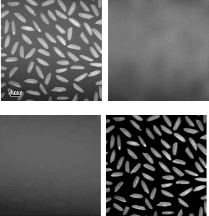
the distances between them, and that the background variation is gradual, as it usually
is for illumination nonuniformities. In the example of Figure 2.37b this technique
is marginally successful, but irregularities in the background remain because the
features are too close together for their complete elimination.
A superior method for removing the features to leave just the background uses
the rank filter. Rather than averaging the values from the pixels within the features
into the background, replacing each pixel with its darkest (or lightest) neighbor will
(a) (b)
(c) (d)
FIGURE 2.37 Removing features to create a background: (a) original image of rice grains
dispersed for length measurement (note nonuniform illumination); (b) background generated
by Gaussian smoothing with 15 pixel radius standard deviation; (c) background generated by
replacing each pixel with its darkest neighbor in a 9 pixel neighborhood); (d) subtracting
image (c) from image (a) produces uniform background.
2241_C02.fm Page 114 Thursday, April 28, 2005 10:23 AM
Copyright © 2005 CRC Press LLC
erase light (or dark) features and insert values from the nearby background. This
technique uses much the same computation as the median filter, described above,
and is another example of ranking pixels in a moving neighborhood. As shown in
Figure 2.37c, this method works very well if the features are narrow in at least one
direction. It assumes that the features may vary in brightness but are always darker
(or lighter) than the local background.
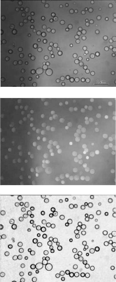
This method can handle rather abrupt changes in brightness, such as can result
from shadows, folds in sections, or changes in surface slope. In the example of
Figure 2.38, the droplets on a glass surface are illuminated from behind and there
is considerable shading, which is not gradual and prevents thresholding to measure
their size distribution and spatial arrangement. In this case the contrast around the
edges of the droplets is darker than the interior or the background. Replacing each
pixel with its brightest neighbor produces a background. Dividing it pixel-by-pixel
into the original image produces a image with uniform contrast, which can be
measured.
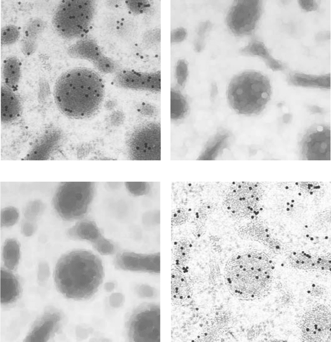
In some cases, it is not sufficient to simply replace pixels by their brighter or
darker neighbors, because it can alter the underlying background structure. A more
general solution is to first replace the feature pixels with those from the background
(e.g., replace dark pixels with lighter ones) and then, when the features are gone,
perform the opposite procedure (e.g., replace light pixels with darker ones) using
the same neighborhood size. This restores the background structure so that it can
be successfully removed, as shown in Figure 2.39. For reasons that will become
clear in the next chapters, these procedures are often called erosion and dilation,
and the combinations are openings and closings.
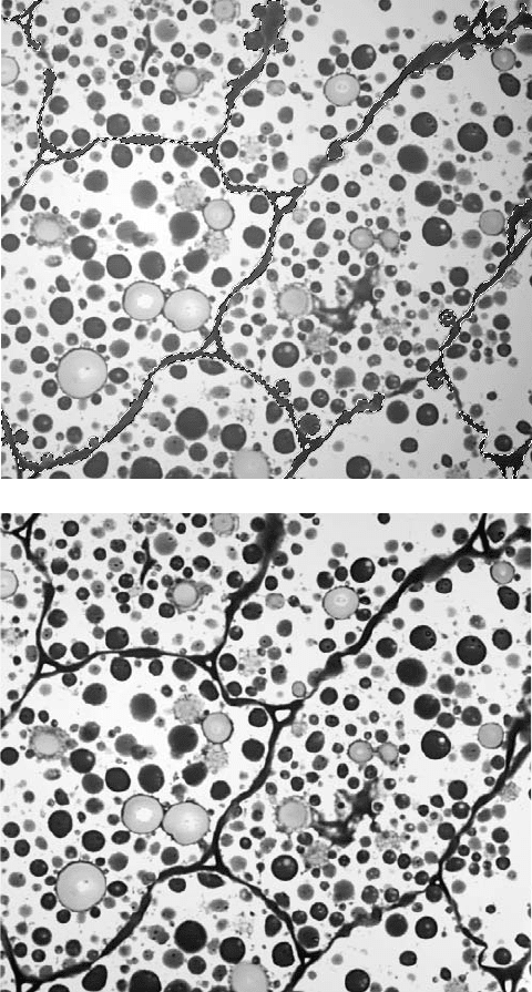
Another method that can be used either manually or automatically is to fit a
smooth polynomial function to selected background regions dispersed throughout
the image. Expressing brightness as a quadratic or cubic function of position in the
image requires coefficients that can be determined by least-squares fitting to a
number of selected locations. In some cases it is practical to select these by hand,
choosing either features or background regions as reference locations that should
all have the same brightness value. Figure 2.40 illustrates the result.
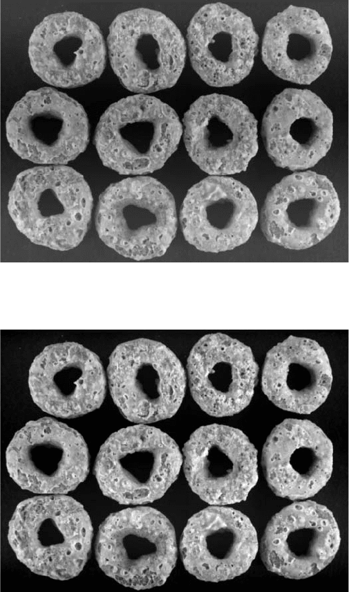
If the reference regions are well distributed throughout the image and are either
the brightest or darkest pixels present, then they can be located automatically and
the polynomial fit performed. The polynomial method requires the variation in
brightness to be gradual, but does not require that the features be small or well
separated as the rank filtering method does. Figure 2.41 shows an example.
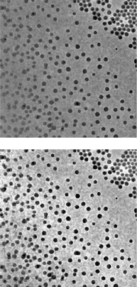
The polynomial fitting technique can be extended to deal with variations in
contrast that can result from nonuniform section thickness. Fitting two polynomials,
one to the brightest points in the image and a second to the darkest points, provides
an envelope that can be used to locally stretch the contrast to a uniform maximum
throughout the image as shown in Figure 2.42.
2241_C02.fm Page 115 Thursday, April 28, 2005 10:23 AM
Copyright © 2005 CRC Press LLC
(a)
(b)
(c)
FIGURE 2.38 Removal of a bright background: (a) original image (liquid droplets on glass);
(b) background produced by replacing each pixel with its brightest neighbor in a 7-pixel wide
neighborhood; (c) dividing image (b) into image (a) produces a leveled image with dark
outlines for the droplets.
2241_C02.fm Page 116 Thursday, April 28, 2005 10:23 AM
Copyright © 2005 CRC Press LLC
(a) (b)
(c) (d)
FIGURE 2.39 Removal of a structured background: (a) original image (gold particles bound
to cell organelles); (b) erosion of the dark gold particles also reduces the size of the organelles;
(c) dilation restores the organelles to their original dimension; (d) dividing the background
from image (c) into the original image (a) produces a leveled imaged in which the gold
particles can be counted.
2241_C02.fm Page 117 Thursday, April 28, 2005 10:23 AM
Copyright © 2005 CRC Press LLC
(a)
(b)
FIGURE 2.40 Selecting background points: (a) original image with nonuniform illumination
(2-mm-thick peanut section stained with toluidine blue to mark protein bodies, see Color
Figure 1.12; see color insert following page 150); the cell walls (marked by the dashed lines)
have been manually selected using the Adobe Photoshop
®
wand tool as features that should
all have the same brightness; (b) removing the polynomial fit to the pixels in the selected
area from the entire image levels the contrast.
2241_C02.fm Page 118 Thursday, April 28, 2005 10:23 AM
Copyright © 2005 CRC Press LLC
(a)
(b)
FIGURE 2.41 Automatic background correction: (a) original image (courtesy of Diana Kit-
tleson, General Mills) with nonuniform illumination (the blue channel was selected with the
best detail from an image originally acquired in color); (b) automatically leveled by removing
a polynomial fit to the darkest pixel values.
2241_C02.fm Page 119 Thursday, April 28, 2005 10:23 AM
Copyright © 2005 CRC Press LLC
(a)
(b)
FIGURE 2.42 Automatic contrast adjustment using two polynomials, one fit to the dark and
one to the light pixels: (a) original (TEM image of latex spheres); (b) processed.
2241_C02.fm Page 120 Thursday, April 28, 2005 10:23 AM
Copyright © 2005 CRC Press LLC
IMAGE DISTORTION AND FOCUS
Another assumption in subsequent chapters on processing and measurement is
that dimensions in an image are uniform in all locations and directions. When images
are acquired from digital or film cameras on light microscopes, or from scanners,
that condition is generally met. Digitized video images often suffer from non-square
pixels because different crystal oscillators control the timing of the camera and the
digitizer. Atomic force microscopes compound problems of different scales in the
slow- and fast-scan direction with nonlinearities due to creep in the piezo drives.
Macro cameras may have lens distortions (either pincushion or barrel distortion;
short focal length lenses are particularly prone to fish-eye distortion) that can create
difficulties.
Electron microscopes have a large depth-of-field that allows in-focus imaging
of tilted specimens. This produces a trapezoidal distortion that alters the shape and
size of features at different locations in the image. Photography with cameras (either
film or digital) under situations that view structures at an angle produces the same
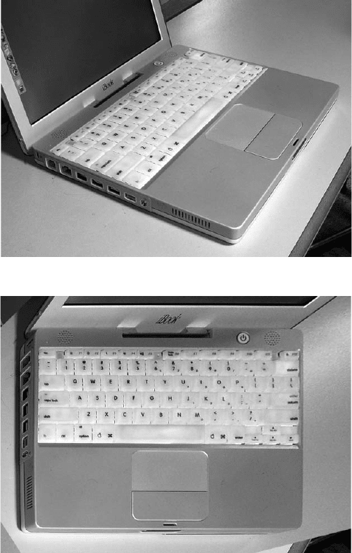
foreshortening difficulties. If some known fiducial marks are present, they can be
used to rectify the image as shown in Figure 2.43, but this correction applies only
to features on the corrected surface.
Distortions in images become particularly noticeable when multiple images of
large specimens are acquired and an attempt is made to tile them together to produce
a single mosaic image. This may be desired because, as mentioned in the discussion
of image resolution, acquiring an image with a large number of pixels allows
measurement of features covering a large size range. But even small changes in
magnification or alignment make it very difficult to fit the pieces together.
This should not be confused with software that constructs panoramas from a
series of images taken with a camera on a tripod. Automatic matching of features
along the image edges, and sometimes knowledge about the lens focal length and
distortion, is used to distort the images gradually to create seamless joins. Such
images are intended for visual effect, not measurement or photogrammetry, and
consequently the distortions are acceptable. There are a few cases in which tiling
of mosaics is used for image analysis, but in most cases this is accomplished by
having automatic microscope stages of sufficient precision that translation produces
correct alignment, and the individual images are not distorted.
Light microscope images typically do not have foreshortening distortion because
the depth of field of high magnification optics is small. It is possible, however, to
acquire a high magnification light microscope image of a sample that has a large
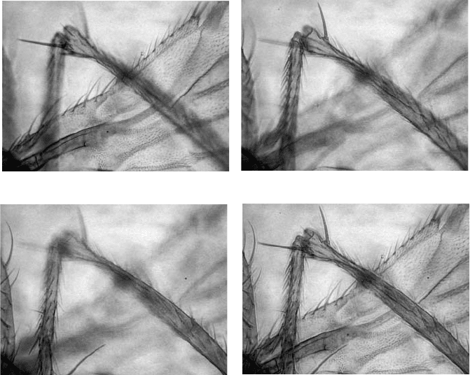
amount of relief. No single image can capture the entire depth of the sample, but if
a series of pictures is taken that cover the full z-depth of the sample, they can be
combined to produce a single extended-focus image as shown in Figure 2.44.
The automatic combining of multiple images to keep the best-focused pixel
value at each location requires that the images be aligned and at the same magnifi-
cation, which makes it ideal for microscope applications but much more difficult to
use with macro photography (shift the camera, do not refocus the lens). The software
uses the variance of the pixel values in a small neighborhood at each location to
2241_C02.fm Page 121 Thursday, April 28, 2005 10:23 AM
Copyright © 2005 CRC Press LLC
select the best focus. For color images, this can be done separately for the red, green,
and blue channels to deal with slight focus differences as a function of wavelength.
When an entire image is imperfectly focused, it is sometimes practical to correct
the problem by deconvolution. This operation is performed using the Fourier trans-
form of the image, and also requires the Fourier transform of the point spread
function (psf). This is the image of a perfect point with the same out-of-focus
(a)
(b)
FIGURE 2.43 Example of correction for trapezoidal distortion: (a) original foreshortened
image; (b) calculated rectified view; note that the screen and the edges of the computer, which
do not lie in the plane of the keyboard, are still distorted.
2241_C02.fm Page 122 Thursday, April 28, 2005 10:23 AM
Copyright © 2005 CRC Press LLC
condition as the image of interest. In a few cases it is practical to measure it directly,
as shown in Figure 2.45. In astronomy, an image of a single star taken through the
same optics becomes the point spread function. In fluorescence microscopy, a single
microbead may serve the same function. It is slightly more complicated to obtain
the psf by obtaining a blurred image of a known structure or test pattern, deconvolve
it with the ideal image to obtain the psf, and then use that to deconvolve other
images. That technique is used with the AFM to characterize the shape of the tip
(which causes blurs and distortion in the image).
In many cases the most straightforward way to perform deconvolution is to
estimate the point spread function, either interactively or iteratively. The two most
common types of blur are defocus blur in which the optics are imperfectly adjusted,
and motion blur in which the camera or sample are moving, usually in a straight
line (Figure 2.46). These produce point spread functions that are adequately modeled
in most cases by a Gaussian or by a line, respectively. Adjusting the standard
deviation of the Gaussian or the length and orientation of the line produces the point
spread function and the deconvolution of the image.
Deconvolution is hampered by the presence of random noise in the image. It is
very important to start with the lowest noise, highest bit depth image possible. The
noise in the deconvolved result will be much greater than in the original because
(a) (b)
(c) (d)
FIGURE 2.44 Example of extended focus: (a, b, c) images of the leg and wing of a fruit fly
at different focal depths; (d) combination keeping the in-focus detail from each image.
2241_C02.fm Page 123 Thursday, April 28, 2005 10:23 AM
Copyright © 2005 CRC Press LLC
