Russ J.C. Image Analysis of Food Microstructure
Подождите немного. Документ загружается.

the results to someone else who has not studied the original images, is something
of an art. There is no best way to do it, although there are often a lot of not so good ways.
Channel merging is also a standard way to present stereo pair images. Colored
glasses with the red lens on the left eye and either green or blue on the right eye
are often used to view composite images which are prepared by placing the corre-
sponding eye views into the color channels.
Separating the channels can also be an aid to visual perception of the information
in an image. The presence of color is important in human vision but not as important
as variations in brightness. The reason that the various compression methods
described above reduce the spatial and tonal resolution of the color information more
than the brightness is that human vision does not generally notice that reduction.
Broadcast television and modern digital video recording use this same trick, assign-
ing twice the bandwidth for the brightness values as for the color information.
Blurring the color information in an image so that the colors bleed across boundaries
does not cause any discomfort on the part of the viewer (which may be why children
do not always color inside the lines).
Seeing the important changes in brightness in an image, which often defines
and locates important features, may be easier in some cases if the distracting vari-
ations in color, or the presence of a single dominant color, are removed. In the color
original of meat shown in Figure 2.2 the predominant color is red. Examining the
red channel as a monochrome (grey scale) image shows little contrast because there
is red everywhere. On the other hand, the green channel (or a mixture of green and
blue, as used in the example) shows good contrast. In general a complementary hue,
opposite on the color wheel, will reveal information hidden by the presence of a
dominant color (just as a photographer uses a yellow filter to enhance the visibility
of clouds in a photograph of blue sky). Of course, in this example an equivalent
result could have been obtained by recording the image with a monochrome camera
through a green filter, rather than acquiring the color image and then extracting the
green channel.
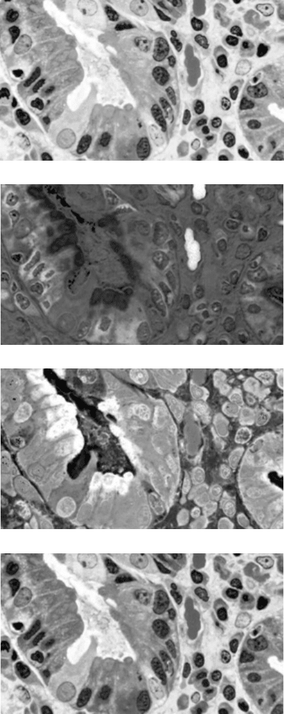
In other cases it is the hue or saturation channels that are most interesting. When
colored stains or dyes are introduced into biological material (Figure 2.16) in order
to color particular structures or localize chemical activity, the colors are selected so
they will be different. That difference is a difference in hue, and examining just the
hue channel will show it clearly. Likewise, the saturation channel intensity corre-
sponds to the amount of the stain in each location. The intensity channel records
the variation in density of the specimen.
There are many ways to extract a monochrome (grey scale) image from a color
image, usually with the goal of providing enhanced contrast for the structures
present. The simplest method is to simply average the red, green, and blue channels.
This does not correspond to the brightness that human vision perceives in a color
scene, because the human eye is primarily sensitive to green wavelengths, less to
red, and much less to blue. Blending the channels in proportions of about 65% green,
25% red, 10% blue will give approximately that result.
But it is also possible to mix the channels arbitrarily to increase the contrast
between particular colors. An optimum contrast grey image can be constructed from
any color image by fitting a regression line through a 3D plot of the pixel color
2241_C02.fm Page 84 Thursday, April 28, 2005 10:23 AM
Copyright © 2005 CRC Press LLC
(a)
(b)
(c)
(d)
FIGURE 2.16 Stained tissue imaged in the light microscope, and the HSI color channels:
(a) color original (see color insert following page 150); (b) hue; (c) saturation; (d) intensity.
2241_C02.fm Page 85 Thursday, April 28, 2005 10:23 AM
Copyright © 2005 CRC Press LLC
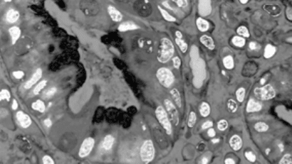
values and mapping the grey scale values along that line (Figure 2.17). In principle
this can be done in any color coordinates, but it is easiest to carry out and to visualize
in RGB.
OPTIMUM IMAGE CONTRAST
Whether images are color or monochrome (many imaging techniques such as
SEM, TEM, AFM, etc. produce only grey scale images), it is desirable to have the
best possible image contrast. Ideally, this can be done by controlling the illumination,
camera gain and contrast, digitizer settings, etc., so that the captured image covers
the full available brightness range and the discrimination between structures is
optimum. In practice, images are often obtained that can be improved by subsequent
processing.
The first tool to understand and use for examining images is the histogram. This
is usually presented as a plot of the number of pixels having each possible brightness
value. For an 8 bit greyscale image, with only 256 possible brightness values, this
plot is compact and easy to view. For an image with greater bit depth, the plot would
be extremely wide and there would be very few counts for most of the brightness
values, so it is common to bin them together into a smaller number of levels (usually
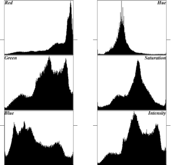
the same 256 as for 8 bit images). For a color image, individual histograms may be
presented for the various channels (RGB or HSI) as shown in Figure 2.18, or other
presentations showing the distribution of combinations of values may be used. It
generally takes some experience to use color histograms effectively, but in most
cases the desired changes in contrast can be effected by working just with the
intensity channel, leaving the color information unchanged.
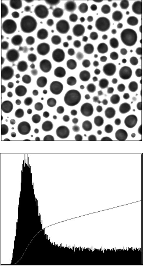
A histogram that shows a significant number of pixels at the extreme white or
black ends of the plot (as shown in Figure 2.19) indicates that the image has been
captured with too great a contrast or gain value, and that data have been lost due to
clipping (values that exceed the limits of the digitizer are set to those limits). This
situation must be avoided because data lost to clipping cannot be recovered, and it
FIGURE 2.17 Mixing the RGB color channels in the color image from Figure 2.16(a) to
produce the optimum grey scale contrast.
2241_C02.fm Page 86 Thursday, April 28, 2005 10:23 AM
Copyright © 2005 CRC Press LLC
is likely that the size and other details of the remaining features have been altered.
This problem frequently arises when particulates are dispersed on a bright or dark
substrate. Adjusting contrast so that the substrate brightness is clipped reduces the
size of the particles.
A histogram that does not cover the full range of intensity values from light to
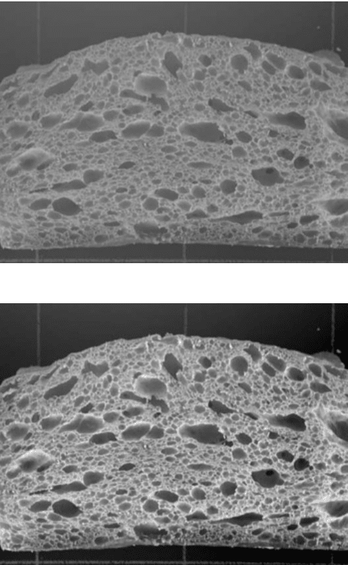
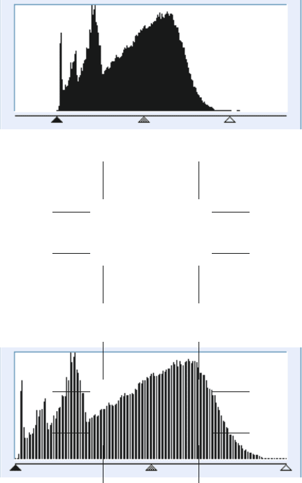
dark indicates that the image has low contrast (Figure 2.20). Simply stretching the
values out to cover the full range will leave gaps throughout the histogram, but
because the human eye cannot detect small brightness differences this is not a
problem for visual examination. There are just as many missing values in the
stretched histogram as in the original, but in the original they were grouped together
at the high and low brightness end of the scale where their absence was noticeable.
Human vision can detect a brightness change of several percent under good viewing
conditions, which translates to roughly 30 brightness levels in a scene. Most modern
computer monitors can display 256 brightness levels, so missing a few scattered
throughout the range does not degrade the image appearance.
(a) (b)
FIGURE 2.18 Histograms for the RGB and HSI channels of the stained tissue image in
Figure 2.16. In each, the horizontal axis goes 0 (dark) to 255 (maximum), and the vertical
axis is the number of pixels.
2241_C02.fm Page 87 Thursday, April 28, 2005 10:23 AM
Copyright © 2005 CRC Press LLC
(a)
(b)
FIGURE 2.19 Bubbles in a whipped food product: (a) image (courtesy of Allen Foegeding,
North Carolina State University, Department of Food Science) in which much of the white
area around the bubbles is pure white (value = 255); (b) histogram, showing a large pixel
count at 255 (white) where the single line in the histogram rises off scale. The cumulative
histogram (grey line) is the integral of the histogram. Since 100% is full scale, the cumulative
plot indicates that about 40% of the image area is occupied by exactly white pixels.
2241_C02.fm Page 88 Thursday, April 28, 2005 10:23 AM
Copyright © 2005 CRC Press LLC
Human vision is not linear, but instead detects a proportional change in bright-
ness. Equal increments in brightness are viewed differently at the bright and dark
ends of the brightness range. Adjusting the overall gamma value of the image allows
detail to be seen equally well in bright and dark regions. One effective way to control
gamma settings is by setting the brightness level (on the 0 to 255 scale) for the
midpoint of the pixel brightness range (the median value, with 50% darker and 50%
lighter). This produces a smooth curve relating the displayed brightness value to the
original value, which can either stretch out dark values to increase detail visibility
in dark regions by compressing bright values so detail there becomes less distin-
guishable, or vice versa.
For the experienced film photographer, setting the midpoint of the brightness
range to a desired value is equivalent to the zone system for controlling the contrast
of prints. Typical film or digital camera images can record a much wider latitude of
brightness values than can be printed as hardcopy, and selecting a target zone for
the image directly affects the visibility of detail in the bright and dark areas of the
resulting image as shown in Figure 2.21.
Photographers with darkroom experience also know that detail is often visible
in the negative that is hard to see in the print, and vice versa. Because of the
nonlinearity of human vision, simply inverting the image contrast often assists in
visual perception of detail in images.
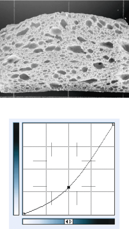
Of course, there is no reason that the curve relating displayed brightness to
original brightness (usually called the “transfer function”) must be a smooth con-
stant-gamma curve. Arbitrary relationships can be used to increase brightness in any
particular segment of the total range, and these may even include reversal of contrast
in one part of the brightness range, equivalent to the darkroom technique of solar-
ization. Figure 2.22 illustrates a few possibilities.
A specific curve can be used as a transfer function for a given image that
transforms the brightness values so that equal areas of the image are displayed with
each brightness value. This is histogram equalization, and is most readily visualized
by replotting the conventional histogram as a cumulative plot of the number of pixels
with brightness values equal to or less than each brightness values (mathematically,
the integral of the usual plot). As shown in Figure 2.23, this curve rises quickly
where there is a peak and slowly where there is not. If the cumulative histogram
curve is used as the transfer function, it spreads out subtle gradients and makes them
more visible. It also transforms the cumulative histogram so that it becomes a straight
line.
Transforms like these that alter the brightness values of pixels in the image are
global, because each pixel is affected the same way. That is, two pixels that started
out with the same value will end up with the same value (although it will probably
be a different one from the original) regardless of where they are located or what
their surroundings may be. The goal is to increase the ability of the viewer to perceive
the contrast in the image. To achieve this, any consistent relationship between
brightness and a physical property of the specimen is sacrificed. Density, dosage
measurements, activity, or other properties that might be calibrated against brightness
cannot be measured after these brightness transformations.
2241_C02.fm Page 89 Thursday, April 28, 2005 10:23 AM
Copyright © 2005 CRC Press LLC
(a)
(b)
FIGURE 2.20 Image of bread slice (courtesy of Diana Kittleson, General Mills): (a) original
low contrast image; (b) contrast stretched linearly to maximum; (c) original histogram showing
black and white limits set to end points; (d) histogram after stretching to full range.
2241_C02.fm Page 90 Thursday, April 28, 2005 10:23 AM
Copyright © 2005 CRC Press LLC
(c)
(d)
FIGURE 2.20 (continued)
2241_C02.fm Page 91 Thursday, April 28, 2005 10:23 AM
Copyright © 2005 CRC Press LLC
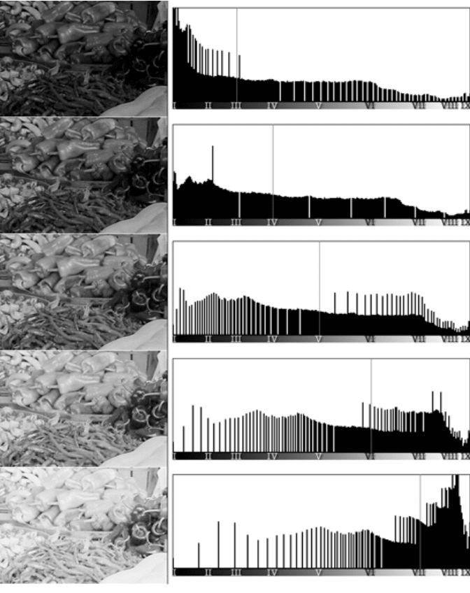
FIGURE 2.21 Adjusting the appearance of an image by setting the target zone (from III to
VII) for the midpoint of the histogram. Note the appearance of gaps and pileup at either end
of the histogram (fragment of the market image in Color Figure 2.3; the histogram represents
the entire image; see color insert following page 150).
2241_C02.fm Page 92 Thursday, April 28, 2005 10:23 AM
Copyright © 2005 CRC Press LLC
(a)
(b)
FIGURE 2.22 Manipulation of the transfer function for the bread slice image from Figure
2.20: (a, b) positive gamma reveals detail inside the dark shadow areas; (c, d) negative gamma
increases the contrast in the bright areas; (e, f) inverting the contrast produces a negative
image; (g, h) solarization shows both positive and negative contrast. The transfer function or
contrast curve used for each image is shown; the horizontal axis shows the stored brightness
values and the vertical axis shows the corresponding displayed brightness.
2241_C02.fm Page 93 Thursday, April 28, 2005 10:23 AM
Copyright © 2005 CRC Press LLC
