Russ J.C. Image Analysis of Food Microstructure
Подождите немного. Документ загружается.

bright and space is very dark, cameras cooled to liquid helium temperatures record
such a range of brightness data, but it requires very specialized detectors. The actual
performance of the detectors in most flat bed scanners is more like 12 bits (2
12
=
4096 values), which is still a great improvement over a simple 8-bit image and plenty
for most of the applications used. Because of the organization of computer memory
into bytes, each of 8 bits, it is usually convenient to store any image with more than
256 values in two bytes per channel, and hence these are usually called “16-bit images.”
In such an image, the measured values are multiplied up to fill the full 16-bit range of
values, with the result that very small differences in value (for instance, the difference
between a brightness value of 10,000 and 10,005) are not significant.
A good way to understand the importance of high bit depth for images is to
consider trying to record elevations of the Earth’s surface. In the Himalayas, altitudes
reach over 25,000 feet, so if we use 8 bits (256 discrete values), each one will
(a)
FIGURE 2.2
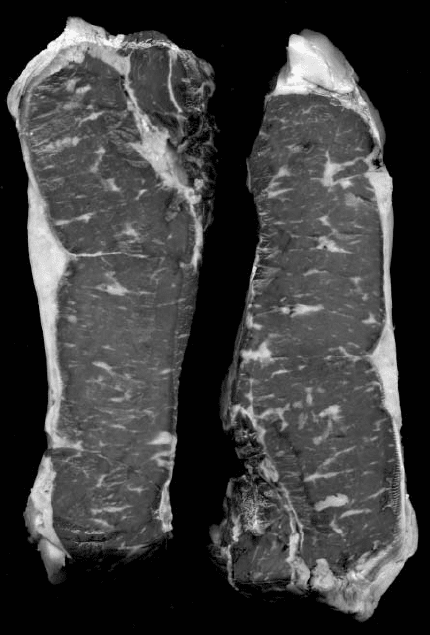
Image of beef steaks from Figure 2.1. Thresholding of the fat (b) allows
measurement of the total volume fraction of fat, and the size, shape, and spatial distribution
of the individual areas of fat in order to quantify the marbling of the cut.
2241_C02.fm Page 54 Thursday, April 28, 2005 10:23 AM
Copyright © 2005 CRC Press LLC

represent about 100 feet, and smaller variations cannot be recorded. With that
resolution for the elevation data, the entire Florida peninsula would be indistinguish-
able from the ocean, and much fine detail would be lost everywhere. Using 12 bits
(4096 values) allows distinguishing elevation differences of slightly over 6 feet, and
now even highway overpasses are detectable. If 16 bit data could be acquired, the
vertical resolution would be about 5 inches. With only 8 bit images, a great amount
of information is lost, although it typically requires image processing to make it
visible since human vision does not detect very slight differences in brightness.
In this text, because images will come from many different sources, some of
which are 8 bit, some 16 bit, and some other values, the convention will be adopted
that a value of 0 is completely dark, a value of 255 is completely bright, and if the
available precision is more than 8 bits it will be represented by a decimal fraction.
In other words, a point on a feature might have a set of brightness values of
R = 158.53, G = 74.125, B = 146.88 (if imaged with an 8 bit device, that would
have been recorded as (R = 158, G = 74, B = 146). Figure 2.3 illustrates the ability
(b)
FIGURE 2.2 (continued)
2241_C02.fm Page 55 Thursday, April 28, 2005 10:23 AM
Copyright © 2005 CRC Press LLC
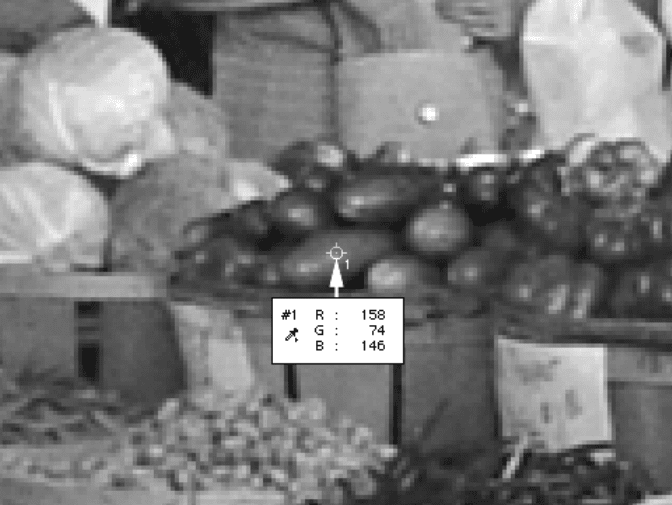
of most programs to read out the color values. How such values correspond to
perceived colors is discussed later in this chapter.
Most flat bed scanners provide 16 bit per channel data, although the software
may truncate these to a lower 8-bit precision unless the user elects to save the full
data (file sizes increase, but storage is constantly becoming faster and less expensive).
Many manufacturers describe scanners that store images with 8 bits in each of the
three color channels as 24-bit RGB scanners, and ones that produce images that
occupy 16 bits per channel as 48-bit RGB scanners, even though the actual dynamic
range or tonal resolution of the image is somewhat less.
Some flatbed scanners provide a second light source (or sometimes a mirror) in
the lid and can be used to scan transparencies, but much better results are obtained
with dedicated film scanners. Mechanically, these are similar to but smaller than the
flatbed scanners. Optically they provide very high spatial resolution, typically in the
3500 to 5000 dpi range, which corresponds to the resolution that film can record.
Most units have built-in autofocusing and color calibration, and require no particular
skill on the part of the operator. Some have special software, either built into the
unit or provided for the host computer, that can selectively remove scratches or other
defects present on the film. Because these can also remove real detail in some cases,
it is usually best to examine the raw data before allowing the software to “enhance”
the result. Most film scanners come with software that includes color calibration
FIGURE 2.3
Reading the color values at a point in an image (the original color image is
shown as Color Figure 2.3; see color insert following page 150). The point measured is on
a neon purple eggplant; the image fragment is enlarged to show individual pixels.
2241_C02.fm Page 56 Thursday, April 28, 2005 10:23 AM
Copyright © 2005 CRC Press LLC
curves for both positive (slide film) and negative (print film) color films, and give
excellent results.
An inexpensive negative scanner combined with an existing film camera back
used with macro lenses or a microscope represents an extremely cost-effective way
to accomplish digital imaging. Typical 35 mm color slide film has a spatial resolution
of about 4500
×
3000 points and a tonal resolution of one part in 4000 (12 bits per
RGB channel). Color negative film (used for prints) has less dynamic range. All of
this can be captured by the scanner, with scan times of tens of seconds per image.
No affordable digital camera produces images with such high spatial or tonal reso-
lution. But the drawback of the film-and-scanner approach is the need to take the
entire roll of pictures and develop them before knowing if you have a good photo,
and before you can perform any measurements on the image. It is the immediacy
of digital cameras rather than any technical superiority that has encouraged their
widespread adoption.
DIGITAL CAMERAS
Digital still cameras (as distinct from video cameras, which use similar technol-
ogy but have much less resolution and much more noise in the images) have taken
over a significant segment of the scientific imaging market. Driven largely by a much
greater consumer marketplace for instant photography, and spurred by the Internet,
these cameras continue to drop in price while improving in technical specifications.
But it is not usually very satisfactory to try to adapt a consumer digital camera to
a scientific imaging task. For these applications, a camera back that can accept
different lenses, or at least attachments that will connect to other optical devices
such as a microscope, is usually more appropriate. (The other major issue with
consumer cameras is the data storage format, which is discussed below.)
The same technical issues that arise for the scanner also apply to cameras. We
would ideally like to have a high spatial resolution along with a high dynamic range
and low noise. High spatial resolution means a lot of detectors in the array. At this
writing, a typical high-end consumer or low-end professional camera has about
5 to 8 million “pixels” and a few professional units have three times that many. A
note of caution here — pixel has several different meanings and applications. Some
manufacturers specify the number of pixels in the stored image as the measure of
resolution, but extract that image by interpolating the signals from a smaller number
of actual detectors on the chip. Many use the word pixel for the number of diodes
that sense light, but choose to ignore the fact that only one fourth of them may be
filtered to receive red light and one fourth for blue light, meaning that the color data
is interpolated and the actual resolution is about half the value expected based on
the pixel count.
Since most digital cameras are intended for, and capable of recording color
images, it is worth considering how this may be accomplished. It is perfectly
possible, for example, to have a camera containing a single, high resolution array
of detectors that respond to all of the visible wavelengths. Actually, silicon-based
detectors are quite inefficient at detecting blue light and have sensitivity well into
the infrared, but a good infrared-cutoff filter should be installed in the optical path
2241_C02.fm Page 57 Thursday, April 28, 2005 10:23 AM
Copyright © 2005 CRC Press LLC
to prevent out-of-focus infrared light from reaching the detector and creating a fuzzy,
low contrast background. If a complete camera system is purchased, either one with
a fixed lens or one from the same manufacturer as the microscope, this filter is
usually included. But if you are assembling components from several sources, it is
quite easy to overlook something as simple as this filter, and to find the results very
disappointing with blurry, low-contrast pictures.
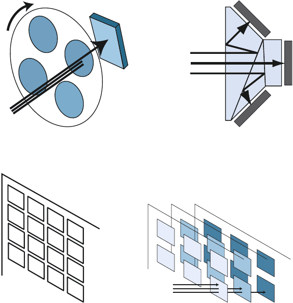
If a single detector array is used, it is then necessary to acquire three exposures
through different filters to capture a color image (Figure 2.4a). A few cameras have
used this strategy, combining the three images electronically to produce a color
picture. The advantages are high resolution at modest cost, and the ability to achieve
color balance by varying the exposure through each filter. The penalty is that the
time required to obtain the full-color picture can be many seconds, the filters must
be changed (either manually or automatically), and during this long time it is
necessary for the specimen to remain perfectly still. Also, vibration or other inter-
ference can further degrade images with long exposure times. And of course, there
is no live color preview with such an arrangement.
At the other extreme, it is possible to use three chips with separate detector
arrays, and to split the incoming light with prisms so that the red, green, and blue
portions of the image fall onto different chips (Figure 2.4b). Combining the signals
electronically produces a full-color image. This method is expensive because of the
cost of the three detectors, electronics, prisms, and alignment hardware. In addition,
the cameras tend to be fragile. The optics absorb much of the incoming light, so
brightly lit scenes are needed. Also, because of the prisms, the satisfactory use of
the three-chip approach is usually limited to telephoto lenses. Short focal length
lenses direct the light through the prisms at different angles resulting in images with
color gradients from top to bottom and/or left to right. Three-chip cameras are used
for many high-end video cameras, but rarely for digital still cameras.
Many experimental approaches are being tried. The Foveon® detector uses a
single chip with three transistors stacked in depth at each pixel location (Figure
2.4d). The blue light penetrates silicon the least and is detected near the surface.
Green and red penetrate farther before absorption, and are measured by transistors
deeper beneath the surface. At present, because these devices are fabricated using
complementary metal oxide on silicon (CMOS) technology, the cameras are fairly
noisy compared to high performance charge coupled device (CCD) cameras, and
combining the three signals to get accurately calibrated color remains a challenge.
The overwhelming majority of color digital still cameras being used for technical
applications employ a single array of transistors on a CCD chip, with a filter array
that allows some detectors to see red, some green, and some blue (there are a few
consumer cameras that use other combinations of color filters). Various filter arrange-
ments are used but the Bayer pattern shown in Figure 2.4c is the most common. It
devotes half of the transistors to green sensitivity, and one quarter each to red and
blue, emulating human vision which is most sensitive in the green portion of the
spectrum.
With this arrangement, it is necessary to interpolate to estimate the amount of
red light that fell where there was no red detector, and so on. That interpolation
reduces resolution to about 60% of the value that might be expected based on the
2241_C02.fm Page 58 Thursday, April 28, 2005 10:23 AM
Copyright © 2005 CRC Press LLC
number of camera transistors, and also introduces problems of incorrect colors at
boundaries (e.g., is the average between a red pixel and a green pixel yellow or
grey?). Interpolation is a complicated procedure that each manufacturer has solved
in different (and patented) ways. Problems, if present, will often show up as zipper
marks along high contrast edges. The user typically has no control over this except
to not expect to measure features with a width of only one or two pixels in the
image. In the measurement chapters we will typically try to limit measurement to
features that cover multiple pixels.
The principal reason for wanting a high number of pixels in a camera detector
is for more than dealing with small features. Usually that can be done best by
applying the appropriate optical magnification, either with a macro lens or micro-
scope objective. If all of the features of interest are the same size, then even a fairly
small image (at current technology levels that is probably a million pixels, say 1200
×
900) is quite useful provided the image magnification is adjusted so the features
are large enough to adequately define size and shape.
(a) (b)
(c) (d)
FIGURE 2.4
Several ways for a digital camera to acquire a color image: (a) sequential images
through colored filters with a single chip; (b) prisms to direct colors to three chips; (c) Bayer
filter pattern applied to detectors on a single chip; (d) Foveon chip with stacked transistors.
R
G
B
R
G
B
Green
Blue
Red
R
G
B
G
R
G
B
G
R
G
B
G
R
G
B
G
B
lue
Green
R
ed
2241_C02.fm Page 59 Thursday, April 28, 2005 10:23 AM
Copyright © 2005 CRC Press LLC
Difficulties arise when the features cover a range of sizes. Large features have
a greater probability of intersecting the edge of the image frame, in which case they
cannot be measured. In the example just given, if the smallest features of importance
are 20 pixels wide, and the largest features are 20% of the image with (say about
200 pixels) then it is possible to satisfactorily image features with a 10:1 size range.
If the features present actually cover a greater range of sizes, multiple images at
different magnifications are required to adequately image them for measurement.
But sometimes we need to measure spatial arrangements, such as how the small
features may be clustered around (or away from) the large ones. In that case there
is really no good alternative to having an image with enough pixels to show both
the small features with enough resolution and the large ones with enough space.
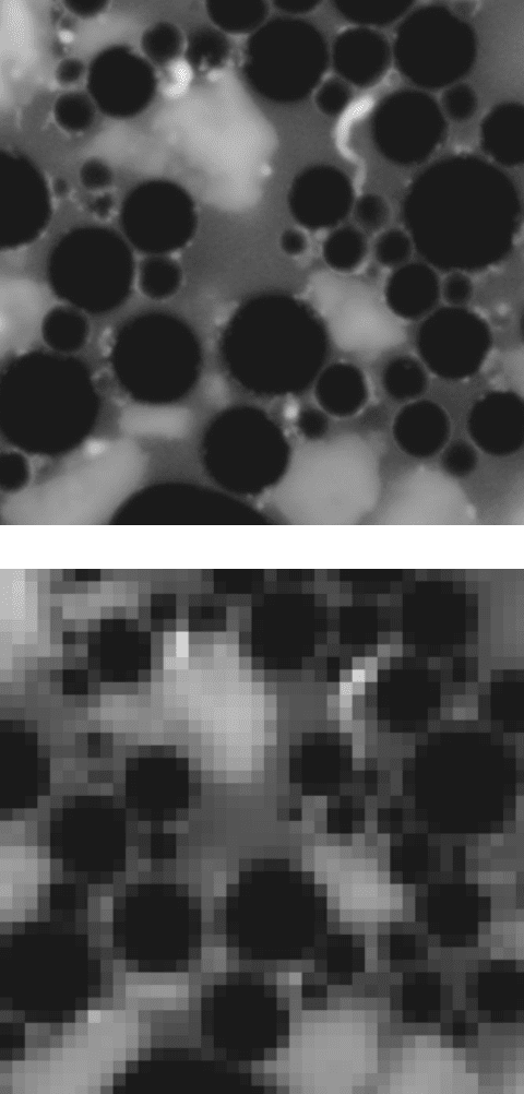
Figure 2.5 illustrates this problem. The fragment of the original image, captured
with a high resolution digital camera, shows both large and small oil droplets and
also details such as the thin layer of emulsifier around the periphery of most droplets.
Decreasing the pixel resolution by a factor of 6, which is roughly equivalent to the
difference between a 3 megapixel digital camera and the resolution of a video
camera, hides much of the important detail and even limits the measurement of
droplet size.
Despite their poor resolution and image noise, video cameras are sometimes
used as microscope attachments. Historically this was because they existed and
(driven by a consumer marketplace) were comparatively inexpensive, produced a
real-time viewable image, and could be connected to “framegrabber” boards for
digitization of the image. Currently, the only reason to use one is for those few
applications that require image capture at a rate of about 30 frames per second. Most
studies do not. Either the specimens are quite stable, permitting longer exposures,
or some dynamic process is being studied which may require much higher speed
imaging. Specially designed cameras with hard disk storage can acquire thousands
of images per second for such purposes.
Human vision typically covers a size range of 1000:1 (we can see meter-sized
objects at the same time and in the same scene as millimeter-sized ones). A 4
×
5
photographic negative can record images with a satisfactory representation of objects
over a 150:1 size range. But even a high performance digital camera with 6 million
pixel resolution (reduced somewhat by interpolation of the color information) is
limited to about 20:1 in size range. That may be frustrating for the human observer
who sees more information in the microscope eyepiece, and is accustomed to seeing
a wider range of object sizes in typical scenes, than the camera can record. This
topic arises again in the context of feature measurement.
The number of camera pixels is most significant for low magnification imaging.
At high magnification, the limit to resolution in the image is typically the optical
performance of the microscope (and perhaps the specimen itself and its preparation).
At low magnification, where a large field of view is recorded, an image with a large
number of pixels provides the ability to resolve small features and provide enough
pixels to define their size and shape.
In addition to the resolution or number of pixels in the camera, the same issues
of well size, noise level and bit depth arise as for scanners. Many consumer cameras,
particularly low-cost ones such as are appearing in telephones and toys, use CMOS
2241_C02.fm Page 60 Thursday, April 28, 2005 10:23 AM
Copyright © 2005 CRC Press LLC
(a)
(b)
FIGURE 2.5
Fragment of an image of oil droplets in mayonnaise, showing a size range of
oil droplets and a thin layer of emulsifier around many: (a) high resolution camera; (b) effect
of a 6
×
decrease in pixel resolution, losing the smaller details.
2241_C02.fm Page 61 Thursday, April 28, 2005 10:23 AM
Copyright © 2005 CRC Press LLC
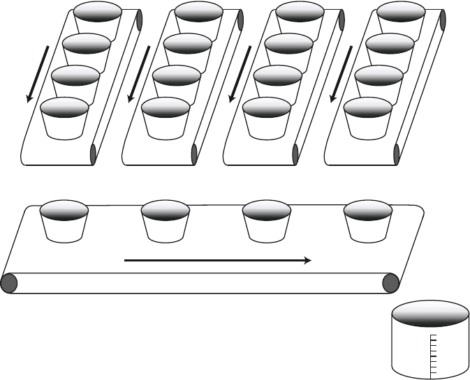
chips. These have the advantage of low cost because they can be fabricated on large
wafers and can include much of the processing electronics right on the chip, whereas
the CCD devices (Figure 2.6) used in higher-end scientific cameras are more expen-
sive to fabricate on smaller wafers, and require separate electronics. CMOS detectors
are also being used in a few prosumer (high-end consumer/low-end professional)
cameras, because it is possible to combine several discrete chips to make one large
detector array, the same size as a traditional 35mm film negative, with about 15
million pixels.
But the CMOS cameras have many drawbacks, including higher amounts of
random noise and fixed pattern noise (a permanent pattern of detector variability
across the image) that limits their usefulness. Also, the extra electronics on the chip
take up space from the light-sensing transistors and reduce sensitivity for low-light
situations. This can be overcome to some extent by using microlenses to collect
light into the detector, but that further increases the point-to-point variations in
sensitivity and fixed pattern noise. It is likely that CCD detectors will remain the
preferred devices for scientific imaging for the foreseeable future.
FIGURE 2.6
Schematic diagram of the functioning of a CCD detector. The individual detec-
tors function like water buckets to collect electrons produced by incoming photons. Shifting
these downwards in columns dumps the charge into a readout row. Shifting that row sideways
dumps the charge into a measuring circuit. The overall result is to read out the image in a
raster fashion, one line at a time.
2241_C02.fm Page 62 Thursday, April 28, 2005 10:23 AM
Copyright © 2005 CRC Press LLC
Even for CCD chips, the drive for lower cost leads manufacturers to reduce the
chip size (at the same time as increasing the number of transistors) in order to get
more devices from a given wafer. That makes the individual transistors smaller,
which reduces light sensitivity and well size. With some of the current small chips
having overall dimensions only a few mm (a nominal one third inch chip has an
actual active area of 3.6
×
4.8 mm, while a one quarter inch chip is only 2.4
×
3.2
mm), the individual transistors are so small that a maximum signal to noise ratio of
200:1 can barely be achieved. That is still enough for a video camera, which is only
expected to produce a dynamic range between 50:1 and 100:1, but not enough for
a good digital still camera.
Most digital still cameras record images with at least 8 bits (256 brightness
values) in each RGB channel. Internally, most of them have a somewhat greater
dynamic range, perhaps 10 bits (1000 brightness values), which the programs in
their internal computers convert to the best 8 bits for output. With many of the
cameras it is also possible to access the full internal data (RAW data) to perform
your own conversion if desired, but except in cases with unusual illumination or
other problems the results are not likely to produce more information than the built-
in firmware. Also, the raw internal data is usually linear but for most cameras the
output is made logarithmic, to achieve a more film-like response, and this reduces
the effective bit depth.
Obtaining more than 8 bits of useful data from a digital camera usually requires
cooling the chip to reduce the noise associated with the readout process. Extreme
cooling as for astronomical imaging is not needed. Peltier cooling of the chip by
about 40 degrees, combined with a somewhat slower readout of the data, is typically
enough to produce images with 10 or even 12 bits (approximately 1000 to 4000
brightness levels) in each channel. Cooling the chip also helps to reduce thermal
noise in the chip itself when exposure times are longer than about one-quarter to
one-half second. Cooled cameras are often used for microscopes when dark field or
fluorescence imaging is needed, whereas 8 bits is probably enough when only bright
field images are captured.
SCANNING MICROSCOPES
Several types of microscopes produce images by raster scanning and are capable
of directly capturing images for computer storage (Figure 2.7). This includes the
scanning electron microscope, the scanning confocal laser microscope, and the
atomic force microscope, as well as other similar scanned probe instruments. These
use very different physical principles to generate the raster scan and the imaging
signal. The confocal microscope uses mirrors to deflect the laser beam, while the
SEM uses magnetic fields to deflect an electron beam, the AFM uses piezoelectric
devices to shift the specimen under a fine-pointed stylus. But they share many of
the same attributes in so far as the resulting image quality is concerned.
The desire for spatial resolution and dynamic range are the same as for any other
image source, with the single exception that these devices produce monochrome
rather than color images. Confocal microscope images are often displayed as color,
2241_C02.fm Page 63 Thursday, April 28, 2005 10:23 AM
Copyright © 2005 CRC Press LLC
