Maier S.A. Plasmonics: Fundamentals and Applications. Майер С.А. Плазмоника: Теория и приложения
Подождите немного. Документ загружается.

Chapter 10
SPECTROSCOPY AND SENSING
The main part of this chapter describes different techniques for spectro-
scopic investigations of localized plasmon resonances in single metal nanopar-
ticles, with a view to applications in sensing. The basic principle of single-
particle sensors is the exploitation of the fact that the spectral position of their
resonances depends on the dielectric environment within the electromagnetic
near field. Applied to biological sensing, adsorption of molecules on a func-
tionalized metal surface leads to spectral changes of the sustained plasmon
modes. Due to the very localized nature and therefore high energy concentra-
tion in the near field of surface plasmons, even molecular monolayers can lead
to discernible spectral changes. This high sensitivity has allowed surface plas-
mon sensors to become established as an analytical sensing technology over
the last two decades.
The most important challenge encountered in almost any biosensor design
is that of ensuring selectivity. In the case of surface-plasmon-based sensors,
this is achieved via functionalization of the metallic surface to ensure only se-
lective binding of the agent to be sensed. We will not focus on this aspect of
sensor design here, but only mention that the surface chemistry of gold de-
serves special attention due to the relative ease of establishing sulfur bonds be-
tween gold atoms and organic molecules. Therefore, gold has emerged as the
metal of choice for almost all practical optical sensing applications, including
those based on surface plasmons. An important consequence is that due to the
permittivity of gold, sensing is usually limited to the visible and near-infrared
part of the spectrum.
We provide an overview of different excitation geometries for the investiga-
tion of localized surface plasmons, which is related to the analogous discussion
of SPP excitation in chapter 3. The second part of this chapter aimes to give
a flavor for different aspects of sensors based on propagating SPPs, relying on
178 Spectroscopy and Sensing
changes to the dispersion relation and the condition of phase-matching upon
refractive index changes at flat metal interfaces. We will limit the discussion
to two prominent excitation geometries, based on prism coupling and coupling
using optical fibers coated with a metal film. However, we will not embark on
a discussion of sensor performance in terms of selectivity and sensitivity. As
a starting point for the exploration of important omitted aspects such as these,
the reader is referred to the review by Homola [Homola et al., 1999].
10.1 Single-Particle Spectroscopy
This section continues the discussion of excitation mechanisms, presented
in chapter 3 for propagating SPPs, by describing different excitation pathways
for localized plasmon resonances in metal nanoparticles. We have seen in the
description of the fundamentals of localized resonances in chapter 5 that the
frequencies of the resonant modes of regular particle ensembles can be deter-
mined using conventional far-field extinction spectroscopy. Upon resonance
the extinction cross sections of individual particles are resonantly enhanced,
and for a sufficient spacing the extinction peak of the ensembles coincides with
the localized plasmon frequency of an individual particle. However, due to
slight differences in particle shape, inhomogeneous broadening of the extinc-
tion line shape can occur. Spectroscopy of single nanoparticles requires more
sensitive detection techniques (due to the large background of radiation directly
passing from the source to the detector), which will be outlined in this section.
The investigation of plasmon resonances of single particles is not only rele-
vant from a fundamental point of view (e.g. the determination of the homoge-
neous linewidth ), but also potentially for practical applications in sensing. In
this context, single metal nanoparticle sensors operate via the detection of fre-
quency shifts of the dipolar plasmon resonance upon binding of molecules to
the nanoparticle surface, which can be detected using spectroscopic techniques
suitable for single-particle investigation.
Let us briefly review: for a spherical particle of a sub-wavelength diameter
d λ
0
, the resonance frequency of the dipole mode for small damping is
given by the Fröhlich condition
ε
ω
sp
=−2ε
m
. (10.1)
Here, ε
(
ω
)
is the dielectric function of the metal, and ε
m
the dielectric con-
stant of the insulating host. Whereas the derivation of (10.1) in chapter 5 has
assumed an infinite extent of the surrounding host medium, the sub-wavelength
localization of the dipolar plasmon mode means that ω
sp
is only determined by
the dielectric environment within the tail of the evanescent near field of the
particle. Changes in ε
m
, induced for example via adsorption of a molecular
monolayer on the particle surface, can then be detected via changes in the
dipolar resonance frequency ω
sp
.
Single-Particle Spectroscopy 179
While sensing in this manner can be easily performed using far-field extinc-
tion spectroscopy if a large amount of particles arranged in a regular array is
used as the sensing template, sensors based on a single metallic nanostructure
are highly desirable. Firstly, interrogation of a single particle does not suf-
fer from the inhomogeneous broadening of the resonant line shape observed
in far-field spectroscopy. This, together with the fact that binding events are
monitored in a local manner, leads to an increased sensitivity, expressed via
observed peak-shift with quantity of agent binding. Also, sensors based on in-
dividual particles of submicron dimensions enable at least in principle a high
integration density of sensing sites for assay-like studies with high through-
put. However, for this vision to come true, a suitable, parallelized addressing
scheme for individual, closely spaced particles has first to be developed.
Proof-of-concept studies of single-particle sensors therefore rely on the spec-
troscopic determination of the plasmon resonance of an individual, sub-wave-
length metallic nanoparticle. In the following, we will present four prominent
optical excitation techniques suitable for this purpose - total internal reflection
spectroscopy, near-field microscopy and dark-field microscopy, and photother-
mal imaging of very small particles with dimensions below 10 nm.
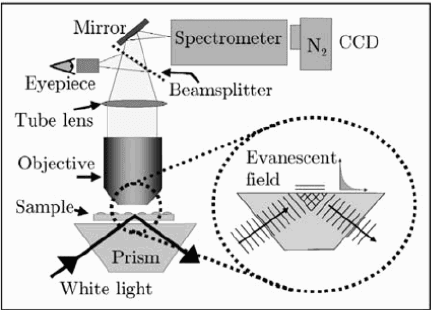
In total internal reflection spectroscopy, metallic nanostructures are deposit-
ed on top of a prism, and excitation takes place using illumination under to-
tal internal reflection conditions. Similar to the excitation of SPPs on a flat
metal film described in chapter 3, the evanescent field above the prism acts
as a local excitation source for modes at the interface, leading to resonantly
enhanced scattering. This way, the frequency of spatially confined modes in
Figure 10.1. Setup for single-particle spectroscopy using evanescent excitation via total inter-
nal reflection at a prism and the monitoring of scattered light. Reprinted with permission from
[Sönnichsen et al., 2000]. Copyright 2000, American Institute of Physics.
180 Spectroscopy and Sensing
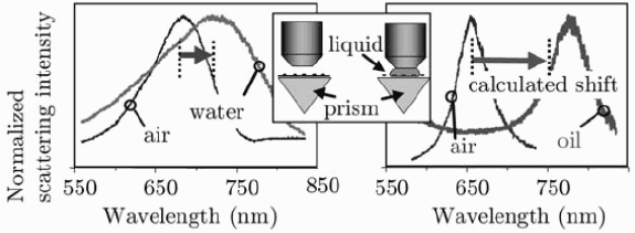
Figure 10.2. Shift of particle plasmon resonance detected using prism excitation. Reprinted
with permission from [Sönnichsen et al., 2000]. Copyright 2000, American Institute of Physics.
metal nanoparticles can be determined using white light illumination and de-
tection of the scattered light via far-field collection from the top (Fig. 10.1).
Examples of plasmon spectra of a single gold particle collected in this man-
ner are shown in Fig. 10.2. As expected from (10.1), the resonance peak of
the dipole plasmon mode red-shifts upon immersion of the particle into a high-
index environment such as water or oil. The expected spectral variation of the
collected intensity can in a first approximation be calculated via the formulae
for the cross sections of first-order Mie theory (5.13), using the appropriate di-
electric data ε(ω) for the metal. For a metal particle on a glass prism immersed
in an external medium, the effective dielectric constant of the host can often
simply be approximated as 1/2
ε
prism
+ε
m
.
Single-particle spectroscopy can also be performed using near-field optical
microscopy, i.e. by placing an apertured fiber tip into the near field of the parti-
cle under study. In its simplest form, spectroscopic information is obtained by
monitoring the spectral intensity distribution of radiation collected in the far
field (either in transmission or reflection) ensuing from local illumination of
the particle with white light. This way, the resonance frequencies and homo-
geneous lineshapes of plasmon modes in single particles can be determined.
Pioneering spectroscopic studies of single particles have been performed using
both transmission near-field optical microscopy with near-field illumination
and far-field collection [Klar et al., 1998], and collection-mode near-field opti-
cal microscopy with prism-coupling illumination as described above, but with
near-field instead of far-field collection [Markel et al., 1999].
In a more recent study, Mikhailovsky and co-workers have shown that trans-
mission mode near-field optical microscopy with local white light illumination
through the sub-wavelength aperture enables a high sensitivity for determining
the plasmon resonance of an individual particle due to phase information en-
coded into the intensity collected in the far field [Mikhailovsky et al., 2004].
This is based on the fact that the light scattered by the particle in the forward
direction interferes either constructively or destructively with the light directly
Single-Particle Spectroscopy 181
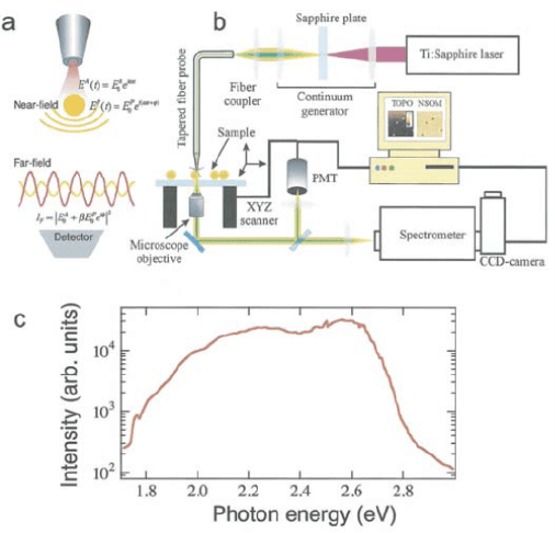
Figure 10.3. Sketch of excitation geometry (a) and experimental setup (b) for white-light illu-
mination mode near-field optical microscopy. (c) Spectrum of the white light supercontinuum
at the output of the fiber tip. Reprinted with permission from [Mikhailovsky et al., 2003]. Copy-
right 2003, Optical Society of America.
collected from the aperture [Batchelder and Taubenblatt, 1989]. Fig. 10.3
shows a sketch of the experimental setup and a spectra of a white light su-
percontinuum passing through an apertured tip. Typical examples of images
of both the topography and the optical near field of gold nanoparticles are pre-
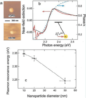
sented in Fig. 10.4a.
An investigation of the scattering and absorption process using the model
of a driven damped harmonic oscillator predicts a contrast reversal of the near-
field image due to the transition from destructive to constructive interference
at ω
sp
(Fig. 10.4b). We recollect from chapter 5 that, in the vicinity of the
resonance, a phase shift φ between the driving field and the response of the
electrons of π occurs, with φ
ω
sp
= π/2. An analysis of near-field images
obtained at different frequencies therefore enables the determination of ω
sp
for
particles of various sizes (Fig. 10.4c).
While near-field optical extinction microscopy provides unprecedented spa-
tial resolution for local spectroscopy, the optical probe placed in the near field
of the particle poses a difficult constraint for practical sensing applications.
Agent binding is additionally often monitored in a liquid environment, which
182 Spectroscopy and Sensing
Figure 10.4. (a) Topography and near-field image of a 50 nm gold sphere. (b) Near-field
extinction spectrum (solid gray curve) compared with interference (black curve) and phase
(dashed curve) spectra for a single 50 nm gold particle calculated using a forced harmonic
oscillator model. (c) Dependence of resonance frequency on particle size inferred from the
spectra. Reprinted with permission from [Mikhailovsky et al., 2003]. Copyright 2003, Optical
Society of America.
poses serious stability problems for the probe movement. Moreover, since
near-field optical microscopy only allows the determination of optical prop-
erties near a surface, in situ measurements of metal nanoparticles within cell
bodies are generally not possible. A more suitable geometry for such purposes
is dark-field optical microscopy, which is a far-field technique where only light
scattered by the nanoparticle is collected. Here, use of a dark-field condenser
prevents the collection of the directly transmitted light. Therefore, in dark-field
images metal nanoparticles appear in bright colors, defined by the resonance
frequency ω
sp
of their scattering cross section (5.13). A typical dark-field im-
age of single gold nanoparticles is shown in Fig. 10.5c. We note that due to the
constraint of the diffraction limit for focusing of the illumination spot, single-
particle sensitivity can only be achieved for well-separated nanoparticles.
An example of the monitoring of a molecular binding event is shown in
Figs. 10.5 and 10.6 [Raschke et al., 2003]. The coating of a gold nanoparti-
cle with a BSA-complex leads to a slight red-shift of ω
sp
, which is increased
upon the selective binding of streptavidin molecules (Fig. 10.5b). The binding
can be monitored in real-time via a recording of the resonance shift with time
(Fig. 10.6), and saturation is achieved upon complete coating of the particle.
Single-Particle Spectroscopy 183
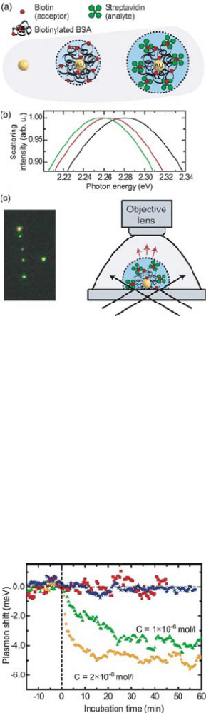
Figure 10.5. (a) Principle of a single nanoparticle biosensor monitoring the selective bind-
ing of streptavidin on an BSA-decoared gold nanoparticle. (b) Mie-theory calculations of the
scattering spectra for the undecorated particle and the particle with BSA and BSA-streptavidin
coating, demonstrating red-shifts of the resonance with each coating layer. (c) Dark-field pic-
tures and sketch of the detection pathway. Reprinted with permission from [Raschke et al.,
2003]. Copyright 2003, American Chemical Society.
A similar study based on single silver nanoparticles demonstrated that a sensi-
tivity on the order of zeptomoles can be achieved, and first applications using
medically-relevant assay studies are emerging [Haes et al., 2004].
Figure 10.6. Resonance shift versus incubation time for streptavidin-BSA binding for differ-
ent streptavidin concentrations C added at t = 0, and control experiments. Reprinted with
permission from [Raschke et al., 2003]. Copyright 2003, American Chemical Society.
184 Spectroscopy and Sensing
Further improvements in sensitivity have been predicted for single-particle
sensors employing resonance line shape design, which can either be achieved
using metallic nanoshells [Raschke et al., 2004], designed particle arrays with
near-field coupling for hot spot generation [Enoch et al., 2004], or by using
a particle-on-extended film approach to couple the particle plasmon to propa-
gating SPPs [Chen et al., 2004]. Also, the use of elongated nanoparticles has
enabled polarization-sensitive orientation sensing [Sönnichsen and Alivisatos,
2005].
The good biocompatability and well-developed surface chemistry of gold
nanoparticles has further lead to their wide use in cellular imaging. In these
studies, the nanoparticles mainly serve as a labeling agent for the tracking of
single molecules or molecular complexes. Optical microscopy techniques such
as the aforementioned dark-field illumination, differential interference contrast
or total internal reflection illumination can be used for image acquisition. First
in vivo studies extracting spectroscopic information akin to the particle-based
studies outlined above are emerging [El-Sayed et al., 2005].
However, dark-field microscopy and other techniques relying on the detec-
tion of scattered light are not suitable for very small metal nanoparticles with
diameters d 40 nm immersed in a background of scatters, such as for ex-
ample a biological cell. This is due to the fact that the scattering cross section
decreases as d
6
with particle diameter as discussed in chapter 5. Thus, the scat-
tering signal of particles in this small size regime is usually completely over-
whelmed by larger scatterers. In order to optically pick out the signature of par-
ticles of these small sizes, a different microscopy method relying on absorption
instead of scattering is required. Since according to Mie theory the absorption
cross section scales with size only as d
3
, sub-10 nm particles can be picked out
of a background of bigger particles using a photothermal imaging technique
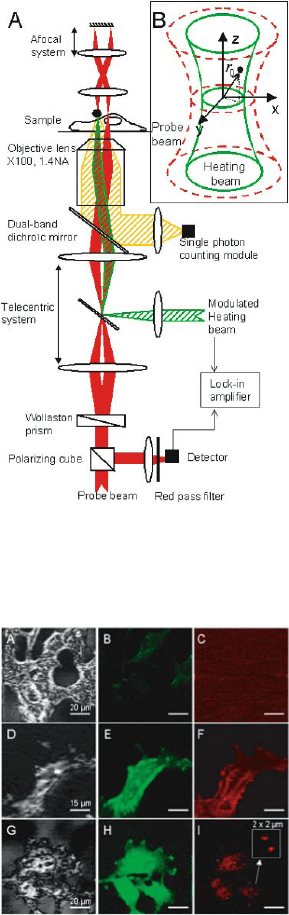
[Boyer et al., 2002]. Fig. 10.7 shows the optical setup used in this imaging
technique, consisting of a heating beam and a second, weaker probe beam
detecting the absorption-induced thermal changes around the metal nanoparti-
cles. The red probe beam is split in two parts of orthogonal polarization, and
both beams are subsequently focused onto the sample to diffraction-limited
spots spaced at a distance on the order of 1 μm from each other. The heating
beam only overlaps with one of the probe beams, resulting in a heat-induced
change in its polarization. Recombination of the two probe beams therefore
leads to an intensity modulation, and via a scanning system an image of the
sample under study can be constructed. In-vivo images acquired using this
technique are shown in Fig. 10.8, and compared with scattering and fluores-
cence images for biological cells with incorporated gold nanoparticles, demon-
strating the improved spatial resolution due to the detection of single particles.
Before moving on, we want to briefly mention another promising technique
for the spectroscopic investigation of localized surface plasmons, based on ex-
Single-Particle Spectroscopy 185
Figure 10.7. Experimental setup for photothermal imaging of very small nanoparticles. For
a description see main text. Reprinted with permission from [Cognet et al., 2003]. Copyright
2003, National Academy of Sciences, U.S.A.
Figure 10.8. Scattering (A, D, G), fluorescence (B, E, H) and photothermal images (C, F, I)
of cells. All cells are transfected with gold nanoparticles functionalized to a membrane protein
(A-F in concentration 10 μg/l and for G-I the concentration is 0.5 μg/l). A-C show cells not
expressing this protein, and D-I cells expressing it and thus binding the particles. The resolution
is highest in pictures F and I obtained with photothermal imaging. Reprinted with permission
from [Cognet et al., 2003]. Copyright 2003, National Academy of Sciences, U.S.A.
186 Spectroscopy and Sensing
citation using electron impact. In cathodoluminescence, photon emission of
the metal nanostructure under investigation is induced via a high-energy elec-
tron beam, and collected using a suitable detection pathway [Yamamoto et al.,
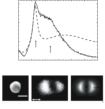
2001]. As an example, Fig. 10.9 (upper part) shows the spectrum of a 140 nm
silver particle excited via grazing-incidence of a 200 keV electron beam, to-
gether with a comparison with theory. Due to the large size of the particle,
the signatures of both a quadrupolar and a dipolar mode are discernible. A
nice feature of this technique is that by scanning of the electron beam over the
particle surface, the spatial profile of the modes can be mapped out via light
collection at the respective peak wavelength (Fig. 10.9, lower part). The same
technique can also be used for the excitation and investigation of propagating
SPPs.
All the aforementioned single-particle spectroscopy techniques are based
on microscopy and thus generally not suitable for field-based sensing, e.g. in a
context of environmental monitoring. Sensors based on localized particle plas-
mon spectroscopy amenable for such applications have been developed in the
context of optical-fiber-based sensing. In a typical geometry, metal nanoparti-
cles are spatially fixed at the end facet of an optical fiber, and the reflected light
λ= 420 nm
E
300 350 400 450 500 550 600
0.00
0.05
0.10
0.15
quadrupole
dipole
Cl intensity [a.u.]
Emission wavelengh [nm]
experiment
theory
SEM CL experiment CL theory
100 nm
Figure 10.9. Cathodoluminescence imaging and spectroscopy of localized surface plasmons.
Upper part: cathodoluminescence (CL) from a 140 nm silver particle induced by the passage
of 200 keV electrons in a grazing trajectory (barely touching the particle surface). Dipolar
and quadrupolar components can be separated in the spectrum. Lower part, from left to right:
SEM image of the particle under consideration; CL rate as a function of the position of the
electron beam, which is scanned over the particle, for an emission wavelength corresponding to
the dipolar feature of the spectrum; theoretical prediction for the latter. Figure courtesy of N.
Yamamoto and F. J. García de Abajo, personal communication.