Hawkes P.W., Spence J.C.H. (Eds.) Science of Microscopy. V.1 and 2
Подождите немного. Документ загружается.


Chapter 7 Cryoelectron Tomography (CET) 585
mark the positions of the features in the input scene. Three different
conventional XCF, phase-only fi ltering (POF), (Horner and Gianino,
1964), and mutual cross-correlation function (MCF; van Heel et al.,
1992). In all those methods a cross-correlation function is computed:
The XCF computes the unmodifi ed cross-correlation function of tem-
plate and micrograph whereas the POF and MCF vary the moduli of
the template to sharpen the cross-correlation peaks. In electron micros-
copy especially, the low frequencies carry an extraordinarily strong
signal, which can be misleading (van Heel et al., 1992). The strength of
the low-frequency contributions can differ considerably within elec-
tron micrographs of amorphous specimens, such as vitrifi ed speci-
mens, since these are typically nonhomogeneous. The POF sets the
moduli of template and input scene to one while the MCF replaces the
moduli by their square roots to reduce the infl uence of the very low-
frequency amplitudes systematically.
Another way of taking into account the strong local variations of the
image contrast has been introduced recently (Roseman, 2000). By nor-
malizing the XCF to the local cross-correlation function (LCF) variance,
the effi ciency of locating local features such as macromolecules within
a micrograph can be increased signifi cantly. Typically, the normaliza-
tion region should correspond to the shape of the molecule. This cal-
culation of the local variance does not lengthen the computing time
unreasonably, since it can be calculated very effi ciently in Fourier space
too (Figures 7–23 and 7–24).
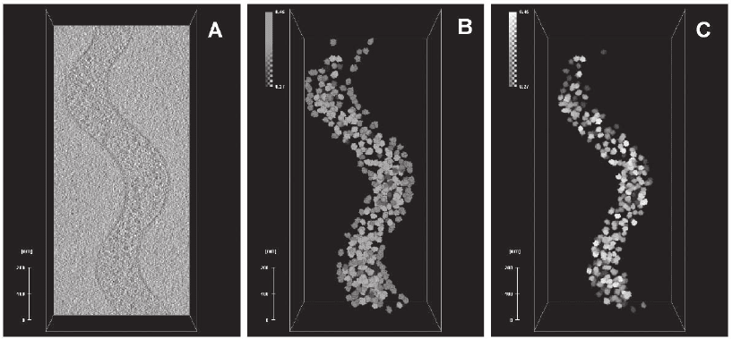
Figure 7–23. Mapping ribosomes in an intact S. melliferum cell. a) x–y slice from the corresponding
tomogram. Image analysis of this portion of the cell is displayed in subsequent panels. b) Locations
and orientation of all ribosomes detected by template matching. Each 70S ribosome is represented by
the averaged density (see Fig. 26) derived from the tomogram. The colour coding indicates the detec-
tion fi delity; green is high, yellow is intermediate, red is low and probably represents false positives.
The brightness of the colour corresponds to correlation peaks heights. c) Final ribosome atlas after
removal of putative false positives (images courtesy of J. Ortiz, MPI of Biochemistry, Martinsried,
CCFs have been introduced in the fi eld of 2D electron microscopy:
Germany; Ortiz et al., 2006). (See color plate.)

586 J.M. Plitzko and W. Baumeister
To evaluate which fi lter is best for the task of locating macromole-
cules, some theoretical considerations are useful. The conventional
XCF has been shown to be optimal with respect to minimization of the
location error (Kumar et al., 1992) and high SNR of the resulting func-
features within micrographs or cryoelectron tomograms yields intoler-
ably high rates of false positives; the XCF tends to locate high-contrast
features rather than low-contrast macromolecules. The same typically
holds for the MCF, whereas the POF overamplifi es noise, which makes
A
96%73%6%detection reliability
143
174
26
presumably correct (~fit)
137127433
CC peaks
50%
33%
16,50%
ribosomes expected in
CC range (~fit)
CC>0.3430.343 CC>0.3
CC range
III
II
I
B
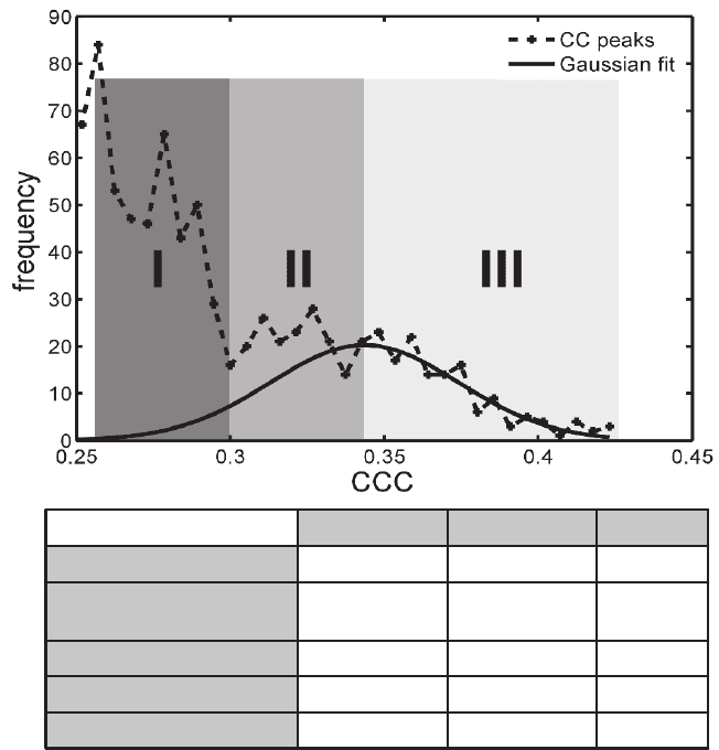
Figure 7–24. a) Histogram of experimentally obtained CCCs (dashed line). Ideally, the histogram
should be Gaussian, refl ecting the variances in SNR of individual ribosomes in the tomogram, and
thresholding of the CCC should allow discriminating between correctly identifi ed ones and false
positives. Non-specifi c correlation with high contrast features such as the cell membrane, leads to a
relative large number of false positives. For an objective setting of thresholds, the Gaussian fi t (con-
tinuous line) was divided into three probability sectors larger than the mean CCC value (I, dark grey);
lower than the mean value up to one standard deviation (II, intermediate grey); lower than the mean
value and between one and two standard deviations (III, light grey). The spreadsheet in b) illiustrates
the Gaussian fi t of the experimental distribution of CCCs and the fi delity of the experimental detection
of 70S ribosomes. About 80% of the particles are detected with more than 85% reliability (images
0.3 CC>0.257
tion (VanderLugt, 1964). However, use of the XCF for locating small
≥
≥
courtesy of J. Ortiz, MPI of Biochemistry, Martinsried, Germany; Ortiz et al., 2006).

Chapter 7 Cryoelectron Tomography (CET) 587
it unfavorable. Therefore, local normalization is appropriate. However,
regarding the SNR of the resulting correlation function, the LCF is
inevitably worse than XCF or MCF; confi nement of the correlation to
N voxels raises the noise level, which should be proportional to N
−1/2
.
The size of the normalization region should correspond to the envelope
of the template under scrutiny. Choosing the local region larger will
increase the SNR of the fi lter output at the cost of increasing the chance
of false positives simultaneously, in particular if particles tend to be
very dense within the input scene. Whenever justifi ed circular (2D) or
spherical (3D) normalization regions (Θ) will be used because this
accelerates the computation considerably compared to approaches
using asymmetric regions. If the template has one rotational degree of
freedom perpendicular to the specimen plane within the input scene,
the problem of matching a 2D template in a 2D image can be formu-
lated as determining the maxima of a cross-correlation function as a
function of spatial variable r and rotation angle ϕ:
XCF I R T
rrrr
r
,ϕϕ
=⋅
(
)
+
′′
′
∑
.
Here R
ϕ
denotes the rotation operator. By expanding the template in
circular harmonics, exhaustive scanning of the polar angle ϕ can be
avoided (Kunath and Sack-Kongehl, 1989). In 3D, the problem of fi nding
a macromolecule in a tomogram that matches a 3D template by cross-
correlation becomes six-dimensional: Using the Euler angles ϕ, ψ and
ϑ to describe the orientation, the maxima of the CCF
r
.
;ϕψϑ
are to be
determined. Again, the translational dependence can be covered using
FFT, but it is normally necessary to perform an explicit scanning in
angular space to cover those parameters. An exhaustive search is com-
putationally very time demanding. However, because of the emerging
facilities of parallel computing this task can easily be faced: Since the
CCF can be computed individually for different orientations, the entire
calculation can be distributed over different processors with hardly
any administrative overlap (Frangakis and Hegerl, 2002; Rath et al.,
2003).
In cryo-ET the partial sampling of the object’s Fourier space, the
“missing wedge,” has to be considered further. This can be done by
confi ning the correlation to the sampled region in Fourier space or
equivalently by convoluting the object with the point-spread function
(Frangakis and Hegerl, 2002). In the framework of the locally normal-
ized CCF the missing wedge affects the local normalization. The
missing wedge can be taken into account in Fourier space by multipli-
cation of the template with a suitable binary function or equivalently
by convoluting with a suitable PSF in real space. In this notation, the
confi ned cross-correlation function can be defi ned as
CCF
IT PSF
II
r
rr r r
r
rrr r
r
ϕψϑ
ϕψϑ
ϕψϑ
=
⋅⊗ ⋅
()
⋅−
()
+
′′ ′
′
′
+
′′
′
∑
∑
Θ
Q
2
where I
¯
r
′ represents the local mean value within Θ. A natural way of
avoiding the limitation of beam damage in tomography is averaging
of copies of structural elements occurring in a tomogram. Saxberg and

588 J.M. Plitzko and W. Baumeister
Saxton (1981) already concluded from their results on resolution limita-
tion in electron tomography due to beam damage that averaging is a
practical way of increasing the effective electron doses and should be
considered to achieve high resolutions. The importance of those tech-
niques to derive information on the molecular level was reemphasized
recently (Steven and Aebi, 2003).
According to the dose fractionation theorem a tomogram describes
a volume element with the same statistical signifi cance as a projection
with the same dose (Hegerl and Hoppe, 1976; McEwen et al., 1995).
Therefore, tomograms should be just as well suited to posterior averag-
ing procedures for obtaining high-resolution information as compared
to single-particle averaging. In practice, both approaches have their
pros and cons, which are discussed below.
Averaging of particles from tomograms can be performed in the
same way as single-particle averaging. Projection matching is crucial
in these techniques: The individual particle projections are iteratively
aligned to projections of a reference from different angles by means of
cross-correlation. The reference is thus the average of the particles
using the orientations and displacements of the previous iteration. The
iterative refi nement of orientation and displacements improves the
resolution of the average until convergence is achieved. The analogous
procedure in cryo-ET could be termed “tomogram matching.” Subto-
mograms containing the roughly centered particles of interest need to
be extracted from the entire tomogram and aligned using procedures
as sketched in Figure 7–25. The aim of those tomogram matching pro-
cedures is to optimize the orientations, i.e., the Euler angles ϕ, ψ, and
ϑ, and the displacement shifts of the individual particles. A fi rst real-
ization of this approach was reported by Walz et al. (1997). Application
of this implementation on tomograms of purifi ed protein complexes
resulted in the generation of a pseudo-atomic map of the thermosome
in the open conformation (Nitsch et al., 1998).
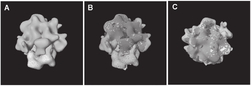
Figure 7–25. a) Averaged structure of the 70S ribosome derived from the dataset above (average from
300 individual particles to ∼4 nm resolution). The map highlights the 30S subunit in yellow. b) Docking
of high resolution structures of the 70S ribosome into the map shown in A. c) “Crown view” of B
(images courtesy of J. Ortiz, MPI of Biochemistry, Martinsried, Germany). (See color plate.)
Chapter 7 Cryoelectron Tomography (CET) 589
However, cryo-ET is conventionally not used for this purpose for
several major reasons: (1) It is much easier to acquire the data by spend-
ing the entire dose on only one projection; this makes acquisition of
large amounts of individual molecules much easier. (2) The SNR of a
projection of a particle is better than the SNR of a tomogram of the
same particle acquired with the same electron dose. (3) There is only
one level of alignment: In tomography the projections need to be
aligned to a common origin prior to aligning the individual particles
with respect to each other. Both steps inevitably introduce errors that
sum up.
But averaging of cryo-ET data can offer some advantages that are
worth the additional efforts in some cases. The most important reason
is the ability to image nonpurifi ed samples (Figure 7–26). CET is cur-
rently the only technique that can be used for quaternary structure
determination of fragile or even transient complexes. The degree of
alteration that biological macromolecules undergo under physiological
conditions is largely undetermined since there was no imaging tech-
nique that could resolve them in the context of a cell. Apart from this,
averaging of subtomograms also offers one principal advantage com-
pared to averaging from projections: It is fundamentally easier to
determine the orientations of an individual copy from 3D data than
from projection data.
4 Perspectives: New Strategies and Developments
The achievements made by cryo-ET over the past decade have proven
the possibilities and the feasibility of this technique for quasi in vivo
studies of the ultrastructure and larger supramolecular assemblies
within whole cells. However, based on the initial developments in
cryo-ET, major improvements in instrumentation and sample prepara-
tion have to be made in the future to exploit its full potential.
The ultimate goal in structural biology is to investigate the
structure–function relationship of molecular complexes and supramo-
lecular assemblies in their native environment, e.g., in large cells or
even tissue, across several dimensions, from the micrometer level
to the subnanometer level, and if possible within one single experi-
ment, to realize the complete view into the inner space of cells and its
constituents. Even at the present practical level cryoelectron tomo-
grams of organelles and cells contain an imposing amount of informa-
tion. They are, essentially, 3D images of entire proteomes, and they
should ultimately enable us to map the spatial relationships of the full
complement of macromolecules in an unperturbed cellular context.
However, it is obvious that new strategies in sample preparation,
advancements in instrumentation, and innovative image analysis tech-
niques are needed to make this dream come true, at least to some
extent.
In principle there are three major routes to increase the image
quality, and especially the 3D image quality, which are intrinsically
linked to each other: (1) the technological developments comprising
590 J.M. Plitzko and W. Baumeister
advanced electron optics, highly resolved high-sensitive detection,
improved tilting devices, and the geometry of the sample holder, (2)
the sample preparation and thus the quality of the sample, and (3)
the design of new software algorithms and acquisition schemes to
guarantee a quantitative, objective, and exhaustive analysis of the
recorded data.
TEM or EM is still a very young method if compared to light micro-
scopy. While the latter was developed and improved over centuries,
EM has a history of less than 80 years. Microscopes can now easily can
reach a resolution in 2D of less than 1 Å, clearly orders of magnitude
better than what light microscopy can offer. However, the electron
optical system, determined mainly by the objective lens system and
characterized by the spherical and chromatic aberration coeffi cients, is
far from being ideal and is still inferior to light optical devices. Simpli-
fi ed, the performance of present objective lens systems in EM can be
described as an attempt to focus through the bottom of a champagne
bottle. This way, the quality of the recorded micrograph in EM suffers
greatly from the severe shortcomings of the objective lens system, e.g.,
the higher the spherical aberration coeffi cient C
s
, the better the result-
ing image contrast, but the lower the resolution. However, the spherical
aberration coeffi cient is linked directly to the “spacing” inside the
objective lens system and thus determines the maximum achievable
tilt range for an angular acquisition. Recent developments in C
s
correc-
tion technology for TEM (and even for SEM) demonstrated the possi-
bilities and the fi nal image improvement, especially in material science
studies. However, EM investigations of biological samples at cryotem-
peratures are characterized by a very low image contrast, due to the
very weak scatterers, mainly low Z elements, such as carbon, oxygen,
and hydrogen.
Thus high defocus values are more or less mandatory, to increase
the image contrast. Unfortunately, by tuning the objective lens to very
low defocus values of a couple of micrometers, low-frequency infor-
mation is enhanced, while high-frequency information is almost
completely obscured if not totally lost. Thus C
s
correction without
any additional equipment is not an option in any cryo-EM study,
because the actual improvement in image quality is restricted to a
defocus regime in the range of a couple of tenths of nanometers, thus
orders of magnitude higher than what would be needed for a high-
contrast (low-dose) cryoimage. Nevertheless, it might be possible to
increase the contrast without defocusing the objective lens, by using
so-called phase plates in combination with C
s
correctors. The benefi t
of C
s
correction would then become clearly visible, because even high-
frequency information would be accessible. Clearly the design and the
application of phase plates are not new, but the technological prob-
lems in former times could not be addressed to enable their routine
use. Two of the major problems were contamination and stability.
While placed in the back focal plane, very close to the sample, con-
tamination caused charging and drift problems. Moreover, because
the stability was low, it was mandatory to readjust the phase plate in
respect to the central beam, which, in terms of required accuracy, is
Chapter 7 Cryoelectron Tomography (CET) 591
possible today only with piezo-controlled stages. However, present
achievements and possibilities in nanostructuring and nanotechnol-
ogy made it possible to utilize the already existing designs of phase
plates, addressing the aforementioned problems, and place them in a
normal EM environment.
The prospects are good and in the near future experiments will be
done to show not only the feasibility of this approach, but also the fi nal
image improvement if, for example, used in combination with C
s
cor-
rection. However, the total sum of elastically scattered electrons will
be reduced and thus the large fraction of inelastically scattered elec-
trons has to be removed or even utilized for fi nal image formation.
“Removal” can be done with imaging energy fi lters, which are now
routinely incorporated into the EM setup. They are especially helpful
in studies in which thick cells or sections are used, because the larger
the material layer to be penetrated by the electron beam, the larger the
fraction of inelastically and multiple-scattered electrons. They will
have a different energy, and thus they will be slightly out of focus,
blurring the recorded image. However, if the chromatic electrons are
removed, the image contrast can be defi nitely improved, but the number
of elastically scattered electrons (forming the image) will be just a frac-
tion of the total sum of electrons leaving the sample. In most cases the
amount is only a half or a third of the total applied electron dose to
the sample, which contributes to fi nal image contrast. This way, the
detection has to be very sensitive, so that literally every electron will
count. Today, typically CCD cameras are used, which detect the signal
indirectly and which add noise to the recorded image. Moreover the
signal is spread over several pixels, decreasing the lateral resolution
and thus the transfer of the recorded image. Direct detection would be
benefi cial and favorable, because every electron would contribute to
the fi nal signal, with no readout noise and the best possible lateral
resolution. Developments will utilize detectors based on CMOS tech-
nology, which will guarantee a noise-free detection and, if back-
thinned, a highly resolved recording. However, the radiation hardness
of these devices, for 300-keV electrons, is at the moment insuffi cient for
routine use in EM, but improvements are being made to increase the
lifetime of these detection devices, so that they can be used almost
continuously.
Since we cannot increase the total applied dose to the sample, not
even by cooling to liquid helium temperature, the only option we do
have is to increase the performance of the detection device and the
electron optics. If, for example, an almost ideal detector, could be
utilized, the dose in a recorded image can be as low as possible
and thus the number of acquired projections in an angular regime
can be as large as possible. This way the fi nal resolution would be
defi nitely improved. Moreover, if high-end electron optical systems
are used, e.g., C
s
or even C
c
correctors (which would even utilize the
fraction of inelastically scattered electrons for image formation)
in combination with phase plates and energy fi lters, not only the
low-frequency information, responsible for the fi nal image contrast,
but also the high-frequency information, containing the fi ne struc-
592 J.M. Plitzko and W. Baumeister
tural details, could be harvested. Moreover, with just one single 2D
image, it would be possible to obtain phase and amplitude informa-
tion separately.
In addition to the improved quality of the single 2D projection, in
phase plate C
s
-corrected imaging, ET is still hampered by an uneven
sampling in Fourier space and thus by the fact that information in the
direction of the tilt axis cannot be accessed. This missing information
(missing wedge), due to the limited tilt range, can be decreased by
utilizing dual-axis acquisition schemes to a missing pyramid. Clearly,
with the 90° in-plane rotation of the sample and the acquisition of two-
tilt series of the same sample area, it is possible to increase the amount
of information by more than 20% and thus reduce the resulting arti-
facts related to single-axis-acquisition schemes. The resolution is not
improved, however, it is defi nitely more isotropic. Especially for sub-
sequent automatic detection of macromolecules and proteins within
the tomographic volume, e.g., by template matching, errors (false-
positives) can be minimized, and thus the quality of the detection
can be vastly improved.
At the present resolution of 4–5 nm only very large complexes (ribos-
omes, 26S proteasomes) can be detected reliably within cryoelectron
tomograms of whole cells. However, an improvement in resolution to
2 nm will allow us to detect medium sized complexes in the range of
200–400 kDa. Some of the problems in the interpretation of tomograms
will disappear once a resolution of 2 nm is obtained. At the moment,
with a resolution of 4–6 nm, docking of high-resolution structures to
yield pseudo-atomic maps of molecular complexes is computationally
demanding and time consuming. However, this procedure of template
matching and docking will become easier if a resolution of 2 nm can
be routinely obtained. The computational methods described will aid
and complement the information provided by other approaches and
information, such as localization, labeling studies, and the binding
properties of molecules. While tomograms with a resolution of 2 nm
are a realistic prospect, major technical innovations (see above) will be
required to go beyond this.
CET of whole cells allows us to investigate the structure–
function relationship of molecular complexes and supramolecular
assemblies in their native environment. It thus results in a fundamental
change in the way we approach biochemical processes that underlie
and orchestrate higher cellular functions. In the past, molecular inter-
actions were studied mostly in a collective manner, whereas now we
have the tools to visualize the interactions between individual mole-
cules in their unperturbed functional environments. Although they
share common underlying principles, no two cells or organelles are
identical, owing to the inherent stochasticity of biochemical processes
in cells as well as their functional diversity. Therefore, it will be a major
challenge to extract generic features from the maps, such as the modes
of interaction between molecular species. The ultimate goal, the dis-
covery of general rules that underlie cellular processes, has to go
beyond observing qualitative features and has to be based on stringent
analytical criteria (Lucic et al., 2005).
Chapter 7 Cryoelectron Tomography (CET) 593
References
Al-Amoudi, A., Dubochet, J., Gnaegi, H., Lueth, W. and Studer, D. (2003). An
oscillating cryo-knife reduces cutting-induced deformation of vitreous ultra-
thin sections. J. Microsc. 212(1), 26–33.
Al-Amoudi, A., Studer, D. and Dubochet, J. (2005). Cutting artefacts and cutting
process in vitreous sections for cryo-electron microscopy. J. Struct. Biol.
150(1), 109–121.
Alberts, B. (1998). The cell as a collection of protein machines: Preparing the
next generation of molecular biologists. Cell 92, 291–294.
Adrian, M., Dubochet, J., Lepault, J. and McDowall, A.W. (1984). Cryo-electron
microscopy of viruses. Nature 308, 32–36.
Agard, D.A. (2005). Private communications.
and modifi ed contrast transfer theory in EFTEM. Ultramicroscopy 81(3–4),
203–222.
Baumeister, W. and Typke, D. (1993). Electron crystallography of proteins: State
of the art strategies for the furure. MSA Bull. 23, 11–19.
Beck, M., Forster, F., Ecke, M., Plitzko, J.M., Melchior, F., Gerisch, G.,
Baumeister, W. and Medalia, O. (2004). Nuclear pore complex structure
1387–1390.
Boehm, J., Frangakis, A.S., Hegerl, R., Nickell, S., Typke, D. and Baumeister, W.
(2000). Toward detecting and identifying macromolecules in a cellular
context: Template matching applied to electron tomograms. Proc. Natl. Acad.
Boehm, J., Lamber, O., Frangakis, A.S., Letellier, L., Baumeister, W. and Rigaud,
J.L. (2001). FhuA-mediated phage genome transfer into liposomes: A cryo-
electron tomography study. Curr. Biol. 11, 1168–1175.
Bottcher, B., Wynne, S.A. and Crowther, R.A. (1997). Determination of the fold
of the core protein of hepatitis B virus by electron cryomicroscopy. Nature
386, 88–91.
Bouwer, J.C., Mackey, M.R., Lawrence, A., Deerinck, T.J., Jones, Y.Z., Terada,
M., Martone, M.E., Peltier, S. and Ellisman, M.H. (2004). Automated most-
probable loss tomography of thick selectively stained biological specimens
with quantitative measurement of resolution improvement. J. Struc. Biol.
148(3), 297–306.
Bracewell, R.N. (1956). Strip integration in radio astronomy. Aust. J. Phys. 9,
198–217.
Braunfeld, M.B., Koster, A.J., Sedat, J.W. and Agard, D.A. (1994). Cryo auto-
mated electron tomography—towards high resolution reconstructions of
plastic embedded structures. J. Microsc. 172(2), 75–84.
Brenner, S. and Horne, R.W. (1959). A negative staining method for high resolu-
tion electron microscopy of viruses. Biochim. Biophys. Acta 34, 60–71.
Brink, H.A., Barfels, M.M.G., Burgner, R.P. and Edwards, B.N. (2003). A sub-50
meV spectrometer and energy fi lter for use in combination with 200 kV
monochromated (S)TEMs. Ultramicroscopy 96(3–4), 367–384.
Carazo, J.M. and Carrascosa, J.L. (1987). Restoration of direct Fourier three-
dimensional reconstructions of crystalline specimens by the method of
convex projections. J. Microsc. 145(2), 159–177.
Barnard, D.P., Turner, J.N., Frank, J. and McEwen, B.F. (1992). A 360° single-axis
tilt stage for the high-voltage electron microscope. J. Microsc. 167, 39–48.
Bernhard, W. (1965). Ultramicrotomie à basse température. Ann. Biol. 4, 5–19.
of nuclear pore complexes in action captured by cryo-electron tomography.
Angert, I., Majorovits, E. and Schroeder, R.R. (2000). Zero-loss image formation
Sci. USA 97, 14245–14250.
*Beck, M., Lucic, V., Förster, F., Baumeister, W. and Medalia, O. (2007). Snapshots
and dynamics revealed by cryoelectron tomography. Science 306(5700),
Nature 449, 611–615.
594 J.M. Plitzko and W. Baumeister
Coene, W.M.J., Thust, A., Op de Beeck, M. and Van Dyck, D. (1996). Maximum-
likelihood method for focus-variation image reconstruction in high
resolution transmission electron microscopy. Ultramicroscopy 64(1–4),
109–135.
Comolli, L.R. and Downing, K. (2005). Dose tolerance at helium and nitrogen
temperature for whole cell electron tomography. J Struct. Biol. 152,
149–156.
Conway, J.F., Trus, B.L., Booy, F.P., Newcomb, W.W., Brown, J.C. and Steven,
A.C. (1993). The effects of radiation damage on the structure of forzen
hydrated HSV-1 capsids. J. Struct. Biol. 111(3), 222–233.
Cormack, A.M. (1963). Representation of a function by its line integrals, with
some radiological applications. J. Appl. Phys. 34(1530), 2722–2727.
Cormack, A.M. (1980). Early two-dimensional reconstruction and recent topics
stemming from it. Nobel lecture 1979. Medi. Phys. 7(4), 277–282.
Crowther, R.A. and Klug, A. (1971). ART and science or conditions for three-
dimensional reconstruction from electron microscope images. J. Theor. Biol.
32, 199–203.
Crowther, R.A., DeRosier, D.J. and Klug, A. (1970). The reconstruction of a
three-dimensional structure from its projections and its applications to elec-
tron microscopy. Proc. R. Soci. London 317, 319–340.
Danev, R. and Nagayama, K. (2001). Complex observation in electron micro-
scopy. II. Direct visualization of phases and amplitudes of exit wave func-
tions. J. Phys. Soc. Jpn. 70(3), 696–702.
Deans, S.R. (1983). The Radon Transform and Some of Its Applications (John Wiley
& Sons, New York).
De Carlo, S., Adrian, M., Kalin, P., Mayer, J.M. and Dubochet, J. (1999). Unex-
pected property of trehalose as observed by cryo-electron microscopy.
J. Microsc. 196(1), 40–45.
DeRosier, D.J. and Klug, A. (1968). Reconstruction of three dimensional struc-
tures from electron micrographs. Nature 217, 130–134.
Dierksen, K., Typke, D., Hegerl, R., Koster, A.J. and Baumeister, W. (1992).
Towards automated electron tomography. Ultramicroscopy 40, 71–87.
Dierksen, K., Typke, D., Hegerl, R. and Baumeister, W. (1993). Towards auto-
matic electron tomography. II. Implementation of autofocus and low-dose
procedures. Ultramicroscopy 49(1–4), 109–120.
Dierksen, K., Typke, D., Hegerl, R., Walz, J., Sackmann, E. and Baumeister, W.
(1995). Three-dimensional structure of lipid vesicles embedded in vitreous
ice and investigated by automated electron tomography. Biophys. J. 68,
1416–1422.
Downing, K.H. and Hendrickson, F.M. (1999). Performance of a 2k CCD camera
designed for electron crystallography at 400 kV. Ultramicroscopy 75(4),
215–233.
Downing, K.H. and Mooney, P.E. (2004). CCD camera with electron decelerator
for intermediate voltage electron microscopy. Microsc. Microanal. 10(Suppl.
2), 1378–1379.
Downing, K.H., Favia, P. and Mooney, P.E. (2000). Electron decelerator for
improved CCD performance in intermediate voltage electron microscopy. Pro-
ceedings of the 58th Annual Meeting of the Microscopy Society of America, 734–735.
Dubochet, J. and McDowall, A.W. (1981). Vitrifi cation of pure water for elec-
tron microscopy. J. Microsc. 124, RP3–4.
Dubochet, J., Adrian, M., Chang, J.J., Homo, J.-C., Lepault, J., McDowall, A.W.
and Schultz, P. (1988). Cryo-electron microscopy of vitrifi ed sections. Quart.
Rev. Biophys. 21(2), 129–228.
Egerton, R.F. (1996). Electron Energy-Loss Spectroscopy in the Electron Microscope
(Plenum Press, New York).
