Hawkes P.W., Spence J.C.H. (Eds.) Science of Microscopy. V.1 and 2
Подождите немного. Документ загружается.

Chapter 7 Cryoelectron Tomography (CET) 575
tives and hybrids of these three different acquisition models, especially
modifi ed to serve specifi c purposes and applications. The full-tracking
full-focusing scheme (described above) is the most common, due to the
fact that it can be literally used universally, e.g., with almost any kind
of sample and microscope and holder system. However, the time
required for the recording of a tilt series can be very long, up to several
hours, depending on the acquisition parameters.
The precalibration model was developed originally for side-entry
systems. It mainly includes the calibration of the tilting stage synchro-
nized with the specimen holder. The basic idea is that the image move-
ment is determined prior to data collection. The movement of the stage
is measured with a calibration sample (typically fi ducial markers on a
carbon foil) both in the xy plane (image shifts) and the z direction
(defocus change) for the range of tilt angles needed to acquire a tilt
series. Typically, the calibration is started at a high tilt angle (e.g., either
−65° or +65°, depending on the characteristics of the stage) and contin-
ues to the outermost tilt on the opposite side. For every tilt angle an
image is acquired and the image feature is recentered automatically
(as described above) while the defocus change is measured (not cor-
rected) only with the beam-tilt method. In this way, the absolute image
shifts in xy and z (defocus) can be determined and stored in a calibra-
tion fi le, which then provides the basis for the xy and z shift compensa-
tion during actual data acquisition. Moreover, it is also possible to
mathematically model (least-squares fi t) the displacements during
tilting, based on the calibration curves now available, to predict the
behavior of the stage. However, there are some restraints on this course
of action: the calibration strongly depends on the initial setup of the
microscope (illumination settings, image shift settings, distance from
eucentricity, initial xy position of the stage), sample being used, and
thermal stability of the cryoholder utilized. Thermal drift of the speci-
men is not included in the calibration. For higher magnifi cations, espe-
cially, it has to be addressed separately, for example, with an additional
tracking step by cross-correlation. Additionally, a signifi cant misalign-
ment of the optic axis relative to the tilt axis can necessitate large cor-
rections in both translation and focus, which in turn can lead to image
rotation and magnifi cation changes. Since the image rotation and a
possible change in magnifi cation are not covered by the pre-calibration
procedure, the acquisition is severely hampered. However, prealigning
the optic axis to the tilt axis by invoking an appropriate amount of
image shift can circumvent this possible source of error (Ziese et al.,
2002). Based on the measured calibration curve this “optimized posi-
tion” (where the optic axis coincides with the tilt axis) can be deter-
mined and adjusted. Overall, it can be said that even in case of thermal
instabilities, which will lead to rather small displacements, not every
change in tilt angle will require a tracking step and thus the acquisition
of a tilt series by precalibration is defi nitely sped up enormously (fi ve
times faster than the full-tracking method).
The prediction-based scheme assumes that the sample follows a
simple geometric rotation and that the optical system can be character-
ized in terms of an offset between the optic and mechanical axes of
576 J.M. Plitzko and W. Baumeister
the microscope (Zheng et al., 2004). In contrast to the precalibration
and full-tracking procedures where the acquisition begins at one end
of the tilt range, the prediction scheme starts at zero tilt. In this way, it
is possible to model and compensate for any errors in sample geometry
simultaneously. The image movement in the xy and z direction due to
a stage tilt can be dynamically predicted without precalibration.
Mathematically, one data point, characterized by the tilt angle and the
x translation, is suffi cient to calculate the offset between the object and
tilt axis (z
0
). However, least-squares fi tting from multiple data points
is typically used to minimize the consequences of errors introduced
when measuring the x translation. In this way, it is possible to estimate
and refi ne “on-the-fl y” the focus offset z
0
during the course of data
collection, with an accuracy of 100 nm.
The microscope optical system (beam/image shift and focus) is auto-
matically adjusted to compensate for the predicted and refi ned image
movement prior to taking the projected image at each tilt angle. As a
consequence, it is not necessary either to record additional images for
tracking and focusing during the course of data collection or to spend
valuable setup time in a lengthy precalibration of stage motions.
However, thermal drift is not taken into account in the dynamic predic-
tion procedure, which will affect the precision of the prediction and
thus the quality of the recorded tilt series.
Nevertheless, the acquisition of tilt series of cryosamples even
with the given advances in automation is not yet performed on a high
throughput basis. This is mainly due to the fact that side-entry cryo-
holders are typically used, which are still subject to thermal instabili-
ties, and that the accuracy and reproducibility of mechanical tilting
still require compensation with elaborate automation procedures (see
above). However, recent advances in instrumentation, like the integra-
tion of the cooling system into the microscope column, the simplifi ca-
tion of the sample transfer and mounting procedure, multispecimen
holders, and the development of piezo-controlled stages will widen the
prospects of high-throughput approaches in the near future.
3.5 Alignment, Reconstruction, and Visualization
3.5.1 Alignment
To obtain the 3D image from a set of acquired projections two initial
steps have to be carried out: fi rst the individual micrographs need to
be aligned to a common coordinate system. The second step is the
actual 3D reconstruction of the tomographic volume. The compensa-
tion of the specimen movement during the data acquisition process
will keep the feature focused in most cases but the compensation for
the xy displacement is not suffi cient for a subsequent reconstruction,
which makes a second, more precise alignment necessary. Primarily,
the alignment procedure has to determine the angle of the tilt axis and
the lateral shifts. Other changes, such as magnifi cation changes or
image rotation, due to large defocus changes during the acquisition,
have to be determined and compensated for as well, if present in the
recorded tilt series.
Chapter 7 Cryoelectron Tomography (CET) 577
Alignment of the individual micrographs in cellular tomography is
normally done by high-density markers, so-called fi ducial markers.
Typically, spherical gold beads with a diameter of ∼10 nm are added to
the sample solution or directly onto the carbon foil prior to the vitrifi ca-
tion process. They can be easily recognized within a single 2D micro-
graph as “black dots,” due to their high Z number and thus their
pronounced elastic scattering. The coordinates of the markers on each
projection are determined manually or automatically. To minimize the
alignment error as a function of the lateral translations and the tilt axis
angle, an alignment model can be calculated based on least-squares
procedures (Lawrence, 1992). Their locations are then adjusted to a 3D
coordinate system. Apart from translations, this procedure for the
determination of a common origin often accounts for possible image
rotations or magnifi cation changes (Lawrence, 1992; Luther, 1988).
However, the larger the number of determined parameters, the more
gold beads need to be located to achieve a suffi cient signifi cance level
during the minimization of the residual. Therefore, another alignment
approach is by means of cross-correlating the entire image (Taylor et
al., 1997) or part of the images. Sequentially the images are compared
and compensated for image shifts throughout the series. This proce-
dure can be repeated iteratively to achieve a higher accuracy of the
alignment. However, this approach inevitably requires some approxi-
mation of the shape of the objects to interpolate data from different tilt
angles. Moreover, distinct features will dominate the outcome of the
cross-correlation, which might not be suitable for proper alignment.
Therefore it is often useful to apply image fi lters (Fourier fi lter, Sobel
fi lter, etc.) prior to the cross-correlation, to enhance features that
are expected to correlate well. The major problem in using cross-
correlation-based alignment procedures in combination with low-dose
cryoimages of vitrifi ed samples is the very low SNR of these images
and the very weak contrast, since cross-correlation methods are typi-
cally very noise sensitive. Therefore they work well only for a limited
class of specimens, e.g., high-contrast or paracrystalline objects, or in
cases where fi ducial markers are not present, or diffi cult to add, as seen
in tilt series from cryosections. The marker alignment method is defi -
nitely more general because it can even cope with very low SNRs and
is therefore more common in cryo-ET applications.
3.5.2 Reconstruction
Although the fi rst practical formulation for applied tomography was
achieved in the 1950s (Bracewell, 1956), Johan Radon fi rst outlined the
mathematical principles behind the technique in 1917 (Radon, 1917;
English translation in Deans, 1983). The paper defi nes the Radon trans-
form R as the mapping of a function f(x, y), describing a real space object
D, by the projection, or line integral, through f along all possible lines L:
Rf f x y ds
L
=
()
∫
,
where ds is the unit length of L. A discrete sampling of the Radon
transform is geometrically equivalent to the sampling of an experimen-
578 J.M. Plitzko and W. Baumeister
tal object by some form of transmitted signal: a projection. The conse-
quence of such equivalence is that the reconstruction of an object
f(x, y) from projections Rf can be achieved by implementation of the
inverse Radon transform. All conventional reconstruction algorithms
are approximations, with varying accuracy, to this inverse transform.
It is mathematically convenient to use polar coordinates (r, φ), related
to Cartesian axes for an explicit description of the Radon transform.
The Radon transform operation converts the coordinates of the experi-
mental data into “Radon space,” where l is a line perpendicular to the
projection direction and θ denotes the angle of the projection. Thus a
point in real space (r, φ) becomes a line in Radon space (l, θ) with the
relation l = r ⋅ cos (θ − φ). This relationship between real space and
Radon space allows a more explicit examination of the experimental
situation. A single projection of a one-dimensional (1D) object, for
example, a point in real space (a discrete sampling of the Radon trans-
form) is a 2D line at constant θ in Radon space. Thus a series of projec-
tions at different angles will provide an increased sampling of the
Radon space. Given a suffi cient number of (n − 1)-dimensional projec-
tions (of an n-dimensional object) from different views, an inverse
Radon transform should reconstruct the n-dimensional object. However,
in ET the experimental sampling (l, θ) is discrete (limited amount of
projections in a restricted angular range), therefore the inversion will
be imperfect and the problem of an undistorted and complete recon-
struction, especially in ET, becomes evident: to achieve the best recon-
struction of the object it is necessary to obtain as many projections as
possible in an angular regime as large as possible.
In practice the reconstruction from projections is aided by the under-
standing of the relationship of a projection in real space and Fourier
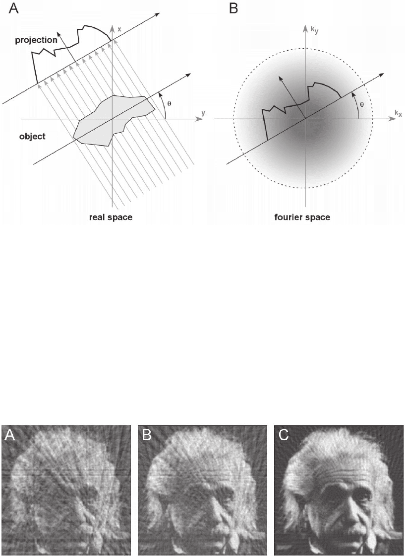
space. The “projection-slice theorem” states that a 2D projection at a
given angle is a central section through the 3D Fourier transform of
this object (Figure 7–19). If a series of projections is acquired at different
tilt angles, each projection will correspond to part of an object’s Fourier
transform, thus sampling the object over the full range of frequencies
in a central section. The shape of most objects will be described only
partially by the frequencies in one section, but by taking multiple pro-
jections at different angles many sections will be sampled in Fourier
space. This will describe the Fourier transform of an object in many
directions, allowing a fuller description of an object in real space. In
principle a suffi ciently large number of projections taken over all angles
will provide a complete description of the object. Therefore tomo-
graphic reconstruction is possible from an inverse Fourier transform
of the superposition of a set of Fourier transformed projections: an
approach known as direct Fourier reconstruction. This was the
approach formulated by Bracewell (1956) and was used for the fi rst
tomographic reconstruction from electron micrographs (DeRosier and
Klug, 1968). It is also known as “Fourier synthesis” and used for atomic
structure determination by electron (Henderson et al., 1990) and X-ray
crystallography (Perutz et al., 1960).
The direct interpretation of the projection-slice theorem would
suggest a reconstruction algorithm based in Fourier space (Hoppe and

Chapter 7 Cryoelectron Tomography (CET) 579
Hegerl, 1980). Despite the fact that the fi rst 3D reconstruction by
DeRosier and Klug (1968) was carried out in Fourier space, it is common
to use real-space-based reconstruction algorithms, because the practi-
cal implementation of Fourier-space reconstruction is not as simple as
an inverse transform. The projection data are always sampled at dis-
crete angles leaving regular gaps in Fourier space (Figure 7–20). But
the inverse transform intrinsically requires a continuous function and
therefore interpolation is required to fi ll the gaps in Fourier space
(Crowther et al., 1970). However, the quality of a Fourier-space recon-
struction is greatly affected by the type of interpolation implemented,
Figure 7–19. ‘Projection-slice theorem’. a) An object is shown in real space (x, y) at the origin, and one
of its projection images is presented fromed by tilted parallel beams. b) The Fourier transform of the
projection is a section through the origin of the Fourier space (k
x
, k
y
) tilted by θ.
Figure 7–20. Illustration of the effect of sampling at discrete angles in a 2 dimensional example. a)
For 18 projections over the full angular range at 10° increment. b) For 36 projections at 5° increment
and c) for 72 projections at 2.5° increment which is almost identical to the original image.
580 J.M. Plitzko and W. Baumeister
as examined by Smith et al. (1973). Although elegant, Fourier recon-
struction methods have the disadvantage of being computationally
intensive and rather diffi cult to implement. Therefore they have been
superseded by faster real-space alternatives since interpolations are
easier to implement there.
By far the most widely utilized algorithm is weighted backprojection
(Radermacher, 1992). Algebraic reconstruction techniques (ART) are
also well established, despite the fact that they were subject to criticism
in their beginnings (Crowther and Klug, 1971; for a detailed review of
reconstruction algorithms we refer to Frank, 1996). The theory of back-
projection relies on a simple principle: a point in space may be uniquely
described by any three “rays” passing through that point. However, as
the object increases in complexity more “rays” are required to describe
it uniquely. A projection of an object is the inverse of such a “ray,” and
will describe some of the complexity of the object at hand. Therefore by
inversing the projection, smearing out the projection into an object space
at the angle of projection, a “ray” is generated that will uniquely describe
an object in the projection direction, a process known as backprojection.
With suffi cient projections, from different angles, the superposition of
all the backprojected “rays” will reproduce the shape of the original
object—a reconstruction technique known as direct backprojection.
Direct backprojection is used for reconstruction in classical computer-
assisted tomography (CT; Herman, 1980), and it was the technique used
by Hart (1968) for the “polytropic montage.” It was also mentioned as
an alternative to Fourier methods by Crowther et al. (1970).
However, reconstructions made by direct backprojection are excep-
tionally blurred, showing distinct enhancement of low frequencies,
while fi ne spatial details are reconstructed poorly. This problem is an
effect of the uneven sampling of spatial frequencies in the series of
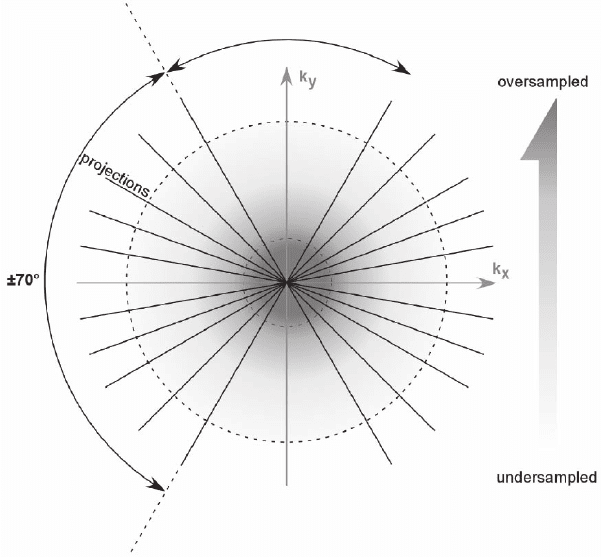
original projections (Figure 7–21). In 2D each of the acquired projec-
tions is a line intersecting the center of the Fourier space. Assuming a
regular sampling of Fourier space in each projection, far more sam-
pling points are located in the center of Fourier space than in the
periphery. The outcome of this is an “undersampling” of the high
spatial frequencies and an “oversampling” of the low spatial frequen-
cies of the object, which will subsequently result in a “blurred” recon-
struction. Therefore, to remove the blurring in real space and to restore
the correct “frequency balance” in Fourier space, it is necessary to
apply weighting schemes. The weighted backprojection consists of two
steps: First the aligned projections are weighted in Fourier space by a
function that characterizes the different sampling density in Fourier
space. Assuming an infi nitely small tilt increment the weighting has
to be proportional to the amount of the spatial frequency orthogonal
to the tilt axis (“analytical” weighting); more precise weighting (“exact”
weighting) has to consider the shape function of the object to approxi-
mate the sampling density in Fourier space, normally using a sine
function, which is retained only up to its fi rst zero crossing (Harauz
and van Heel, 1986; Hoppe and Hegerl, 1980). The second step is the
backprojection of the weighted micrographs into the reconstruction
body, most frequently by trilinear interpolation.

Chapter 7 Cryoelectron Tomography (CET) 581
ART formulates the Radon transform as a system of algebraic equa-
tions. The inversion of this algebraic system is performed iteratively
until self-consistency is achieved. The solutions of both reconstruction
methods should be similar; however, ART determines the correct
weighting parameters automatically whereas weighted backprojection
requires a priori knowledge about the specimen to provide a correct
weighting parameter for exact weighting. Despite this fact weighted
backprojection is used in most software packages for 3D reconstruction
due to its computational speed. However, the development of parallel-
ized implementations as described in Fernandez et al. (2002) might
establish ART as an alternative.
Several methods have been introduced to increase the quality of
tomographic reconstructions, i.e., improving the SNR of the data and
fi lling unsampled regions of the object’s Fourier space with consistent
data. All of these methods incorporate additional constraints to restrict
the solution of the reconstruction. Application of solvent fl attening in
single particle analysis (van Heel, 2000), projection onto convex sets
(Carazo and Carrascosa, 1987), or the Gerchberg–Fienup algorithm
(Spence et al., 2003) in electron crystallography can improve the
Figure 7–21. Illustration of the sampling ‘density’ problem in Fourier space.
The large number of sampling points at low frequencies (darker area) are in
contrast to the periphery (high frequencies), very only few sampling points
are included in the single projections (brighter area). This sampling imbalance
will result in ‘blurred’ reconstructions, which can be overcome by weighting
schemes.
582 J.M. Plitzko and W. Baumeister
resulting reconstructions considerably. Despite this fact, in most cases
collection of suffi cient data is preferred since confi ning the basis set of
the reconstruction can cause artifacts. In cryo-ET there is typically no
a priori information available; furthermore the data are not fully con-
sistent because some features occurring at high tilt angles are not
present in low tilt angles, i.e., the “unit cell is only partially defi ned”
(Hoppe and Hegerl, 1980). Therefore, refi nement techniques are com-
monly not used in cryo-ET, despite unproven claims of resolution
improvement close to the subnanometer regime (Sandin et al., 2004).
To extend the interpretability of tomograms beyond the fi rst zero of
the CTF it would be necessary to correct for the effects of the CTF as
is done in single-particle analysis. Corrections based on exit-wave
reconstruction, which rely on very few projections, have been pro-
posed to extend the achievable resolution of tomograms (Han et al.,
1996). For practical implementation of a CTF correction for tomography
the lateral focus gradient, particularly for high tilt angles, needs to be
incorporated, which make exit-front reconstructions considerably more
complicated. A simpler restoration method, which requires only one
micrograph per tilt and incorporates the lateral focus gradient, has
been realized recently (Winkler and Taylor, 2003). However, this res-
toration method is designed primarily for thin specimens. In cryo-ET,
CTF corrections have not been established yet. This is primarily due
to the fact that the SNR of the individual micrographs is still too low,
particularly because of the poor MTF of the CCD cameras, which pro-
hibits a precise determination of the CTF.
3.5.3 Visualization and Image Analysis
The interpretation of tomograms at the ultrastructural level requires
decomposition of a tomogram into its structural components, e.g., the
segmentation of intracellular membranes or the assignment of organ-
elles. Currently, a manual assignment of features is commonly used
because human anticipation is still superior in most cases to available
segmentation algorithms, although machine-based segmentation is in
principle more objective (Frangakis and Hegerl, 2002; Volkmann, 2002).
Instead of addressing the ultrastructure (Ladinsky et al., 1999), cryo-ET
provides the basis for interpreting tomograms even at the molecular
level. However, the analysis and 3D visualization are hampered by a
very low SNR. To increase the SNR, so-called denoising algorithms
have been developed (reviewed in Frangakis and Foerster, 2004). These
algorithms aim to identify noise and remove it from the tomogram, but
in practice, they also remove a certain fraction of the signal, resulting
in data with reduced information but higher SNR. The simplest denois-
ing techniques used diverse linear fi ltering operations, such as a simple
low-pass fi ltering in Fourier space. Better signal preservation can be
achieved by nonlinear fi ltering algorithms, such as nonlinear anisotro-
pic diffusion (NAD) (Frangakis and Hegerl, 2001; Fernandez and Li,
2003) or bilateral fi ltering (Jiang et al., 2003b). NAD is particularly
useful for the visualization of the ultrastructural features because it
can enhance features such as membranes. In addition, these fi lters
preserve the signal, without major alterations, which is especially suit-
Chapter 7 Cryoelectron Tomography (CET) 583
able for visualization at the molecular resolution level. Denoising based
on the wavelet transformations (Stoschek and Hegerl, 1997) maintains
the high-resolution content, but requires immense computational
efforts, thus currently favoring the former approaches.
CET enables us to resolve large macromolecular complexes such
as the 26S proteasome inside intact cells (Medalia et al., 2002). High-
resolution structures of numerous macromolecules are available from
X-ray crystallography or nuclear magnetic resonance (NMR). The
purpose of many ongoing structural genomics projects is to generate
a comprehensive library of protein structures. Therefore, molecular
recognition is the task of locating a priori known structures in the
context of cells or other complex biological samples rather than clarify-
ing structurally novel features, e.g., as intended with denoising
methods. Mapping of macromolecules on the basis of their structural
signature requires the quantitative comparison of tomogram data with
a library of macromolecular structures. Ideally, the search of tomo-
grams should be exhaustive and would reveal a cellular atlas of resolv-
able macromolecules. Simulations and experiments with “phantom
cells,” i.e., liposomes encapsulating macromolecules, indicated that
such an approach is feasible (Boehm et al., 2000; Frangakis et al., 2002).
The information addressing the spatial relationship of different com-
plexes fosters and complements other proteomic methods and will be
indispensable for structural proteomic approaches (Sali et al., 2003).
The most common molecular detection algorithm is a locally normal-
ized, matched fi lter, introduced in a different context (Roseman, 2000,
2003). It was modifi ed to account for the missing-wedge effect (Fran-
gakis et al., 2002) and applied to cryotomograms (Rath et al., 2003).
These applications demonstrated that it is feasible to identify large
macromolecular complexes (>500 kDa) within cryotomograms with
high fi delity. Furthermore, the high computational demand of template
matching is signifi cantly reduced by parallelization. The information
an individually resolved macromolecule contains is limited because of
the low electron dose and the missing-wedge effect. Averaging tech-
niques aim to overcome the dose limitation of resolution by explicit
averaging of different reconstructions from the same particle. Iterative
algorithms are used to align subtomograms of arbitrarily oriented
copies of a particle and obtain a consistent average. Medium resolution
(2–3 nm) structures of the thermosome and tricorn could be obtained
from cryotomograms of purifi ed complexes (Walz et al., 1997b; Nitsch
et al., 1998). Generally, the resolution from cryo-ET is inferior to
conventional single-particle reconstructions, which use 2D projections
of particles recorded on fi lm to obtain 3D structures. Considering
the aforementioned dose fractionation theorem (see Section 3.2), the
higher information content of tomograms should favor averaging of
tomograms over the averaging from projections. However, the low
resolution of CCD cameras compared with that of fi lm, the alignment
error of the micrographs prior to 3D reconstruction, and the current
inability to correct reliably for the CTF argue against averaging of
tomograms. Nevertheless, cryo-ET can provide medium-resolution
structures of protein complexes without using extensive purifi cation

584 J.M. Plitzko and W. Baumeister
procedures (Foerster et al., 2005). Furthermore, structures obtained by
cryo-ET can be used as starting models that are refi ned by single-
particle techniques or can be combined with other higher resolution
structural techniques to provide comprehensive descriptions of molec-
ular complexes.
3.5.4 Identifi cation Strategies of Macromolecular Complexes
A very common approach to the task of locating features of known
structure in an input scene is “matched fi ltering.” Matched fi ltering is
basically the computation of a suitable cross-correlation function (CCF)
of the template, a macromolecule of known structure, and the input
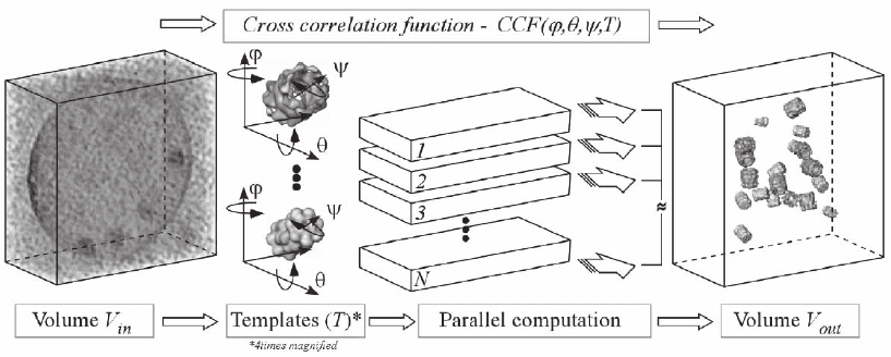
scene, the cryo-electron tomogram (Figure 7–22). Cross-correlation
functions are straightforward to compute as a function of spatial
parameters r, since they can be calculated using use the fast Fourier
transform (FFT) (F):
CCF I T F F I F T
r
rr r
r
=⋅=
()
⋅
()
[]
+
′′
−
′
∑
1
*
.
The gray values of the input scene are denoted I
r
and the template’s
gray values are T
r
. The maxima of this cross-correlation function should
Figure 7–22. Detection and identifi cation of individual macromolecules in cellular tomograms is
based on their structural signature. Because of the crowded nature of the cytoplasm and ‘contamina-
tion’ with noise, an interactive segmentation and feature extraction is not feasible. It requires sophis-
ticated pattern recognition techniques to exploit the information contained in the tomograms. A
volume rendered presentation of a 3D image is presented on the left. Even though some high-density
features may be visible, an unambiguous identifi cation of individual structures would be diffi cult if
not impossible given the residual noise. An approach, which has proven to work, is based on template
matching. Templates of the macromolecules under scrutiny are obtained by a high- or medium resolu-
tion technique (X-ray crystallography, NMR, electron crystallography or single particle analysis).
These templates (4 times magnifi ed in this fi gure; 20S proteasome and thermosome) are then used
to search the entire volume of the tomogram systematically for matching structures by three-
dimensional cross-correlation and the result is refi ned by multivariate statistical analysis. In principle
the 3D image has to be scanned for all possible Eulerian angles ϕ,ψ and θ around three different axes,
with templates of all the different protein structures one is interested in e.g. the thermosome (blue)
and proteasome (yellow). The search procedure is computationally very demanding but can be paral-
lelized with respect to the different angular combinations in a highly effi cient manner. Finally, the
position and orientation of the different complexes can be mapped directly in the 3D image. (See color
plate.)
