Hawkes P.W., Spence J.C.H. (Eds.) Science of Microscopy. V.1 and 2
Подождите немного. Документ загружается.


Chapter 7 Cryoelectron Tomography (CET) 555
work [C
p
∼ 2, if compared to liquid nitrogen studies (Fujiyoshi, 1998)].
However, the benefi t of liquid helium cooling is still a subject of discus-
sion, especially for cryo-ET, where cells embedded in thick ice layers
are typically used. Unfortunately, the cryoprotection can be determined
quantitatively only by measuring the fading of the electron diffraction
patterns using 2D protein crystals. The transfer of these fi ndings to
frozen-hydrated specimens of, for example, cellular structures, where
in most cases no crystalline arrays are present, remains diffi cult and
therefore these values can be regarded as only a rough estimate. More-
over, the experiments made at helium temperature indicate a change
in the density of the vitrifi ed ice (after an initial illumination of a couple
of e
−
/Å
2
), from a low-density amorphous phase at liquid nitrogen tem-
perature to a high-density form at liquid helium temperature, which
could be responsible for the decreased contrast observed at around 9 K
Downing, 2005; Wright, 2006).
Regarding the complexity of the biological system at hand, every
sample material will show a slightly different behavior during the
investigation at low temperatures. High amounts of sugars, for example,
either as ingredients of the buffer solution (DeCarlo et al., 1999) or the
RNA content of the macromolecules, tend to increase the acceptable
dose. Overall it can be said that electron damage at lower temperatures
induces a gradual loss of high-resolution features, much slower than
in room temperature experiments, and a reduced mass loss. However,
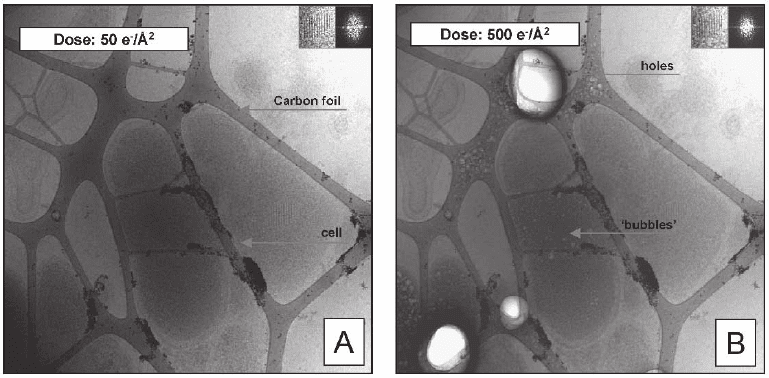
at “very high doses” (>500 e
−
/Å
2
) extensive mass loss, the formation of
bubbles, and various other dimensional changes can be seen in all
cases (Figure 7–10).
Figure 7–10. Electron dose and radiation damage shown for vitrifi ed samples. a) Cryo-electron micro-
graph of a ice embedded prokaryotic cell on lacey carbon fi lm exposed to 50 e
−
/Å
2
. b) The same cell
after an exposure to 500 e
−
/Å
2
. The adverse effect of increased electron exposure manifest at the carbon
foil and within the cellular structure in the formation of bubbles and furthermore in the smelting of
the ice (Images courtesy of S. Nickell, MPI of Biochemistry, Martinsried, Germany).
(Heide, 1984; Schweikert et al., 2007; Plitzko et al., 2004; Comolli and
556 J.M. Plitzko and W. Baumeister
A second measure among the physical methods to improve the sta-
bility of the sample in the electron beam is the use of intermediate
voltage instruments. The ionization probability is, in a fi rst-order
approximation, inversely proportional to the acceleration voltage. Thus
increasing the acceleration voltage will increase the penetration depth
and thus reduce the infl uence of multiple scattering. Unfortunately,
transmission electron microscopes with, e.g., 1250 kV acceleration
voltage are huge instruments extending over almost three stories of a
regular building. The cost for the instrument and for all subsequent
resources is immense, making it almost impossible to place it in a
normal laboratory environment. Furthermore, the higher the voltage
used the lower the recorded image contrast, which is undesirable in
the case of ice-embedded biological samples, where scattering occurs
mainly from low Z-elements. Therefore, only a few high-voltage EMs
have been built and installed around the world at selected places. It is
more practical to operate EMs in the intermediate voltage range,
between 200 and 400 kV. Instruments with 300-kV guns are the most
common and they represent a good compromise in costs and resulting
performance.
A last measure to reduce the exposure to electron radiation is
the application of so-called “low-dose” acquisition schemes (Dierksen
et al., 1992; Rath and Marko, 1997; see Section 3.4.7). Only during the
time of recording electron micrographs is the electron beam allowed
to illuminate the specimen. In all other cases the beam remains blanked.
Additionally, the microscope can be preset in different states, to reduce
the amount of time needed for changing the magnifi cation or the
adjustment of other electron optical parameters, during the process of
screening, focusing, and fi nal image acquisition. It is obvious that this
“low-dose” approach became possible only with the introduction of
computer-controlled instruments with the necessary stability and
reproducibility in the illumination system.
During the acquisition of a tilt series in electron tomography, where
typically 100–200 images are recorded, it is essential that the total
applied dose stays below a critical value, which is given by the specifi c
sample at hand. Most biological materials can tolerate an exposure of
no more than 10 e
−
/Å
2
to 100 e
−
/Å
2
, at which point major changes will
have become evident in the structure of the specimen. According to
the dose-fractionation theorem (Hegerl and Hoppe, 1976), the inte-
grated dose of a conventional 2D image is likewise suffi cient for a 3D
reconstruction, if the resolution and the statistical signifi cance between
the two are identical. It is therefore feasible to distribute the total
applied dose among as many statistically “noisy” 2D images as possi-
ble. A total applied dose of 50 e
−
/Å
2
distributed among 100 2D images
over an angular range would correspond to 0.5 e
−
/Å
2
per image. Thus
the resulting single images will be of a very noisy quality. Depending
on the detection device utilized the statistical fl uctuations from one
picture element (pixel) to another can be much greater than the inher-
ent change in image intensity from one pixel to another, i.e., the statisti-
cal fl uctuations may exceed the inherent contrast. While the structure
of interest is well preserved, it is impossible to observe the structural

Chapter 7 Cryoelectron Tomography (CET) 557
detail because of the noisy quality of the image, associated with the
very low SNR. Therefore it is very important that the quality and the
performance of the detection device are suffi cient to record images,
even at very low doses (see Section 3.4.5). While the radiation sensitiv-
ity of biological samples restricts the exposure to the electron beam,
the detection device fundamentally limits the resolution of the recorded
tomogram. Investigations on ice-embedded biological samples under
low-dose conditions can literally be described as “cloak-and-dagger
operations” and only by means of appropriate data acquisition schemes
and image analysis methods, i.e., tomographic methods, can the struc-
tural “secret” of the biological entity be disclosed.
3.3 Tilting Geometry
3.3.1 Single-Axis Tilting
The quality of a tomographic reconstruction is furthermore dependent
on the tilting geometry within an electron microscope. In contrast to
computer-assisted tomography (CT), tilting the specimen through the
complete angular regime of 180° within the EM is not possible. For the
collection of a single-axis tilt series, the specimen is typically tilted
around ±70° in very small increments between 0.5° and 3° and an
image of the same object area is recorded for every tilt angle. This
restricted tilt range is due to the limited spacing within the objective
lens polepieces, the slab geometry of the holder, and moreover the
planar geometry of the object itself. At higher tilt angles (e.g., ±90°) the
electron beam is blocked by the holder and eventually by the bars of
the copper grid, depending on the location of the structure within a
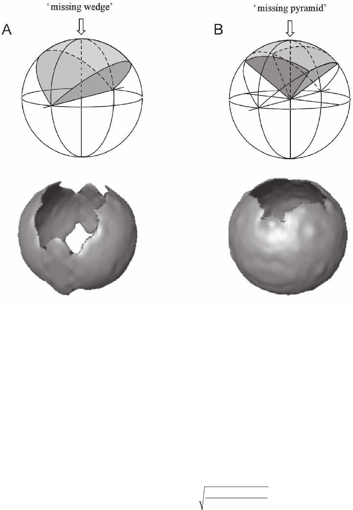
mesh of the grid. This angular gap can be nicely illustrated in recipro-
cal space (respectively, Fourier space), where a wedge-shaped “blind”
region is formed, the so-called “missing wedge” (Figure 7–11A).
However, the requirement for a distortion-free 3D reconstruction, all
projections of the sample over the complete tilt range (±90°) (Barnard
et al., 1992), is not fulfi lled, thus leading to imperfections in the recon-
structed object. There are mainly two types of distortions that can
be distinguished. First the resolution of the 3D reconstruction will be
direction dependent. The resolution parallel to the tilt axis d
x
will be
determined by the resolution of the instrument itself. Perpendicular to
the direction of the tilt axis, the resolution d
y
will be directly related to
the angular increment α
0
and thus to the number of projections N. The
relationship between resolution d
y
, increment α
0
, and the amount of
projections N for a spherical object with diameter D is given by the
Crowther theorem (Hoppe, 1969; Crowther et al., 1970), based on the
projection slice theorem (Radon, 1917):
d
D
N
y
=⋅π
The reconstruction of a 200 nm-thick cell (assuming that the cell is
spherical), with a resolution of 2 nm, requires 320 projections with an
angular increment of 0.6°. The Crowther expression assumes that the
N projections cover the full angular space of ±90°. Therefore the angular

558 J.M. Plitzko and W. Baumeister
gap has to be included to determine the maximum achievable resolu-
tion in a tomogram, which directly implicates the second type of dis-
tortion that can be observed in ET: features will become elongated in
the direction of the missing wedge. A spherical object, for example, a
gold bead, will be elongated in its 3D reconstruction. This elongation
can be described with the elongation factor e
yz
, which includes the
maximum tilt angle α (Radermacher and Hoppe, 1980):
e
yz
=
+⋅
−⋅
ααα
ααα
sin cos
sin cos
The resolution in the third direction d
z
, the “depth direction,” is there-
fore further degraded to
d
z
= d
y
⋅ e
yz
To provide the maximum amount of 3D information, as many projec-
tions as possible should be acquired over as wide a tilt range as pos-
Figure 7–11. The diagrams show schematically the sectors in the Fourier domain, which remain
unsampled owing to the limited tilt range (here ± 60°). a) In single-axis tilting there is a ‘missing
wedge’. b) In dual-axis tilting a ‘missing pyramid’. The artefacts resulting from the missing informa-
tion are shown below for an ideal thin-walled vesicle. In the single-axis case, the vesicle walls perpen-
dicular to the tilt axis and the poles of the vesicle are poorly represented in the reconstruction. In the
two-axis case, only the poles of the vesicle are affected by the missing information. In both cases, the
same threshold level has been applied.
Chapter 7 Cryoelectron Tomography (CET) 559
sible. In practice the molecular resolution is not limited by the Crowther
criterion but by the structural preservation of the specimen and the
signal recording and the noise (see above). Moreover, the size and the
shape of the recorded object are poorly defi ned, especially in cellular
tomography, where the cellular structure itself consists of objects of
various sizes. However, it remains a useful guide for the expected reso-
lution for a 3D reconstruction.
3.3.2 Dual-Axis Tilting
To reduce the missing information space in single-axis tomography, the
acquisition of a second single-axis tilt series, where the tilt axis is
rotated in-plane by 90°, can offer some remedy. Dual-axis tilting will
reduce the “missing wedge” to a “missing pyramid” and thus increase
the amount of information up to almost 20% (Figure 7–11B). Although
the maximum resolution is not necessarily increased, the achievable
resolution is defi nitely more isotropic. Moreover, structures that are
“hidden” in the single-axis case, because of their orientation relative to
the tilt axis and their location in regard to the missing wedge, will
emerge in a dual-axis acquisition scheme.
However, dual-axis tilting for cryoapplications is demanding in two
ways. First, special cryoholders have to be built, to enable the physical
90° rotation at liquid nitrogen temperatures within the EM (Nickell
et al., 2003; Gatan, Pleasanton, USA). Second, for the 3D reconstruc-
tion both tilt series have to be combined as accurately as possible,
which demands refi ned alignment routines (Penczek et al., 1995;
dual-axis tilt series requires either lowering the dose further or reduc-
ing the number of projections to preserve the structure under inves-
tigation, to comply with the requirements stated above. Dual-axis tilt
cryoholders are now commercially available, which allows either a
discrete or semicontinuous in-plane rotation by 90° (Gatan, Pleasan-
ton, CA). The continuous rotation, implemented in side-entry cryohol-
ders, simplifi es the “tracking” (see Section 3.5.7) at lower magnifi cations
of the sample during the in-plane rotation because the sample stays
inside the objective. The discrete rotation is done “outside” the objec-
tive lens system but within the vacuum of the system, making it
sometimes tedious to recenter the region of interest after the in-plane
rotation.
Despite the missing information in Fourier space (wedge or pyramid),
a more fundamental limitation in a tilting experiment within the EM
is the increasing specimen thickness at larger tilt angles. Because of
the planar (slab) geometry of the sample–holder arrangement, the
transmission path of the electrons will increase. Simple geometric cal-
culations show that the transmission path of the electrons for a speci-
men initially 100 nm thick will double at 60° (200 nm), almost triple at
70° (290 nm), and the projected thickness will be increased more than
fi ve times at 80° (590 nm). Thus at very high tilt angles the image con-
trast is dramatically reduced because of the increase in multiple scat-
tering. For specimens with an initial thickness of more than 500 nm the
projected thickness at high tilt angles would even prevent the trans-
Mastronarde, 1997; Nickell et al., 2007). It is clear that acquisition of a
560 J.M. Plitzko and W. Baumeister
mission of electrons in commonly used 300 kV instruments! However,
to preserve the contrast at even higher tilts for imaging thick speci-
mens such as vitrifi ed cells, which can easily reach dimensions of a
couple of hundred nanometers, energy fi ltering can offer some remedy
(see Section 3.4.6).
3.4 Instrumentation and Automation
Among the keys to the successful investigation of biological structures
at cryotemperatures and especially for the application of cryo-ET are
certainly the transmission electron microscope (TEM), the preparation
of the specimens (see Section 3.1), and sophisticated acquisition and
automation procedures (see Section 3.4.7). We therefore have dedicated
one section of this chapter to the equipment and the technical improve-
ments made, and to a description of the necessary technical require-
ments for 3D structural analysis with the EM.
3.4.1 Acceleration Voltage and Emission
TEMs are now operated typically in an acceleration voltage regime
between 100 and 1250 kV. With increasing voltage the penetration depth
of the electrons increases, allowing us to image samples a couple of
micrometers thick; at the same time the inelastic cross section is
reduced, thus directly resulting in a longer resistance against beam
damage (Wilson et al., 1992; Martone et al., 1999, 2000; O’Toole et al.,
1999). However, high-voltage EMs are less frequently used for biologi-
cal studies, mainly because of the decreased image contrast due to the
reduced cross section. The gain in penetration power is limited, because
the elastic σ
el
as well as the inelastic cross section σ
in
are in a good
approximation proportional to 1/β
2
(Scherzer, 1970), where β is the
ratio of electron velocity to the velocity of light (β = v/c). Thus, increas-
ing the acceleration voltage from 100 to 300 kV results in an improve-
ment of a factor of 2, while an additional increase from 300 kV to 1.2 MV
will result in only a 1.5 times increase (Koster et al., 1997). As men-
tioned earlier, it is more practical to operate EMs in the intermediate
voltage range, between 200 and 300 kV, because this represents a good
compromise in cost and resulting performance and, additionally, EMs
are available in combination with FEGs.
FEGs were implemented in the late 1980s. Compared to thermionic
emitters FEGs possess several advantages, notably a very small energy
spread, usually in the range of ∼0.8 eV, combined with an increased
brightness. Both properties lead to an improved envelope of the CTF,
since the increased temporal and spatial coherence reduces the
damping of the envelope function (Frank, 1973). This is especially
advantageous for ET of ice-embedded specimens at intermediate
resolution (∼1–2 nm) because a relatively large defocus (several micro-
meters) can be selected without sacrifi cing good contrast transfer. FEG
instruments at 200 and 300 kV are today’s “workhorses” in many dif-
ferent laboratories from life and material sciences. But LaB
6
120-kV
systems are widespread as well and are adequate whenever high
resolution is not of major interest, e.g., for sample screening and
assessment.
Chapter 7 Cryoelectron Tomography (CET) 561
3.4.2 Electron Optics
Like regular light microscopes the EM is built up from three major lens
systems: the condenser, the objective, and the projective lenses.
The performance of these electromagnetic lenses almost exclusively
determines the quality of the recorded image. Most important are the
properties of the objective lens system, likewise known from light
microscopy, especially the value of C
s
, the spherical aberration coeffi -
cient. The objective lens infl uences the transfer of electrons, and
together with the illumination system (FEG or LaB
6
) and the chosen
focus, is described by the CTF.
The opening in the center of the objective lens system, where the
electron beam passes, affects the magnitude of C
s
. In simple terms, the
bigger this gap the higher C
s
, the higher the contrast, but the lower
the resulting resolution. However, the sample holder with the speci-
men is located inside the objective lens system and therefore the gap
dimensions cannot be too small. In particular, to tilt the holder, e.g., for
an angular acquisition, a considerable amount of space is required to
allow tilts to higher angles, or the sample holder has to be designed in
a way to enable tilting even within very small gaps (Fischione Instru-
ments, Inc., Export, PA; Gatan, Inc., Pleasanton, CA). The objective lens
spacing in current EM systems is in the range of a couple of millime-
ters, thus resulting in C
s
values from 0.7 mm (lowest) up to 6 mm
(highest). For life science applications typically objectives with a C
s
of ∼2 mm are used, because they offer enough space for tilting the
specimen and at the same time the possibility of recording high-
resolution images.
Ongoing instrumental improvements include, for example, the use
of C
s
correctors, which, especially in materials science, already revealed
new possibilities and real image improvement (Haider et al., 1998;
Lentzen et al., 2002; Jia et al., 2003; Freitag et al., 2005). However, in
biological EM we have to cope with very weak scatterers and so, for
reasonable image contrast, we have to defocus in the range of a couple
of micrometers even for very thin objects. Since this low-frequency
information is essential for cryo-EM investigations, the advantage of
C
s
correction is almost canceled (Plitzko et al., 2005). However, there is
reason to believe that C
s
correctors might be benefi cial for biological
EM and especially cryo-EM if used in combination with phase plates
(Unwin, 1972; Danev and Nagayama, 2001; Majorovits and Schroeder,
2002; Lentzen, 2004; Marko, 2004).
Another important fact is the change in the direction and angle of
the tilting axis at different magnifi cations due to a rotation of the image
within the electron optics of the microscope. Previously this change
was obvious at every magnifi cation step. It is now partly compensated
by so-called “rotation-free lens series,” which guarantee an almost
“fi xed” (in angle and direction) tilt axis in distinct magnifi cation ranges.
This is crucial, especially in low-dose acquisition schemes, where a fast
transition and reproducible change between states (see Section 3.4.7)
and sample areas at different magnifi cations are necessary, e.g., in
dual-axis experiments to reposition the area of interest after the in-
plane rotation.

562 J.M. Plitzko and W. Baumeister
3.4.3 Tilting Device: Goniometer Tilt Stage
As already mentioned, tilting the holder inside the microscope is a
fundamental requirement for ET. The tilting device, normally located
outside the EM column, is called the goniometer tilt stage and in some
instances the “compustage,” because of its power to be controlled by
computers. However, tilting is now done in an exclusively mechanical
way, thus resulting in a major practical diffi culty: the mechanical
imperfections and the inability to set the eucentric height of the speci-
men precisely. The eucentric plane is normal to the optic axis; this way
a point on the optic axis should not move laterally when tilted around
the holder axis. As a result, specimens can experience signifi cant shifts
in the x, y, and z directions during the course of angular acquisition.
Shifts in the z direction cause severe focus changes, and in an uncor-
rected case, complicate the 3D reconstruction or even render a recon-
struction meaningless.
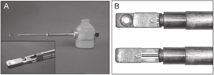
Typically, side entry holders are used—at the rear part of which a
small Dewar for liquid nitrogen is located (Figure 7–12). The Dewar is
connected to a temperature conduction rod, transferring the tempera-
ture of ∼180°C to the specimen mounted at the outermost end of the
holder tip. The cryoholder, almost 30 cm in length, is tilted by the goni-
ometer around its vertical axis. It is easy to imagine that tilting must
be very inaccurate. Especially at high tilt angles above ±45° imprecise
tilting will add up to displacements in the micrometer range. However,
at a resolution of 10 Å a tolerable deviation would be in the range of 3 Å,
thus resulting in a discrepancy of three to four orders of magnitude
between the desired resolution and the actual precision of the tilting
device. The best current stages can tilt and move the sample within an
accuracy of 0.5 µm. To partially compensate for the still large displace-
ments, automation is mandatory during the acquisition of a tilt series
composed typically out of 100 micrographs. Therefore sophisticated
acquisition and correction schemes have been developed, to keep the
feature of interest at all times focused and centered (see Section 3.4.7).
Figure 7–12. Photographs of a standard side entry cryo-holder (Model 626 Gatan, Pleasanton, CA,
USA). a) The holder is almost 30 cm in length and equipped with a nitrogen dewar in the back. b)
Images of the tip of the holder with open (top) and closed ‘shutter’ (bottom). The stationary cryoshield
(‘shutter’) protects the grid from mechanical damage and from frost accumulation during transfer
from the specimen loading workstation (not shown) into the TEM.
Chapter 7 Cryoelectron Tomography (CET) 563
Developments have already been made to test and implement piezo-
controlled stages into the EM environment to allow nanopositioning
in the order of ∼1 nm, which might enable accuracy during tomo-
graphic acquisition to be improved dramatically (Lengweiler et al.,
2004; JEOL Ltd., Tokyo, Japan).
3.4.4 Cooling System
Since all investigations in cryo-EM have to be made under cryo condi-
tions, i.e., under constant cooling with liquid nitrogen, special holders
and/or special cryostages have to be designed. Typically, side entry
holders are used—fi tted with a small Dewar for liquid nitrogen (Gatan
Inc., Pleasanton, CA). The requirements for mechanical and tempera-
ture stability are severe and it is quite clear that the dimensions of the
holder tip must be smaller than the polepiece gap of the objective.
Moreover, special cryoshields have to be installed to guarantee
contamination-free investigations. They are seated next to the tip of
the holder inside the microscope and connected to the cooling trap of
the microscope for constant cooling with liquid nitrogen.
Side-entry cryoholders are routinely used, but recently stages incor-
porating the cooling system within the electron microscope have been
designed (“Polara” Tecnai F30 by FEI Company, Eindhoven, Nether-
lands, and Jeol 3200FSC by JEOL Ltd., Tokyo, Japan). By including the
cooling system for the sample holder into the microscope the mechani-
cal stability and the temperature stability of the holder are improved
enormously, and additionally these systems allow cooling to very low
temperatures, even to liquid helium temperature (∼10 K). Furthermore
the temperature can be kept stable over longer time periods; instead
of hours in the side-entry case, days or even weeks are now possible.
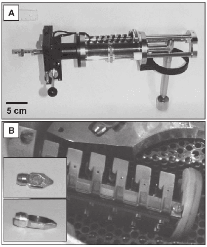
Moreover the stages designed for these cooling systems can hold
several samples, unlike the side-entry holder where only one individ-
ual sample can be mounted at a time (Figure 7–13). Additionally, pro-
totypes have been developed and tested holding up to 100 different
samples, to enable high-throughput applications in the future (Gatan
Inc., Pleasanton, CA). The exchange of samples is faster and the transfer
of the samples to the microscope is done under vacuum conditions, to
minimize the chance of contamination.
3.4.5 Detector
Temperature and mechanical stability of the stages, holders, and goni-
ometers as well as the overall performance of the electron optics are
crucial for high-resolution data acquisition, however, the quality of the
detector system used is of vital importance, especially in cryoapplica-
tions under low-dose conditions (Downing and Hendrickson, 1999;
Fan and Ellisman, 2000). Previously photographic plates were utilized,
making a fast assessment of the collected data impossible; however,
today microscopes are equipped with TV rate systems and digital
cameras (Krivanek and Mooney, 1993; Faruqi and Subramanian, 2000),
so-called CCD cameras (Janesick, 2001). With the help of the CCD
camera it is possible to easily calculate online the Fourier transforma-
tion of the image, for example, to determine the focus or to readjust the
microscope, e.g., astigmatism and coma correction. In principle they

564 J.M. Plitzko and W. Baumeister
are built up from a scintillator (either a single crystal or a polycrystal-
line coating), “decelerating” the electrons and thus creating photons
by cathodoluminescence. The scintillator is coupled to a fi ber optic
array, which will transfer the light optical signal to the CCD sensor,
which is built up from a series of metal oxide semiconductor (MOS)
capacitors, where the light induces the creation of electron hole pairs
in the active area.
CCD cameras are now available in dimensions between 1024 × 1024
and 4096 × 4096 pixels with a pixel size ranging from 15 to 30 µm. Thus
they are still smaller than the typical photographic plate dimensions
(∼10,000 × 8000 pixels assuming a pixel size of 10 µm for a typical plate).
However, CCDs possess an excellent linearity and a large dynamic
range [i.e., ratio between maximum signal and the root mean square
(RMS) of the noise level]. Because of the excellent linearity the intensity
distribution of the recorded image is directly proportional to the
number of primary electrons hitting the detection device. However, the
lateral resolution is worse than in photographic plates, because of the
signal spreading within the whole CCD assembly. This spreading is
mainly caused by multiple light scattering of the photons created and
by electron backscattering within the scintillator. Thus a point-like
input signal produces an output that is spread over several pixels of
the CCD chip. In real space this will result in a “blurring” of the
recorded image, which is similar to a Gaussian smoothing of the image.
In Fourier space it can be observed as a damping of the high spatial
frequencies. The extent of this spread can be quantitatively analyzed
by inspection of the modulation transfer function (MTF), which is the
Fourier transform of the point spread function (PSF), to measure the
spatial frequency response (Weickenmeier et al., 1995; Ruijter, 1995).
Figure 7–13. Multispec-
imen chamber and car-
tridges for the Polara
Te c n a i F3 0 (F EI c om p a ny,
lands). a) The multi-
allows the storage of up
to 6 samples, which are
samples are clamped or
screwed into cartridges
(insets) and the transfer
is done under cryo- and
vacuum conditions.
Eindhoven, The Nether-
specimen chamber
nary cryoshields. b) The
of the complete system
all protected by statio-
